Review
. 2008:2008:723539.
doi: 10.1155/2008/723539.

Antigen-induced immunomodulation in the pathogenesis of atherosclerosis

Affiliations
Review

Antigen-induced immunomodulation in the pathogenesis of atherosclerosis

Natalia Milioti et al. Clin Dev Immunol. 2008.

Abstract

Atherosclerosis is a chronic inflammatory disorder characterised by the accumulation of monocytes/macrophages, smooth muscle cells, and lymphocytes within the arterial wall in response to the release of proinflammatory molecules. Such accumulation results in the formation of the atherosclerotic plaque, which would eventually evolve to complications such as total artery occlusion, rupture, calcification, or aneurysm. Although the molecular mechanism responsible for the development of atherosclerosis is not completely understood, it is clear that the immune system plays a key role in the development of the atherosclerotic plaque and in its complications. There are multiple antigenic stimuli that have been associated with the pathogenesis of atherosclerosis. Most of these stimuli come from modified self-molecules such as oxidised low-density lipoproteins (oxLDLs), beta2glycoprotein1 (beta2GP1), lipoprotein a (LP(a)), heat shock proteins (HSPs), and protein components of the extracellular matrix such as collagen and fibrinogen in the form of advanced glycation-end (AGE) products. In addition, several foreign antigens including bacteria such as Porphyromonas gingivalis and Chlamydia pneumoniae and viruses such as enterovirus and cytomegalovirus have been associated with atherosclerosis as potentially causative or bystander participants, adding another level of complexity to the analysis of the pathophysiology of atherosclerosis. The present review summarises the most important scientific findings published within the last two decades on the importance of antigens, antigen stimulation, and adaptive immune responses in the development of atherosclerotic plaques.


Figures

Figure 1
Figure 1
Inflammatory basis of atherosclerotic plaque formation. Led by inflammatory signals derived from the damaged endothelium, monocytes and lymphocytes migrate into the vessel wall. Monocytes differentiate into macrophages that recognise and phagocytiose oxidised LDL particles. The protein component of the LDL particle is processed and presented in the form of peptides by macrophages (also by dendritic cells) to T-lymphocytes in the context of the major histocompatibility complex class II (MHC-II). Other self or foreign antigens that may gain access to the vascular wall can also trigger similar mechanisms. It is believed that most of these lymphocytes differentiate in situ, under the influence of the specific antigen stimulation, into effector T-cells, but this has yet to be demonstrated. Upon activation, both macrophages and lymphocytes release a range of proinflammatory molecules including chemokines which stimulate the migration of smooth muscle cells (SMCs) from the media. SMCs contribute to foam cell and fibrous cap formation. This process is facilitated by cytokines such as IFNγ and TNFα secreted by proatherogenic Th1 cells and also IL-12 secreted by macrophages and foam cells. Eventually foam cells die by apoptosis in situ leaving nondegradable cholesterol crystals that form the lipid core of the plaque.
Figure 2
Figure 2
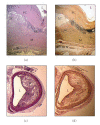
Morphological features of advanced atherosclerotic plaques. (a) and (b) show sections from a human carotid artery; (c) and (d) are sections from an apoE deficient mouse brachiocephalic artery. Sections (a) and (c) have been stained with haematoxylin and eosin and sections (b) and (d) with van Gieson staining (used to demonstrate the increase of collagen deposition and development of elastic fibres, a characteristic feature of the atherogenic process. A positive staining is depicted by a brown colour). L: lumen of the vessel; SR: shoulder region (it is believed to contain large numbers of proinflammatory cells including macrophages and lymphocytes, and it is the site related with the onset of the development of the atherosclerotic plaque); FC: fibrous cap (It also contains large numbers of mononuclear infiltrate and smooth muscle cells that have migrated from the media layer (M) and proliferated in response to the local inflammatory stimuli. It is also characterised by high collagen deposition and little or no endothelial cells); LC: lipid core (it contains mainly macrophage and smooth muscle cell-derived foam cells, apoptotic cells, and cholesterol crystals. Older lesions may also display signs of calcification). Contrasting differences can be recognised in the anatomic development of atherosclerotic plaques between human and mouse including the hypertrophy associated with the proliferation of the smooth muscle cells in the media layer and the fibrous cap. In humans, some lesions may also contain signs of intraplaque haemorrhage. Signs of plaque rupture are usually best recognised in mouse (reviewed in [13, 14]).
Figure 3
Figure 3
Schematic representation of the low-density lipoprotein particle (LDL). The LDL particle has a size of approximately 21–24 nm and is the main transporter of unesterified cholesterol, cholesterol esters, and triglycerides in the blood. It contains an outer layer composed of phospholipids and unesterified cholesterol in which a single protein is embedded, the apolipoprotein B-100 (apoB-100). These components are more susceptible to oxidation by free radicals in the subendothelial space during inflammation. They are also targets for the recognition of the LDL by scavenger receptors, proteoglycans, and low-density lipoprotein receptor (LDLr). The core of the particle contains primarily cholesterol esters and triglycerides. In atherogenesis, a large number of IgM antibodies are created in response to oxidative stress-modified phospholipids, whereas IgG antibodies and T-cell clones are generated against apoB-100.

Similar articles

Cited by

References

    1. Shashkin P, Dragulev B, Ley K. Macrophage differentiation to foam cells. Current Pharmaceutical Design. 2005;11(23):3061–3072. - PubMed
    1. Shi W, Wang X, Shih DM, Laubach VE, Navab M, Lusis AJ. Paradoxical reduction of fatty streak formation in mice lacking endothelial nitric oxide synthase. Circulation. 2002;105(17):2078–2082. - PubMed
    1. Pan JH, Sukhova GK, Yang JT, et al. Macrophage migration inhibitory factor deficiency impairs atherosclerosis in low-density lipoprotein receptor-deficient mice. Circulation. 2004;109(25):3149–3153. - PubMed
    1. Lucas AD, Greaves DR. Atherosclerosis: role of chemokines and macrophages. Expert Reviews in Molecular Medicine. 2001;3(25):1–18. - PubMed
    1. Fan J, Watanabe T. Inflammatory reactions in the pathogenesis of atherosclerosis. Journal of Atherosclerosis and Thrombosis. 2003;10(2):63–71. - PubMed