. 2008 Jul;118(7):2651-60.
doi: 10.1172/JCI34753.

Therapeutic suppression of translation initiation modulates chemosensitivity in a mouse lymphoma model

Affiliations

Therapeutic suppression of translation initiation modulates chemosensitivity in a mouse lymphoma model

Marie-Eve Bordeleau et al. J Clin Invest. 2008 Jul.

Abstract

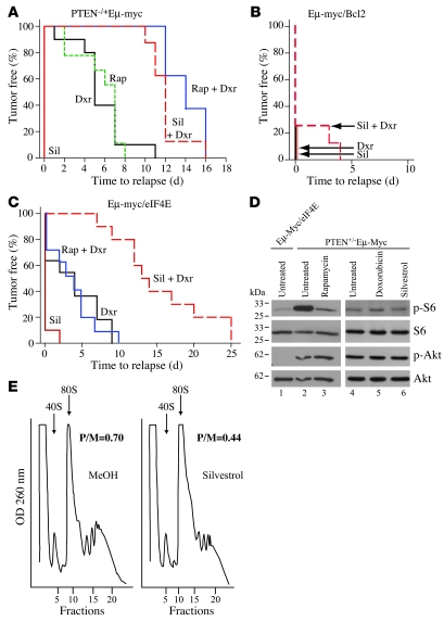
Disablement of cell death programs in cancer cells contributes to drug resistance and in some cases has been associated with altered translational control. As eukaryotic translation initiation factor 4E (eIF4E) cooperates with c-Myc during lymphomagenesis, induces drug resistance, and is a genetic modifier of the rapamycin response, we have investigated the effect of dysregulation of the ribosome recruitment phase of translation initiation on tumor progression and chemosensitivity. eIF4E is a subunit of eIF4F, a complex that stimulates ribosome recruitment during translation initiation by delivering the DEAD-box RNA helicase eIF4A to the 5' end of mRNAs. eIF4A is thought to prepare a ribosome landing pad on mRNA templates for incoming 40S ribosomes (and associated factors). Using small molecule screening, we found that cyclopenta[b]benzofuran flavaglines, a class of natural products, modulate eIF4A activity and inhibit translation initiation. One member of this class of compounds, silvestrol, was able to enhance chemosensitivity in a mouse lymphoma model in which carcinogenesis is driven by phosphatase and tensin homolog (PTEN) inactivation or elevated eIF4E levels. These results establish that targeting translation initiation can restore drug sensitivity in vivo and provide an approach to modulating chemosensitivity.


Figures

Figure 1
Figure 1. CBFs inhibit cap-dependent translation.
(A) Chemical structure of FA and silvestrol. (B) FA and silvestrol inhibit cap-dependent translation. Top: Schematic representation of the FF/HCV/Ren mRNA. Bottom: Dose-dependent inhibition of cap-dependent protein synthesis by FA and silvestrol in Krebs-2 translation extracts programmed with FF/HCV/Ren mRNA (10 μg/ml). Luciferase activity (RLU) results are expressed relative to values obtained in the presence of vehicle (MeOH) control. Results are expressed as mean ± SEM of 2 experiments. (C) A representative autoradiograph from in vitro translations performed in Krebs-2 extracts with [35S]methionine and programmed with FF/HCV/Ren mRNA. The position of migration of FF and Ren proteins is indicated on the right.
Figure 2
Figure 2. CBFs inhibit translation initiation and stimulate eIF4A RNA-binding activity.
(A) CBFs prevent 80S complex formation. [32P]-labeled CAT mRNA (200,000 cpm) was incubated with cycloheximide (CHX) in the presence of FA or vehicle (MeOH) in rabbit reticulocyte lysates. Reactions were resolved by centrifugation through glycerol gradients. Following centrifugation, fractions were collected and the amount of radioactivity determined by liquid scintillation counting. The total counts recovered from each gradient and the percentages of mRNA bound in 80S complexes were: CAT mRNA/CHX + MeOH: 35,178 cpm, 19% binding; and CAT mRNA/CHX + FA: 37,655 cpm, 2% binding. (B) CBFs stimulate cross-linking of eIF4A to mRNA. Initiation factor preparations were cross-linked to [32P]-cap-labeled mRNA in the absence (lane 2) or presence of ATP (lanes 1 and 3–6), 0.6 mM m7GDP (lane 3), 0.6 mM GDP (lane 4), MeOH (lane 5), or 50 μM FA (lane 6). Following nuclease digestion, samples were resolved by SDS-PAGE, and the gel was subjected to autoradiography. The position of migration of eIF4E, eIF4A, and eIF4B is indicated and is based on their known behavior in this assay (30). (C) CBFs stimulate the RNA-binding activity of eIF4AIf. [32P]-cap-labeled mRNA was cross-linked to recombinant eIF4AIf in the presence of MeOH (lane 1), 50 μM FA (lane 3), or 50 μM silvestrol (lane 2). Following nuclease digestion, samples were resolved by SDS-PAGE, and the gel was subjected to autoradiography. (D) CBFs stimulate the RNA-binding activity of eIF4Ac. [32P]-cap-labeled mRNA was cross-linked to eIF4F in the presence of MeOH (lane 1) or 50 μM FA (lane 2). Following nuclease digestion, samples were resolved by SDS-PAGE, and the gel was subjected to autoradiography. Cross-linking of a higher-molecular-mass species (>175 kDa) is indicated by “eIF4G ?” since it may reflect RNA-bound eIF4G.
Figure 3
Figure 3. Effects of CBFs on protein, RNA, and DNA synthesis in vivo.
(A) Dose-dependent inhibition of protein synthesis in vivo by CBFs. HeLa cells were incubated with the indicated concentrations of FA and silvestrol for 1 hour, with [35S]methionine added 10 minutes before the end of the incubation. The rate of [35S]methionine (35S]-Met) incorporation is expressed relative to that of cells treated with vehicle (MeOH). Results are expressed as mean ± SEM of 2 experiments. (B) CBFs primarily impact protein synthesis in vivo. HeLa cells were incubated with 5 μM FA, 0.4 μM silvestrol, or vehicle (MeOH) for 1 hour. The rate of incorporation of each radioisotope tracer into TCA-insoluble material is expressed relative to that in MeOH-treated cells. Results are expressed as mean ± SEM of 2 experiments. (C) Inhibition of translation by CBFs is reversible. HeLa cells were incubated for 1 hour with 10 μM anisomycin, 5 μM FA, 0.4 μM silvestrol, or MeOH. Cells were then washed with PBS and incubated with medium lacking compound for the indicated times. Ten minutes before harvesting, [35S]methionine was added to the culture. The rate of [35S]methionine incorporation into TCA-insoluble material is expressed relative to that in MeOH-treated cells. Results are expressed as mean ± SEM of 3 experiments. Anisomycin acts a positive control, since recovery of protein synthesis from inhibition with this compound occurs within an hour of its removal from cells (65). (D) CBFs inhibit cap-dependent translation in vivo. Top: Schematic representation of pcDNA/Ren/HCV/FF expression vector. Bottom: Effect of FA on cap-dependent and HCV IRES–mediated translation in 293 cells transfected with pcDNA/Ren/HCV/FF. Luciferase activity is expressed relative to that in MeOH-treated cells and is the mean ± SEM of 2 experiments. (E) Silvestrol does not induce eIF2α phosphorylation. HeLa cells were incubated for 2 hours in the presence of vehicle (DMSO), thapsigargin (2 μg/ml), or silvestrol (400 nM), after which extracts were analyzed by Western blotting. (F) Silvestrol induces apoptosis at concentrations higher than those required to inhibit protein synthesis. Jurkat cells were incubated with the indicated silvestrol concentrations for 13 hours, after which the rate of [35S]methionine incorporation or the percentage of living cells was measured. Results are expressed as mean ± SEM of 2 experiments.
Figure 4
Figure 4. Silvestrol induces eIF4A association into RNAse-sensitive heavy sedimenting complexes in vivo.
(A) Effect of silvestrol on Jurkat cell polyribosomes. Jurkat cells were exposed to vehicle (MeOH) or 0.2 μM silvestrol for 60 minutes. Cell extracts were prepared and treated with micrococcal nuclease (MN) where indicated. Reactions were resolved on 10%–50% sucrose gradients by centrifugation in an SW40 rotor at 150,000 g for 2 hours. Fractions were collected from the gradients and monitored with an ISCO UA-6 UV detector. (B) Western blots demonstrating the position of migration of eIF4A, eIF4E, and eIF4G1 in sucrose fractions collected from untreated or MN-treated lysates prepared from cells exposed to MeOH or 0.2 μM silvestrol.
Figure 5
Figure 5. Silvestrol alters chemosensitivity in Pten+/–Eμ-Myc and Eμ-Myc/eIF4E tumors in vivo.
(A) Silvestrol sensitizes Pten+/–Eμ-Myc tumors to the effects of doxorubicin in vivo. Kaplan-Meier plot showing tumor-free survival of mice bearing Pten+/–Eμ-Myc tumors following treatment with doxorubicin (Dxr, solid black line; n = 10), rapamycin (Rap, dashed green line; n = 9), rapamycin and doxorubicin (Rap + Dxr; solid blue line; n = 8), silvestrol (Sil, solid red line; n = 10), or silvestrol and doxorubicin (Sil + Dxr, dashed red line; n = 8). (B) Silvestrol does not alter drug response in Eμ-Myc/Bcl-2 tumors in vivo. Kaplan-Meier plot showing tumor-free survival of mice bearing Eμ-Myc/Bcl-2 tumors following treatment with Dxr (n = 9), Sil (n = 7), or Sil + Dxr (n = 8). (C) Silvestrol sensitizes Eμ-Myc/eIF4E tumors to the effects of Dxr in vivo. Kaplan-Meier plot showing tumor-free survival of mice bearing Eμ-Myc/eIF4E tumors following treatment with Rap + Dxr (n = 10), Dxr (n = 11), Sil (n = 10), or Sil + Dxr (n = 10). (D) Western blot analysis of Eμ-myc/eIF4E (lane 1) and Pten+/–Eμ-Myc lymphomas (lanes 2–6). Lysates prepared from Eμ-myc/eIF4E or Pten+/–Eμ-Myc lymphomas from untreated (lanes 1, 2, and 4) and rapamycin- (lane 3), doxorubicin- (lane 5), and silvestrol-treated (lane 6) animals were subjected to immunoblotting for analysis of phosphorylated and total ribosomal S6 protein (p-S6 and S6) and Akt (p-Akt and Akt). (E) Silvestrol inhibits translation in Pten+/–Eμ-Myc tumors in vivo. Mice bearing Pten+/–Eμ-Myc tumors were injected with MeOH or silvestrol (0.2 mg/kg). Cytoplasmic extracts were prepared from tumors 4 hours later and resolved on 10%–50% sucrose gradients by centrifugation in an SW40 rotor at 150,000 g for 2 hours. Fractions were collected and monitored using an ISCO UA-6 UV detector. Plotted are results of 1 representative experiment of 3 that showed similar results. The positions in the gradients of 40S and 80S ribosomes are labeled, and the polysome/monosome (P/M) ratios are indicated.
Figure 6
Figure 6. Silvestrol synergizes with doxorubicin to induce apoptosis in Pten+/–Eμ-Myc lymphoma cells in vivo.
(A) Representative micrographs (original magnification, ×200) of Pten+/–Eμ-Myc lymphoma sections stained with H&E and TUNEL. C57BL/6 mice bearing well-palpable tumors were injected with vehicle, silvestrol, or rapamycin. Twenty-four hours later, the mice were injected again with silvestrol or rapamycin alone or in combination with doxorubicin. Three and 6 hours after treatment, tumors were extracted and stained. (B) Amount of tumor cells positive for TUNEL staining per 1,000 cells following the treatments described in A. The cell count was obtained from 4 different fields taken from 2 different sections. Results are expressed as mean ± SD. In vehicle-treated cells, there were 36 ± 1 TUNEL-positive cells/1,000 cells. (C) Western blot analysis of Pten+/–Eμ-Myc lymphomas treated as described in A. Tumor cells were extracted and lysed and the amount of cleaved poly(ADP-ribose) polymerase (c-PARP) and tubulin determined by Western blotting.

Similar articles

Cited by

References

    1. Raught, B., Gingras, A.-C., and Sonenberg, N. 2000.Regulation of ribosome recruitment in eukaryotes. Cold Spring Harbor Laboratory Press. Cold Spring Harbor, New York, USA. 245–293.
    1. Pestova T.V., et al. Molecular mechanisms of translation initiation in eukaryotes. Proc. Natl. Acad. Sci. U. S. A. 2001;98:7029–7036. doi: 10.1073/pnas.111145798. - DOI - PMC - PubMed
    1. Dorrello N.V., et al. S6K1- and betaTRCP-mediated degradation of PDCD4 promotes protein translation and cell growth. Science. 2006;314:467–471. doi: 10.1126/science.1130276. - DOI - PubMed
    1. Yang H.S., et al. The transformation suppressor Pdcd4 is a novel eukaryotic translation initiation factor 4A binding protein that inhibits translation. Mol. Cell. Biol. 2003;23:26–37. doi: 10.1128/MCB.23.1.26-37.2003. - DOI - PMC - PubMed
    1. Avdulov S., et al. Activation of translation complex eIF4F is essential for the genesis and maintenance of the malignant phenotype in human mammary epithelial cells. Cancer Cell. 2004;5:553–563. doi: 10.1016/j.ccr.2004.05.024. - DOI - PubMed

Publication types

MeSH terms