. 2008 Jul;118(7):2506-15.
doi: 10.1172/JCI35243.

Phosphorylation of GSK-3beta by cGMP-dependent protein kinase II promotes hypertrophic differentiation of murine chondrocytes

Affiliations

Phosphorylation of GSK-3beta by cGMP-dependent protein kinase II promotes hypertrophic differentiation of murine chondrocytes

Yosuke Kawasaki et al. J Clin Invest. 2008 Jul.

Erratum in

  • J Clin Invest. 2008 Aug;118(8):2986

Abstract

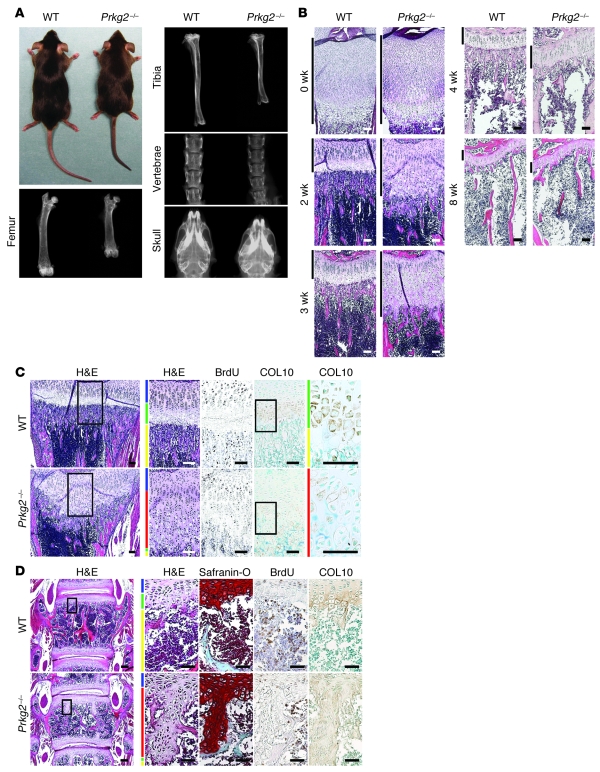
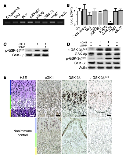
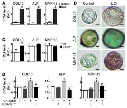
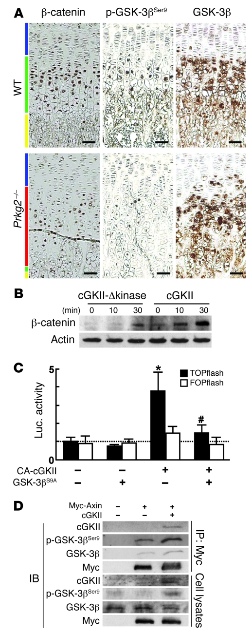
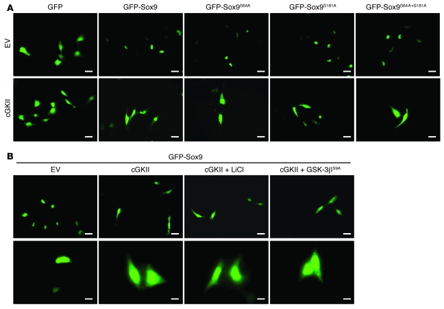
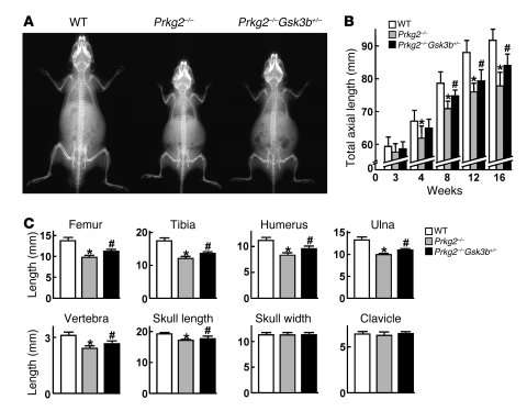
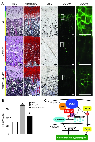
cGMP-dependent protein kinase II (cGKII; encoded by PRKG2) is a serine/threonine kinase that is critical for skeletal growth in mammals; in mice, cGKII deficiency results in dwarfism. Using radiographic analysis, we determined that this growth defect was a consequence of an elongated growth plate and impaired chondrocyte hypertrophy. To investigate the mechanism of cGKII-mediated chondrocyte hypertrophy, we performed a kinase substrate array and identified glycogen synthase kinase-3beta (GSK-3beta; encoded by Gsk3b) as a principal phosphorylation target of cGKII. In cultured mouse chondrocytes, phosphorylation-mediated inhibition of GSK-3beta was associated with enhanced hypertrophic differentiation. Furthermore, cGKII induction of chondrocyte hypertrophy was suppressed by cotransfection with a phosphorylation-deficient mutant of GSK-3beta. Analyses of mice with compound deficiencies in both protein kinases (Prkg2(-/-)Gsk3b(+/-)) demonstrated that the growth retardation and elongated growth plate associated with cGKII deficiency were partially rescued by haploinsufficiency of Gsk3b. We found that beta-catenin levels decreased in Prkg2(-/-) mice, while overexpression of cGKII increased the accumulation and transactivation function of beta-catenin in mouse chondroprogenitor ATDC5 cells. This effect was blocked by coexpression of phosphorylation-deficient GSK-3beta. These data indicate that hypertrophic differentiation of growth plate chondrocytes during skeletal growth is promoted by phosphorylation and inactivation of GSK-3beta by cGKII.


Figures

Figure 1
Figure 1. Skeletal abnormality in Prkg2–/– mice.
(A) Gross appearance and radiographs of femurs, tibias, lumbar vertebrae, and skulls of WT and Prkg2–/– littermates at 8 weeks of age. (B) Time course of H&E staining of the growth plates in proximal tibias of the 2 genotypes from 0–8 weeks of age. Vertical black bars indicate the height of the growth plates. (C) H&E staining, BrdU labeling, and in situ hybridization of COL10 of the tibial growth plates in 2-week-old littermates. (D) H&E and Safranin-O stainings, BrdU labeling, and COL10 immunostaining of the growth plates in the fourth lumbar vertebra of 2-week-old littermates. (C and D) Blue, red, green, and yellow bars indicate proliferative zone, abnormal intermediate layer, hypertrophic zone, and primary spongiosa, respectively. Boxed regions in H&E and COL10 panels are shown at higher magnification to the right. Scale bars: 50 μm.
Figure 2
Figure 2. Identification of GSK-3β as a principal phosphorylation target of cGKII during chondrocyte hypertrophy.
(A) RT-PCR of 8 candidate genes that were identified by the serine/threonine kinase substrate array (Supplemental Table 1) in cultured ATDC5 cells in the prehypertrophic or hypertrophic differentiation stage. (B) COL10 promoter activity, as assessed by transfection of the 8 candidate genes or the empty vector (EV) in HuH-7 cells with the luciferase reporter gene construct containing a cloned 4.5-kb promoter fragment of COL10. Data are mean ± SD fold change relative to empty vector. *P < 0.01 versus control. (C) In vitro kinase assay of the phosphorylation of recombinant GSK-3β at Ser9 by recombinant cGKII with or without cGMP. Proteins were incubated in the presence of ATP, and the reaction products were analyzed by IB using the same antibody to Ser9-phosphorylated GSK-3β (p-GSK-3βSer9) as that used in Supplemental Table 1. (D) Phosphorylation of endogenous GSK-3β at Ser9 and GSK-3α at Ser21 by cGKII with or without cGMP in ATDC5 cells. Whole-cell lysates were incubated with recombinant cGKII or cGMP in the presence of ATP, and the reaction products were analyzed as in C. (E) Localization of cGKII, total GSK-3β, and Ser9-phosphorylated GSK-3β, as assessed by immunohistochemistry in the growth plate of proximal tibia in a 2-week-old mouse. Specific stainings were confirmed by immunohistochemistry by respective nonimmune serums (nonimmune control). Blue, green, and yellow bars indicate proliferative zone, hypertrophic zone, and primary spongiosa, respectively. Scale bars: 50 μm.
Figure 3
Figure 3. Regulation of chondrocyte hypertrophy by GSK-3β.
(A) Effects of LiCl on mRNA levels of the hypertrophic markers COL10, ALP, and MMP-13, as assessed by real-time RT-PCR in ATDC5 cells cultured in 3-dimensional alginate beads. (B) Effects of LiCl on the hypertrophic markers, as assessed by immunocytochemistry in primary costal chondrocytes cultured in 3-dimensional alginate beads. For morphological comparison, sections of the representative colonies containing 4 cells were selected. Scale bars: 10 μm. (C) mRNA levels of the hypertrophic markers, as assessed by real-time RT-PCR in cultured costal chondrocytes from WT and Gsk3b+/– mice. (D) mRNA levels of the hypertrophic markers in stable lines of ATDC5 cells retrovirally transfected with the constitutively active form of cGKII (CA-cGKII), GSK-3βS9A, or the control GFP (–). Data are mean ± SD of the relative amount compared with control or WT. *P < 0.01 versus control or WT. #P < 0.01 versus constitutively active cGKII alone.
Figure 4
Figure 4. Mechanism underlying cGKII/GSK-3β signaling in chondrocyte hypertrophy.
(A) Localization of β-catenin, Ser9-phosphorylated GSK-3β, and total GSK-3β, as assessed by immunohistochemistry in the growth plates of the proximal tibias of WT and Prkg2–/– mice at 2 weeks of age. Blue, red, green, and yellow bars indicate proliferative zone, abnormal intermediate layer, hypertrophic zone, and primary spongiosa, respectively. Scale bars: 50 μm. (B) Time course of β-catenin protein level after stimulation by 8-bromo-cGMP, as assessed by IB in the cytosolic fraction of ATDC5 cells with retroviral introduction of cGKII or cGKII-Δkinase. (C) Promoter activity of the β-catenin target TCF, as assessed by luciferase (Luc) assay using TOPflash and FOPflash reporter plasmids in HEK293 cells transfected with constitutively active cGKII, GSK-3βS9A, or the control GFP (–). Data are mean ± SD fold change compared with control (–/–). *P < 0.01 versus control. #P < 0.01 versus constitutively active cGKII alone. (D) Physical association of cGKII and GSK-3β with Axin by IP/IB analysis. HEK293 cells were transfected with Myc-tagged Axin (Myc-Axin) and/or cGKII, and an aliquot of the cell lysates underwent IP with the high-affinity anti–c-Myc antibody–coupled agarose as described in Methods. The IP (Myc) or the whole-cell lysates underwent IB with an antibody to cGKII, Ser9-phosphorylated GSK-3β, GSK-3β, or Myc.
Figure 5
Figure 5. Subcellular localization of Sox9.
(A) Effect of cGKII on subcellular localization of Sox9 and the phosphorylation-deficient mutants at putative phosphorylation sites at Ser64 (Sox9S64A), Ser181 (Sox9S181A), or both (Sox9S64A+S181A). HeLa cells were transfected with GFP, GFP-tagged Sox9 (GFP-Sox9), or the GFP-tagged mutants in combination with cGKII or empty vector. Subcellular localization of Sox9 or the mutants was determined by a fluorescent microscope. (B) Effect of LiCl treatment or GSK-3βS9A transfection on Sox9 subcellular localization in HeLa cells cotransfected with GFP-tagged Sox9 in combination with cGKII or empty vector. Scale bars: 10 μm (A); 20 μm (B, top); 5 μm (B, bottom).
Figure 6
Figure 6. Genetic rescue of growth retardation in Prkg2–/– mice by GSK-3β insufficiency.
(A) Radiographs of WT, Prkg2–/–, and Prkg2–/–Gsk3b+/– littermates at 8 weeks of age. (B) Time course of total axial length (from nose to tail end) of the 3 genotypes from 3 to 16 weeks of age. The recovery by the GSK-3β insufficiency in the Prkg2–/– mice was 43.2%, 31.4%, and 41.9% at 8, 12, and 16 weeks, respectively. (C) Length of bones of the 3 genotypes at 8 weeks of age. Percent recovery was 21.7%, 18.3%, 24.3%, 16.2%, 24.3%, and 42.6% in femur, tibia, humerus, ulna, vertebra, and skull length, respectively. Data are mean ± SD for 4–9 mice per genotype. *P < 0.05 versus WT. #P < 0.05 versus Prkg2–/–.
Figure 7
Figure 7. Genetic rescue of growth plate abnormality in Prkg2–/– mice by GSK-3β insufficiency.
(A) H&E staining, Safranin-O staining, BrdU labeling, and immunohistochemical staining of COL10 in the tibial growth plates of 3-week-old mice of the 3 genotypes. Blue, red, green, and yellow bars indicate proliferative zone, abnormal intermediate layer, hypertrophic zone, and primary spongiosa, respectively. Boxed regions in COL10 panels are shown at higher magnification to the right. Scale bars: 50 μm. (B) Height of the growth plates of the 3 genotypes. The percentage recovery by the GSK-3β insufficiency was 36.0%. Data are mean ± SD of 4 mice per genotype. *P < 0.05 versus WT. #P < 0.05 versus Prkg2–/–. (C) Schematic of the mechanism whereby cGKII promotes growth plate chondrocyte hypertrophy during skeletal growth.

Similar articles

Cited by

References

    1. Kronenberg H.M. Developmental regulation of the growth plate. Nature. 2003;423:332–336. doi: 10.1038/nature01657. - DOI - PubMed
    1. Chusho H., et al. Dwarfism and early death in mice lacking C-type natriuretic peptide. Proc. Natl. Acad. Sci. U. S. A. 2001;98:4016–4021. doi: 10.1073/pnas.071389098. - DOI - PMC - PubMed
    1. Tamura N., et al. Critical roles of the guanylyl cyclase B receptor in endochondral ossification and development of female reproductive organs. Proc. Natl. Acad. Sci. U. S. A. 2004;101:17300–17305. doi: 10.1073/pnas.0407894101. - DOI - PMC - PubMed
    1. Bartels C.F., et al. Mutations in the transmembrane natriuretic peptide receptor NPR-B impair skeletal growth and cause acromesomelic dysplasia, type Maroteaux. Am. J. Hum. Genet. 2004;75:27–34. doi: 10.1086/422013. - DOI - PMC - PubMed
    1. Schulz S. C-type natriuretic peptide and guanylyl cyclase B receptor. Peptides. 2005;26:1024–1034. doi: 10.1016/j.peptides.2004.08.027. - DOI - PubMed

Publication types

MeSH terms

Substances

Grants and funding