Comparative Study
. 2008 Oct;56(13):1414-27.
doi: 10.1002/glia.20708.

Morphine causes rapid increases in glial activation and neuronal injury in the striatum of inducible HIV-1 Tat transgenic mice

Affiliations
Comparative Study

Morphine causes rapid increases in glial activation and neuronal injury in the striatum of inducible HIV-1 Tat transgenic mice

Annadora J Bruce-Keller et al. Glia. 2008 Oct.

Abstract

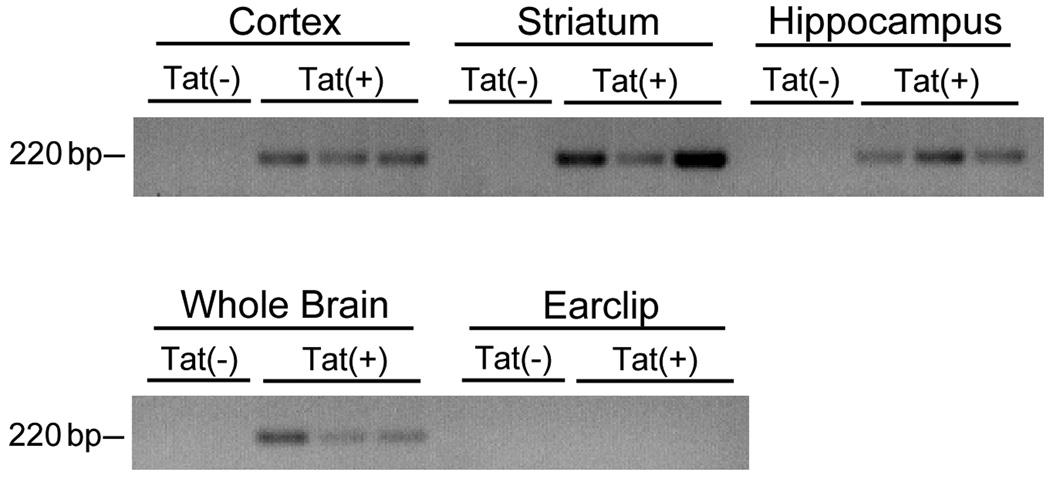
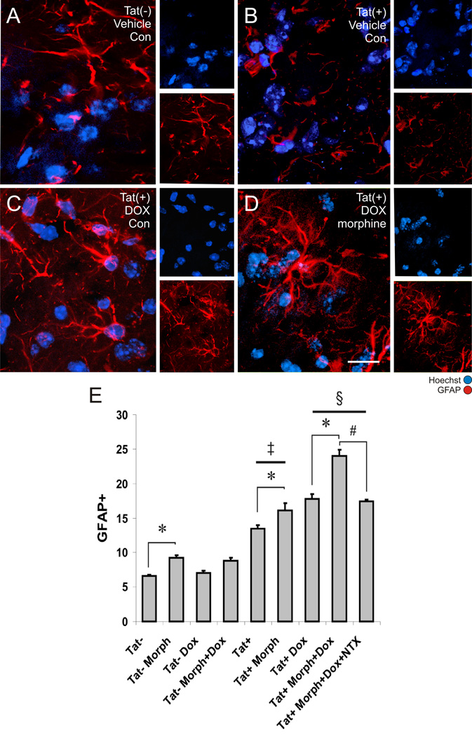
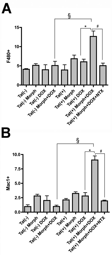
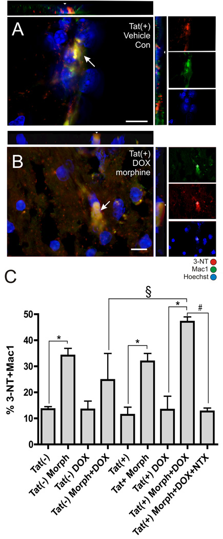
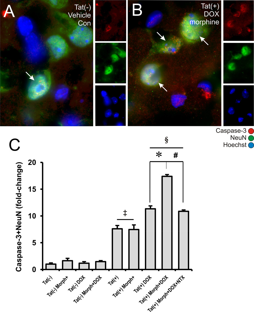
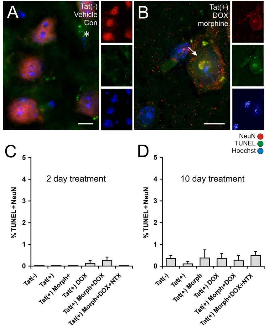
HIV encephalitis (HIVE) is accompanied by brain inflammation, leukocyte infiltration, and glial activation, and HIV patients who abuse opiates are more likely to develop HIVE. To better understand how opiates could alter HIV-related brain inflammation, the expression of astrocyte (GFAP immunoreactivity) and macrophage/microglial (F4/80 or Mac1 immunoreactivity) markers in the striatum, and the percentage of 3-nitrotyrosine (3-NT) positive macrophages/microglia, was determined following a 2-day exposure to morphine (5 mg/kg/day via time-release, subcutaneous implant) and doxycycline in GFAP-driven, doxycycline-inducible HIV-1 Tat transgenic mice. Data show that both morphine and Tat induction via doxycycline increased astrocyte activation, with significant additive increases achieved with combined morphine and doxycycline exposure. By contrast, combined Tat induction and morphine exposure, but neither manipulation alone, significantly increased the proportion of macrophages/microglia present in the striatum of transgenic mice, although morphine exposure was necessary to elevate 3-NT co-detection in Mac1-positive macrophages/microglia. Finally, Tat induction increased the percentage of neurons expressing active caspase-3, and this was even more significantly elevated by co-administration of morphine. In spite of elevations in caspase-3, neuronal TUNEL reactivity was unchanged in all groups, even after 10 days of Tat induction. Importantly, co-administration of naltrexone completely antagonized the effects of morphine. These findings indicate that morphine rapidly and significantly increases the activation of astrocytes and macrophages/microglia in the brains of inducible Tat transgenic mice, supporting the theory that early inflammatory changes in glia could underlie the development of HIVE in opiate-abusing AIDS patients.


Figures

Figure 1
Figure 1. Regional specificity of inducible Tat expression
RT-PCR was used to examine Tat transgene expression in adult Tat(+) and Tat(−) transgenic mice after 2 days of DOX administration. Images depict the presence or absence of Tat mRNA (220 bp) in samples derived from specific brain regions (cortex, striatum, hippocampus) as well as peripheral tissue (earclip) from three Tat(+) and two Tat(−) mice.
Figure 2
Figure 2. Astroglial responses to morphine and the inducible Tat transgene
The proportion of Hoechst-stained striatal cells that were glial fibrillary acidic protein (GFAP) immunoreactive at 2 days following Tat induction was assessed in untreated Tat(−) mice (A), untreated Tat(+) mice (B), Tat(+) mice treated with DOX alone (C), and Tat(+) mice treated with combined DOX and morphine (Morph) after 2 days exposure (D, scale bar = 20 µm). The proportion of GFAP immunoreactive astrocytes in the striata of untreated Tat(+) mice was significantly increased compared to Tat(−) mice irrespective of DOX and morphine treatment [P < 0.01 Tat(+) vs. Tat(−) mice]. Morphine administration significantly increased the proportion of astrocytes in both Tat(−) and Tat(+) mice (*P < 0.05). Tat induction by DOX further increased the proportion of GFAP astroglia compared to Tat+ mice without DOX [§P < 0.05 vs. similarly treated Tat(+) mice without DOX]. Morphine-induced increases in GFAP immunoreactivity in DOX treated Tat(+) mice were reversed by co-administering naltrexone (NTX) (#P < 0.05). Data represent the mean ± SEM of 5–6 mice/group.
Figure 3
Figure 3. Microglial responses to morphine and the inducible Tat transgene
In combination, morphine (Morph) exposure and Tat induction significantly increased the proportion of F4/80 (A) or Mac-1 (B) immunoreactive macrophages/microglia after 2 days [§P < 0.001 vs. DOX and Morph treated Tat(−) mice; *P < 0.001 vs. DOX treated Tat(+) mice without Morph], while neither Tat induction nor Morph alone had any effect. Morphine-induced increases in F4/80 and Mac-1 immunoreactivity in DOX treated Tat(+) mice were reversed by co-administering naltrexone (NTX) (#P < 0.05). Data represent the mean ± SEM of 5–6 mice/group.
Figure 4
Figure 4. 3-NT-positive microglia following morphine treatment in inducible Tat transgenic mice
Mac-1 and 3-NT immunoreactivity were co-localized (arrows) in Hoechst-stained striatum in all groups and shown here for untreated control Tat(+) mice (A) and Tat(+) mice treated with combined doxycycline (DOX) and morphine (Morph) (B) (scale bars = 10 µm; orthogonal views showing co-localization of 3-NT and Mac1 immunoreactivity in xz and yz planes are included). (C) Morphine exposure alone (*P < 0.05 vs. similar conditions without Morph) or combined morphine plus Tat induction (§P < 0.001 vs. DOX and Morph treated Tat(−) mice) significantly increased the proportion of 3-NT positive macrophages/microglia after 2 days. Morphine-induced increases in 3-NT immunoreactivity in DOX treated Tat(+) mice were reversed by co-administering naltrexone (NTX) (#P < 0.05). Data represent the mean ± SEM of 5–6 mice/group.
Figure 5
Figure 5. Effects of Tat induction and morphine exposure on active caspase-3-immunoreactivity in striatal neurons
Although active caspase-3 and NeuN immunofluorescence could be co-localized in some neurons (arrows) in Tat(−) mice receiving placebo implants and normal feed (A), such cells appear to be increased in all Tat(+) groups (B). Significant increases in the proportion of cleaved caspase-3 were seen in (1) untreated Tat(+) mice [P < 0.02 vs. untreated Tat(−) mice]; (2) Tat(+) receiving doxycycline (DOX) [§P < 0.001 vs. Tat(−) mice with DOX]; or (3) with combined morphine (Morph) and DOX treatment [*P < 0.05 vs. DOX treated Tat(+) mice without morphine] (C). Morphine-induced increases in cleaved caspase-3 immunoreactivity in DOX treated Tat(+) mice were antagonized by co-administering naltrexone (NTX) (#P < 0.05) (C). Data represent the mean ± SEM of 5–6 mice/group. Scale bar in A = 10 µm (A-B are equal magnification).
Figure 6
Figure 6. Effects of Tat induction and morphine exposure on TUNEL reactivity in striatal neurons
Tissues from the same mice analyzed for caspase-3 immunostaining after 2 days of treatment (Fig. 5) were analyzed for TUNEL labeling (A). Additional mice were analyzed after 10 days of treatment (B). TUNEL reactivity was rarely detected in NeuN immunoreactive neurons in the striatum of Tat transgenic mice, irrespective of DOX or Morph exposure (A), although an occasional TUNEL positive neuron was seen in Tat(+) mice receiving DOX, or combined DOX and Morph (B). Some TUNEL-positive cells that were not NeuN+ were present in all tissues, and likely represent dying glia (*). The proportion of TUNEL-reactive neurons was unaffected by the Tat transgene or by treatments with DOX and/or Morph following either 2 days (C), or 10 days (D) of exposure. Data represent the mean ± SEM of 5–6 mice/group. Scale bars in A and B = 10 µm.

References

    1. Aldovini A, Debouck C, Feinberg MB, Rosenberg M, Arya SK, Wong-Staal F. Synthesis of the complete trans-activation gene product of human T-lymphotropic virus type III in Escherichia coli: demonstration of immunogenicity in vivo and expression in vitro. Proc Natl Acad Sci U S A. 1986;83:6672–6676. - PMC - PubMed
    1. Anderson CE, Tomlinson GS, Pauly B, Brannan FW, Chiswick A, Brack-Werner R, Simmonds P, Bell JE. Relationship of Nef-positive and GFAP-reactive astrocytes to drug use in early and late HIV infection. Neuropathol Appl Neurobiol. 2003;29:378–388. - PubMed
    1. Anthony IC, Ramage SN, Carnie FW, Simmonds P, Bell JE. Does drug abuse alter microglial phenotype and cell turnover in the context of advancing HIV infection? Neuropathol Appl Neurobiol. 2005;31:325–338. - PubMed
    1. Avgeropoulos N, Kelley B, Middaugh L, Arrigo S, Persidsky Y, Gendelman HE, Tyor WR. SCID mice with HIV encephalitis develop behavioral abnormalities. J Acquir Immune Defic Syndr Hum Retrovirol. 1998;18:13–20. - PubMed
    1. Avison M, Berger JR, McArthur JC, Nath A. HIV meningitis and dementia. In: Nath A, Berger JR, editors. Clinical Neurovirology. New York: Marcel Dekker, Inc.; 2003. pp. 251–276.

Publication types

MeSH terms

Substances