. 2008 Jun;20(6):1538-54.
doi: 10.1105/tpc.108.059741. Epub 2008 Jun 13.

Targeted degradation of the cyclin-dependent kinase inhibitor ICK4/KRP6 by RING-type E3 ligases is essential for mitotic cell cycle progression during Arabidopsis gametogenesis

Affiliations

Targeted degradation of the cyclin-dependent kinase inhibitor ICK4/KRP6 by RING-type E3 ligases is essential for mitotic cell cycle progression during Arabidopsis gametogenesis

Jingjing Liu et al. Plant Cell. 2008 Jun.

Abstract

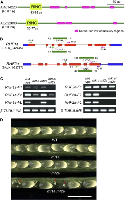
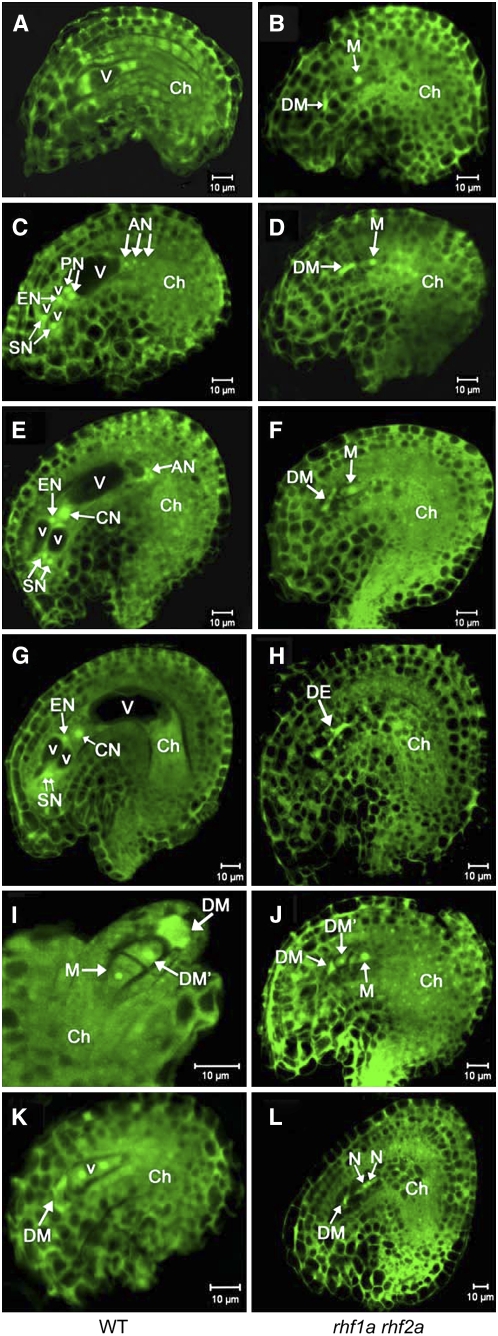
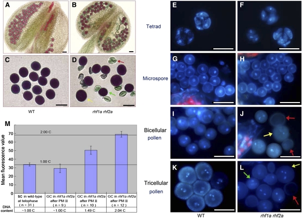
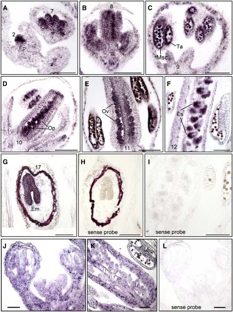
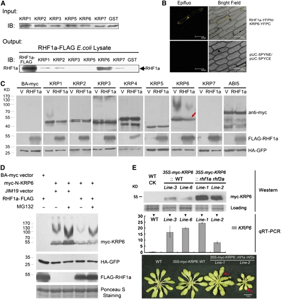
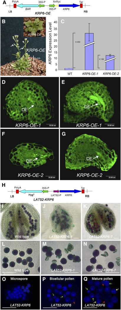
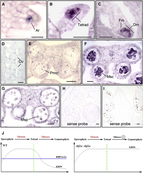
Following meiosis, plant gametophytes develop through two or three rounds of mitosis. Although the ontogeny of gametophyte development has been defined in Arabidopsis thaliana, the molecular mechanisms regulating mitotic cell cycle progression are not well understood. Here, we report that RING-H2 group F 1a (RHF1a) and RHF2a, two RING-finger E3 ligases, play an important role in Arabidopsis gametogenesis. The rhf1a rhf2a double mutants are defective in the formation of male and female gametophytes due to interphase arrest of the mitotic cell cycle at the microspore stage of pollen development and at female gametophyte stage 1 of embryo sac development. We demonstrate that RHF1a directly interacts with and targets a cyclin-dependent kinase inhibitor ICK4/KRP6 (for Interactors of Cdc2 Kinase 4/Kip-related protein 6) for proteasome-mediated degradation. Inactivation of the two redundant RHF genes leads to the accumulation of ICK4/KRP6, and reduction of ICK4/KRP6 expression largely rescues the gametophytic defects in rhf1a rhf2a double mutants, indicating that ICK4/KRP6 is a substrate of the RHF E3 ligases. Interestingly, in situ hybridization showed that ICK4/KRP6 was predominantly expressed in sporophytes during meiosis. Our findings indicate that RHF1a/2a-mediated degradation of the meiosis-accumulated ICK4/KRP6 is essential to ensure the progression of subsequent mitoses to form gametophytes in Arabidopsis.


Figures

Figure 1.
Figure 1.
SALK T-DNA Insertion Mutants of RHF1a and RHF2a. (A) Protein domains of two Arabidopsis RING-finger proteins, RHF1a and RHF2a, showing a RING-finger domain at the N terminus (light green) and several Ser-rich low-complexity regions (magenta). aa, amino acids. (B) T-DNA insertion sites in RHF1a and RHF2a genes. Blue box, untranslated regions; red box, exons; red line, introns. Two neighboring inverted T-DNAs were found inserted in the fourth exon of RHF1a in rhf1a (SALK_042445) and in the seventh exon of RHF2a in rhf2a (SALK_023787) through sequencing the amplified flanking sequence. The green and purple arrows show the positions of primers used to amplify the truncated transcripts in (C). The green arrows indicate the primers used to amplify the F1 fragment. The purple arrows indicate the primers used to amplify the F2 fragment. (C) RHF1a and RHF2a full-length and truncated transcript level in wild-type plants, single insertion mutants, and rhf1a rhf2a double mutants. RNA was extracted from 5-week-old plants. F1, truncated transcript before the T-DNA insertion site; F2, truncated transcript after the T-DNA insertion site; FL, full-length transcript. β-Tubulin8 was used as an internal control. (D) Seed development in the wild-type plant, rhf1a single mutant, rhf2a single mutant, and rhf1a rhf2a double mutant. Aborted ovules (indicated by red arrows) were evident in rhf1a rhf2a mutants. Bar = 1 mm.
Figure 2.
Figure 2.
Ovule Development in Wild-Type Plants and the rhf1a rhf2a Double Mutants Revealed by Confocal Laser Scanning Microscopy Analysis. Ovule cells are visualized by autofluorescence. AN, antipodal nuclei; Ch, chalazal end; CN, central nucleus; DE, degenerated embryo sac; DM, degenerated megaspore; DM', nondegenerated megaspore other than the functional megaspore; EN, egg nucleus; N, nucellus; PN, polar nuclei; SN, synergid nuclei; V, large vacuole; v, small vacuole. Bars = 10 μm. (A) and (B) Wild-type ovule at early FG4, containing a four-nucleate embryo sac (A) compared with the abnormal female gametophytes from the rhf1a rhf2a double mutant pistils at the same stage (B). (C) and (D) Wild-type ovule at FG5 with an eight-nucleate embryo sac (C) compared with the rhf1a rhf2a double mutant ovule at the same stage (D). At this stage in the wild type, cellularization took place and cell differentiation was completed with the formation of two synergid nuclei (SN), an egg nucleus (EN), three antipodal nuclei (AN), and two prominent unfused polar nuclei (PN). (E) and (F) Wild-type ovule with a mature seven-celled embryo sac at FG6 (E) compared with the rhf1a rhf2a double mutant ovule at the same stage (F). During this stage, the two polar nuclei fused to form a diploid central nucleus (CN) in the wild type. (G) and (H) Mature wild-type ovule with a four-celled embryo sac at FG7 (G) and an rhf1a rhf2a double mutant ovule (H). Note that the three antipodal cells have degenerated completely in the wild type. Note also that, in the rhf1a rhf2a double mutant, female gametophytes were arrested at the one-nucleate stage FG1 until the embryo sac (DE) degenerated completely, visualized by its strong autofluorescence (B), (D), (F), and (H). (I) Wild-type ovule at FG1 with one functional megaspore (M) and three degenerating megaspores, of which two micropylar megaspores have degenerated completely (DM) and the other has not degenerated yet (DM'). Note that the outer and inner integuments do not completely enclose the nucellus (N). (J) Ovule from the rhf1a rhf2a double mutant showing a nondegraded megaspore (DM') and a functional megaspore with outer and inner integuments enclosing them. (K) Wild-type ovule at FG2 with two-nucleate embryo sac. (L) Ovule from the rhf1a rhf2a double mutant arresting at the two-nucleate stage. Note that the developmental extent of the ovule in (L) with mature integuments enclosing is much higher than the ovule in (K).
Figure 3.
Figure 3.
Pollen Development in Wild-Type Plants and the rhf1a rhf2a Double Mutants. (A) Alexander's staining of wild-type anthers. (B) Alexander's staining of double mutant anthers containing degenerated pollen. (C) Alexander's staining of wild-type mature pollen grains of uniform size. (D) Alexander's staining of the rhf1a rhf2a double mutant mature pollen grains from the same anther. Note that the double mutant mature pollen is of variable size. Yellow arrow: much larger than normal pollen grain. Red arrow: shrunken pollen. Green arrow: smaller than normal pollen grain. (E) and (F) DAPI staining of tetrads in wild-type (E) and the rhf1a rhf2a double mutant anthers (F). No visible difference was seen between wild-type and double mutant tetrads. (G) and (H) DAPI staining of microspores in wild-type (G) and the rhf1a rhf2a double mutant anthers (H). Note no visible difference between wild-type and double mutant microspores. (I) and (J) DAPI staining of binucleate stage pollen in wild-type (I) and the rhf1a rhf2a double mutant anthers (J). Note that the binucleate pollen are of uniform size in the wild type and of variable size in the double mutant. The yellow arrow indicates larger pollen, and the red arrow indicates abnormal pollen with no DAPI staining. (K) and (L) DAPI staining of mature pollen grains in wild-type (K) and the rhf1a rhf2a double mutant anthers (L). Note that mature pollen grains from the double mutant contain shrunken pollen walls (red arrow), larger (yellow arrow), and smaller pollen grains than normal (green arrow). Bars = 25 μm. (M) Relative DNA content (DAPI fluorescence values) in generative cells (GC) and sperm cells (SC) in wild-type and the rhf1a rhf2a mutant pollen. The mean C values calculated relative to the 1C content of telophase sperm cell nuclei (1C) after the PM II stage are shown. Error bars = se.
Figure 4.
Figure 4.
RNA in Situ Expression of RHF1a and RHF2a in Wild-Type Inflorescences and Siliques. Longitudinal sections ([A] to [G]) of inflorescences were hybridized with RHF1a antisense probe. (A) to (F) Expression of RHF1a in wild-type inflorescence from an early stage to stage 12. (G) Expression of RHF1a in wild-type developing seeds. (H) and (I) Hybridized with RHF1a sense probe. (J) Expression of RHF2a in inflorescence. (K) Expression of RHF2a in developing carpels. (L) Hybridized with RHF2a sense probe. Em, embryos; Es, embryo sac; Fp, floral primordia; Msc, microsporocyte; Op, ovule primordia; Ov, ovule; Ta, tapetum. Bars = 100 μm.
Figure 5.
Figure 5.
RHF1a Primarily Interacts with ICK4/KRP6 and Targets It for Degradation. (A) Pull-down assays of RHF1a and ICKs/KRPs (except for KRP4). Input, protein gel blot using anti-GST antibody, shows comparable protein loading in the GST pull-down assays. Output, Escherichia coli lysates containing RHF1a-FLAG were used in GST pull-down assays with GST-ICK/KRP fusions. RHF1a-FLAG represents lysate from E. coli expressing RHF1a-FLAG. IB, immunoblot detected with anti-FLAG antibody. (B) BiFC assays in vivo. RHF1a-YFPN is able to interact with KRP6-YFPC to generate yellow fluorescent protein (YFP) fluorescence in onion epidermal cells. The expression of pUC-SPYNE and pUC-SPYCE was used as the control. (C) Effects of RHF1a on the degradation of ICKs/KRPs and ABI5 in vivo. N. benthamiana leaves were coinfiltrated with indicated binary vectors. For each experiment, two constructs were infiltrated, one expressing a specific KRP protein (top row of labels) and the other expressing either RHF1a or an empty vector (bottom row of labels). 35S-HA-GFP (green fluorescent protein) and 35S-p19 were also simultaneously coinfiltrated to serve as an internal control and to suppress gene silencing, respectively. Total proteins were extracted and resolved by SDS-PAGE analysis followed by immunoblotting with different antibodies. Anti-myc was used to detect the protein level of ICKs/KRPs or ABI5 (top panel), anti-FLAG was used for detection of FLAG-RHF1a (middle panel), and anti-HA was used to detect the internal control HA-GFP (bottom panel). The red arrow indicates the reduced protein level of ICK4/KRP6. (D) ICK4/KRP6 degradation in a 26S proteasome-dependent manner. Immunoblots were used to detect ICK4/KRP6 levels in the absence or presence of RHF1a and with or without the proteasome inhibitor, MG132 (40 μM), infiltrated into N. benthamiana leaves 12 h before sample collection. Anti-FLAG was used for detection of FLAG-RHF1a, and anti-HA was used to detect HA-GFP as internal controls. Ponceau S staining of the transferred membrane is displayed as a loading control. (E) ICK4/KRP6 protein accumulated much more in the rhf1a rhf2a double mutant plants than in wild-type plants. 35S-KRP6 construct was transformed into wild-type and rhf1a rhf2a double mutant plants, named as 35S-myc-KRP6∷WT and 35S-myc-KRP6∷rhf1a rhf2a, respectively. The analysis with two of the 35S-myc-KRP6∷WT independent lines and two of the 35S-myc-KRP6rhf1a rhf2a independent lines is shown. Proteins from seedlings ∼20 d after germination (DAG) were processed for protein gel blots with anti-myc antibody to detect the ICK4/KRP6 protein level in vivo (top panel). Ponceau S staining of the transferred membrane is displayed as a loading control. Total RNAs from the same transgenic plants were used for quantitative RT-PCR analysis (middle panel). Leaf phenotypes (bottom panel) of wild-type plants, one of the two 35S-myc-KRP6∷WT lines, and two of the 35S-myc-KRP6rhf1a rhf2a lines were shown (bottom panel). Photos were taken from 3-week-old plants. Red arrows indicate serrated leaves.
Figure 6.
Figure 6.
Elevated Expression of ICK4/KRP6 Phenocopies the Gametophytic Defects of rhf1a rhf2a Mutant. (A) Construct of the KRP6-OE plasmid. LB, T-DNA left border; polyA, CaMV 35S poly(A); BAR, resistance gene; 35S-P, CaMV 35S promoter; 6×myc, six myc tags fusion; KRP6, open reading frame of KRP6; Ter, nopaline synthase terminator; RB, T-DNA right border. (B) ICK4/KRP6 overexpression transgenic plants ∼50 DAG. The fertility of the transgenic plants was low as shown by white arrows indicating the extremely short siliques. (C) Real-time quantitative RT-PCR analysis of ICK4/KRP6 expression level in two independent ICK4/KRP6 OE lines. Expression is normalized to a wild-type level of 1. Error bars indicate sd of three real-time PCR experiments of each biological sample. (D) to (G) Confocal laser scanning microscopy analysis of embryo sac development in ovules from the KRP6 OE-1 line ([D] and [E]) and the KRP6 OE-2 line ([F] and [G]) independently. The completely degenerated embryo sacs (DE) were indicated by high autofluorescence regions. (H) Construction of the LAT52-KRP6 plasmid. LAT52P, LAT52 promoter; HygR, hygromycin resistance gene; LB, polyA, 35S-P, Ter, and RB are as in (A). (I) Alexander's staining of wild-type anthers. (J) and (K) Alexander's staining of LAT52-KRP6-1 (J) and LAT52-KRP6-7 (K) transgenic plant anthers. (L) Alexander's staining of wild-type pollen grains with even size. (M) and (N) Alexander's staining of LAT52-KRP6-1 (M) and LAT52-KRP6-7 (N) transgenic plant pollen grains with shrunken pollen walls. (O) DAPI staining of LAT52-KRP6 microspores. (P) DAPI staining of LAT52-KRP6 pollen at bicellular stage with some aberrantly stained pollen (green arrows) and shrunken pollen walls (red arrows). (Q) DAPI staining of LAT52-KRP6 pollen at tricellular stage with microspores (green arrows), bicellular pollen (yellow arrows), and shrunken pollen walls (red arrows) other than the normally stained tricellular pollen grains.
Figure 7.
Figure 7.
The Transcription of ICK4/KRP6 Was Tightly Regulated and Only Detected in Stages before Gametophytic Mitosis by in Situ Hybridization. (A) KRP6 RNA expression in ovule primordia at early stages, showing the KRP6 RNA signal in the female archesporial cell (Ar). (B) and (C) KRP6 RNA expression in ovule primordia at late stages, showing the stronger KRP6 RNA signal in the female tetrad, shown as four neighboring megaspores (B), and later in functional megaspore (Fm) and three other degenerating megaspores (Dm) (C). (D) KRP6 RNA expression in the ovule, showing no or very low KRP6 RNA signal in developing ovules (Ov). (E) Weak KRP6 RNA expression in pollen mother cells (Pmc) in an anther at stage 4. (F) KRP6 expression in anthers at stages 5 to 6, showing that KRP6 RNA signal was predominantly in microsporocyte (Msc). (G) KRP6 expression was not detected in microspores (Msp) in anthers after stage 6. (H) and (I) Control sections hybridized with the sense KRP6 RNA probe, showing no signal detected. (J) A possible working model for RHF1a/2a and ICK4/KRP6 during gametophyte formation in Arabidopsis. The transcript of ICK4/KRP6 was accumulated during meiosis and turned off right before the subsequent gametophytic mitosis. RHF1a/RHF2a-mediated 26S proteasome-dependent proteolysis may serve as a watchdog in Arabidopsis, to keep the level of meiosis-accumulated ICK4/KRP6 below a certain threshold, so that the subsequent gametophytic mitoses can proceed smoothly. Bars = 10 μm in (A) and 20 μm in all other panels.

Comment in

Similar articles

Cited by

References

    1. Alexander, M.P. (1969). Differential staining of aborted and nonaborted pollen. Stain Technol. 44 117–122. - PubMed
    1. Barroco, R.M., De Veylder, L., Magyar, Z., Engler, G., Inzé, D., and Mironov, V. (2003). Novel complexes of cyclin-dependent kinases and a cyclin-like protein from Arabidopsis thaliana with a function unrelated to cell division. Cell. Mol. Life Sci. 60 401–412. - PMC - PubMed
    1. Butaye, K.M., Goderis, I.J., Wouters, P.F., Pues, J.M., Delaure, S.L., Broekaert, W.F., Depicker, A., Cammue, B.P., and De Bolle, M.F. (2004). Stable high-level transgene expression in Arabidopsis thaliana using gene silencing mutants and matrix attachment regions. Plant J. 39 440–449. - PubMed
    1. Christensen, C.A., Subramanian, S., and Drews, G.N. (1998). Identification of gametophytic mutations affecting female gametophyte development in Arabidopsis. Dev. Biol. 202 136–151. - PubMed
    1. Daxinger, L., Hunter, B., Sheikh, M., Jauvion, V., Gasciolli, V., Vaucheret, H., Matzke, M., and Furner, I. (2008). Unexpected silencing effects from T-DNA tags in Arabidopsis. Trends Plant Sci. 13 4–6. - PubMed

Publication types

MeSH terms

Substances