. 2008 Jun;20(6):1437-55.
doi: 10.1105/tpc.108.058891. Epub 2008 Jun 13.

Arabidopsis DDB1-CUL4 ASSOCIATED FACTOR1 forms a nuclear E3 ubiquitin ligase with DDB1 and CUL4 that is involved in multiple plant developmental processes

Affiliations

Arabidopsis DDB1-CUL4 ASSOCIATED FACTOR1 forms a nuclear E3 ubiquitin ligase with DDB1 and CUL4 that is involved in multiple plant developmental processes

Yu Zhang et al. Plant Cell. 2008 Jun.

Abstract

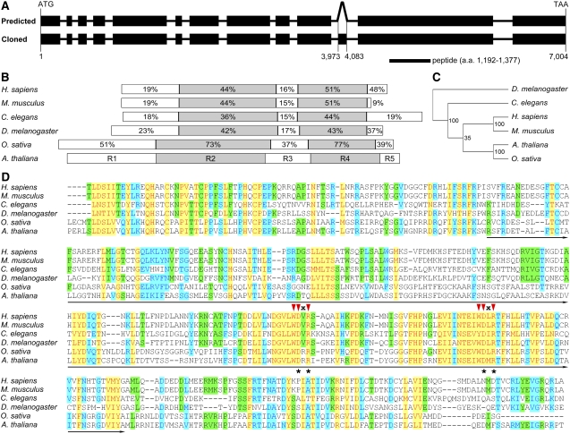
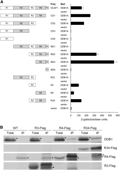
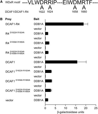
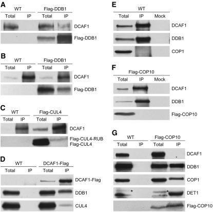
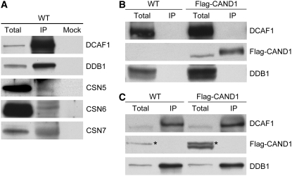
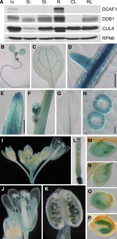
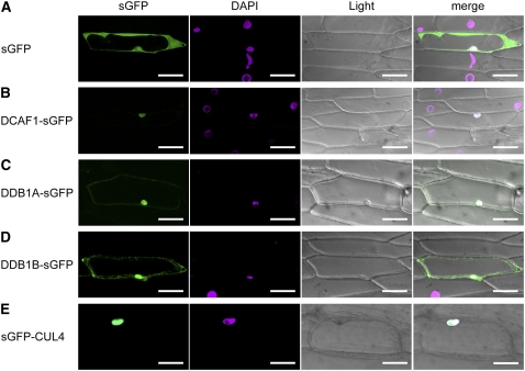
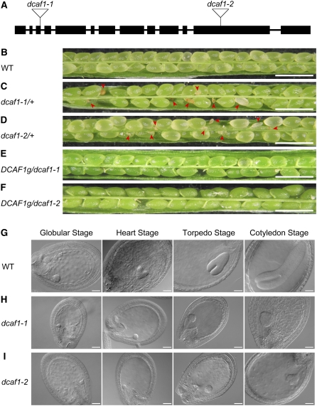
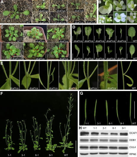
The human DDB1-CUL4 ASSOCIATED FACTOR (DCAF) proteins have been reported to interact directly with UV-DAMAGED DNA BINDING PROTEIN1 (DDB1) through the WDxR motif in their WD40 domain and function as substrate-recognition receptors for CULLIN4-based E3 ubiquitin ligases. Here, we identified and characterized a homolog of human DCAF1/VprBP in Arabidopsis thaliana. Yeast two-hybrid analysis demonstrated the physical interaction between DCAF1 and DDB1 from Arabidopsis, which is likely mediated via the WD40 domain of DCAF1 that contains two WDxR motifs. Moreover, coimmunoprecipitation assays showed that DCAF1 associates with DDB1, RELATED TO UBIQUITIN-modified CUL4, and the COP9 signalosome in vivo but not with CULLIN-ASSOCIATED and NEDDYLATION-DISSOCIATED1, CONSTITUTIVE PHOTOMORPHOGENIC1 (COP1), or the COP10-DET1-DDB1 complex, supporting the existence of a distinct Arabidopsis CUL4 E3 ubiquitin ligase, the CUL4-DDB1-DCAF1 complex. Transient expression of fluorescently tagged DCAF1, DDB1, and CUL4 in onion epidermal cells showed their colocalization in the nucleus, consistent with the notion that the CUL4-DDB1-DCAF1 complex functions as a nuclear E3 ubiquitin ligase. Genetic and phenotypic analysis of two T-DNA insertion mutants of DCAF1 showed that embryonic development of the dcaf1 homozygote is arrested at the globular stage, indicating that DCAF1 is essential for plant embryogenesis. Reducing the levels of DCAF1 leads to diverse developmental defects, implying that DCAF1 might be involved in multiple developmental pathways.


Figures

Figure 1.
Figure 1.
Structure of the Arabidopsis DCAF1 Gene and Sequence Alignment of DCAF1 Homologs. (A) Structure of the Arabidopsis DCAF1 gene and the location of the antigen used for antibody preparation. Exons are presented as filled black rectangles, and introns are presented as solid lines. The top diagram (predicted) depicts the predicted structure of the DCAF1 gene in the AGI database with 11 introns and 12 exons. The bottom one (cloned) presents the structure of the DCAF1 gene cloned in this study, which has an extra exon inside the predicted 10th intron, corresponding to nucleotide 3973 to 4083 of the genomic fragment. The position of the peptide used as antigen for antibody preparation is indicated underneath the gene structure. a.a., amino acids. (B) Schematic comparison of DCAF1 homologs from representative eukaryotic organisms as labeled on the left. Based on the difference in amino acid sequence homology, the DCAF1 proteins are divided into five regions, which, in Arabidopsis, are shown by five differently colored rectangles labeled R1 through R5. The percentages of similarity in the corresponding regions from each homolog to the Arabidopsis DCAF1 are indicated in the respective region. (C) A phylogenetic tree of DCAF1 homologs from the model organisms indicated on the right (see Methods for details on tree generation procedure). The numbers indicate the statistic values of the reliability for each node. (D) Alignment of the R4 region of DCAF1 from the model organisms labeled on the left. The WD40 domain is underlined. The red triangles indicate the two WDxR motifs in the WD40 domain, and “x” stands for an indefinite amino acid. The asterisks indicate the Asn and Arg (on the top) within the WDxR motif, which are mutated into the Ala residue in the point mutation analysis as shown in Figure 3A. The shading mode indicates the level of conservation, with red letters in yellow shading corresponding to a high level of conservation (100%), blue letters in azure shading corresponding to a moderate level of conservation (80%), and black letters in green shading corresponding to a low level of conservation (60%).
Figure 2.
Figure 2.
Direct Interaction between DCAF1 and DDB1. (A) Interaction analysis of DCAF1 and its deletion mutants with DDB1A by Y2H analysis. The left diagrams schematically present a series of deletion mutants corresponding to different regions in DCAF1. Empty bait vector was used as a negative control. The β-galactosidase activities resulting from the interactions are shown in the histogram. Error bars present sd (n = 4). (B) Association analysis of Flag-tagged R3, R4, and R34 (R3 and R4) regions of DCAF1 with DDB1 by in vivo co-IP with the anti-Flag antibody. Seedling total protein extracts prepared from wild-type and 35S:R3-Flag (R3-Flag), 35S:R4-Flag (R4-Flag), and 35S:R34-Flag (R34-Flag) transgenic Arabidopsis plants were incubated with anti-Flag antibody-coupled agarose. The immunoprecipitates (IP) and the total extracts (total) were subjected to immunoblot analysis with antibodies against Flag (for Flag-tagged R3, R4, and R34) and DDB1. The asterisks mark the cross-reacting bands (to the left), and the arrowheads mark the partial degradation product bands (to the left).
Figure 3.
Figure 3.
Point Mutation Analysis of WDxR Motifs in DCAF1. (A) Point mutations introduced in the two WDxR motifs of DCAF1 and its R4 region (DCAF1-R4). The numbers indicate the mutation sites (amino acid residue; D-to-A or R-to-A point mutations) in DCAF1. D1622A and R1624A are situated within the first WDxR motif, whereas D1658A and R1660A occur within the second WDxR motif. (B) Interaction analysis of point mutants of DCAF1 or DCAF1-R4 with DDB1A by Y2H analysis. The previously shown interactions between DCAF1 or DCAF1-R4 and DDB1A (Figure 2A) were used as positive controls. The β-galactosidase activities resulting from the interactions are shown in the histogram. Empty prey vector with DDB1A as bait was used as the negative control. Error bars present sd (n = 4).
Figure 4.
Figure 4.
Evidence for a CUL4-DDB1-DCAF1 Complex in Arabidopsis. (A) and (B) Association of DCAF1 with Flag-DDB1 in vivo. Total seedling protein extracts prepared from wild-type and 35S:Flag-DDB1 (Flag-DDB1) transgenic Arabidopsis were incubated with anti-Flag antibody-conjugated agarose (A) or with anti-DCAF1 antibody and Protein A sepharose (B). The immunoprecipitates (IP) and the total extracts (total) were subjected to immunoblot analysis with antibodies against Flag (for Flag-DDB1) and DCAF1. (C) Association of DCAF1 with RUB-modified Flag-CUL4 in vivo. Total seedling protein extracts prepared from wild-type and 35S:Flag-CUL4 (Falg-CUL4) transgenic Arabidopsis plants were incubated with anti-DCAF1 antibody and Protein A sepharose. The immunoprecipitates (IP) and the total extracts (total) were subjected to immunoblot analysis with antibodies against Flag (for Flag-tagged CUL4 and CUL4-RUB) and DCAF1. (D) Association of DCAF1-Flag with both DDB1 and CUL4 in vivo. Total seedling protein extracts prepared from wild-type and 35S:DCAF1-Flag (DCAF1-Flag) transgenic Arabidopsis were incubated with anti-Flag antibody-conjugated agarose. The immunoprecipitates (IP) and the total extracts (total) were subjected to immunoblot analysis with antibodies against Flag (for DCAF1-Flag), DDB1, and CUL4. (E) DCAF1 does not associate with COP1 in vivo. Total seedling protein extracts prepared from wild-type Arabidopsis were incubated with anti-DCAF1 antibody and Protein A sepharose. Incubation with Protein A sepharose alone was used as mock. The immunoprecipitates (IP) and the total extracts (total) were subjected to immunoblot analysis with antibodies against DCAF1, DDB1, and COP1. (F) DCAF1 does not associate with Flag-COP10 in vivo. Total seedling protein extracts prepared from 35S:Flag-COP10 (Flag-COP10) transgenic Arabidopsis were incubated with anti-DCAF1 antibody and Protein A sepharose. Incubation with Protein A sepharose alone was used as a mock. The immunoprecipitates (IP) and the total extract (total) were subjected to immunoblot analysis with antibodies against DCAF1, DDB1, and Flag (for Flag-COP10). (G) Flag-COP10 does not associate with DCAF1 in vivo. Total seedling protein extracts prepared from wild-type and 35S:Flag-COP10 (Flag-COP10) transgenic Arabidopsis were incubated with anti-Flag antibody-conjugated agarose. The immunoprecipitates (IP) and total extracts (total) were subjected to immunoblot analysis with antibodies against DCAF1, DDB1, COP1, DET1, and Flag (for Flag-COP10). The asterisks mark the cross-reacting bands (to the left).
Figure 5.
Figure 5.
Association Analysis of the CUL4-DDB1-DCAF1 Complex with CRL E3 Activity Regulators. (A) Association of DCAF1 with CSN in vivo. Total seedling protein extracts prepared from wild-type Arabidopsis were incubated with anti-DCAF1 antibody and Protein A sepharose. Incubation with Protein A sepharose alone was used as a mock. The immunoprecipitates (IP) and the total extracts (total) were subjected to immunoblot analysis with antibodies against DCAF1, DDB1, CSN5, CSN6, and CSN7. (B) and (C) DCAF1 does not associate with Flag-CAND1 in vivo. Total seedling protein extracts prepared from wild-type and 35S:Flag-CAND1 (Flag-CAND1) transgenic Arabidopsis were incubated with anti-Flag antibody-conjugated agarose (B) or with anti-DCAF1 antibody and Protein A sepharose (C). The immunoprecipitates (IP) and the total extracts (total) were subjected to immunoblot analysis with antibodies against DCAF1, Flag (for Flag-CAND1), and DDB1. The asterisks mark the cross-reacting bands (to the left).
Figure 6.
Figure 6.
Expression Pattern of the DCAF1 Gene in Arabidopsis. (A) Protein accumulation of DCAF1 in different Arabidopsis organs. Total soluble protein extracts from different organs were examined by immunoblot analysis using antibodies against DCAF1, DDB1, and CUL4. The anti-RPN6 antibody was used as a sample equal loading control. (B) to (P) Temporal and spatial expression patterns of GUS reporter gene in DCAF1:GUS transgenic Arabidopsis. GUS activity was examined in a 7-d-old seedling (B), cotyledons (C), the primary root and lateral root (D), root tip (E), stem and axillary bud (F), rosette leaf (G), guard cell (H), inflorescence (I), mature flower (J), anther and pollen (K), silique (L), embryo at globular stage (M), embryo at heart stage (N), embryo at torpedo stage (O), and embryo at cotyledon stage (P). In, inflorescence; Si, silique; St, stem; R, root; CL, cauline leaf; RL, rosette leaf. Bars = 100 μm in (D), (E), and (M) to (P) and 10 μm in (H).
Figure 7.
Figure 7.
Subcellular Localization of DCAF1, DDB1, and CUL4. The following fluorescence proteins were transformed into and transiently expressed in onion epidermal cells: sGFP (A), DCAF1-sGFP (B), DDB1A-sGFP (C), DDB1B-sGFP (D), and sGFP-CUL4 (E). Signals from sGFP, 4′,6-diamidino-2-phenylindole (DAPI), bright-field (light), and the merge of the three signals (merge) are shown. Bars = 100 μm.
Figure 8.
Figure 8.
Characterization of dcaf1 Mutants. (A) Schematic representation of T-DNA insertions in the Arabidopsis DCAF1 gene. Exons are represented by filled black rectangles, and introns are represented by solid lines. The T-DNA insertion sites of the two mutant alleles are indicated by open inverted triangles, with the assigned allele name of each insertional mutation labeled above. (B) to (F) Stereomicroscopy images of siliques obtained from self-pollinated wild-type (B), dcaf1-1/+ (C), dcaf1-2/+ (D), DCAF1g/dcaf1-1 (E), and DCAF1g/dcaf1-2 (F) parental plants. Red arrowheads indicate abnormal ovules. Bars = 1 mm. (G) to (I) DIC images of cleared ovules obtained from self-pollinated wild-type (G), dcaf1-1/+ (H), and dcaf1-2/+ (I) parental plants. The four embryonic developmental stages (globular, heart, torpedo, and cotyledon) are shown from left to right. Bars = 50 μm.
Figure 9.
Figure 9.
Multifaceted Developmental Defects of dcaf1cs Mutants. (A) Two-week-old wild-type and dcaf1cs plants. Bars = 0.5 cm. (B) Abnormal flowers from dcaf1cs plants. (C) Four-week-old wild-type and dcaf1cs plants. Red arrowheads indicate the primary shoots that were starting to bolt at the transition from vegetative to reproductive growth. Bars = 1 cm. (D) Rosette leaves from 3-week-old wild-type and dcaf1cs plants. Bars = 0.5 cm. (E) Abnormal development of stem, node, internode, lateral shoot, axillary bud, and cauline leaves from dcaf1cs plants, with a wild-type plant as the control. (F) Comparison of wild-type and dcaf1cs adult plants. The numbers indicate independent dcaf1cs lines. Bar = 5 cm. (G) Comparison of wild-type and dcaf1cs siliques. The numbers indicate siliques from independent dcaf1cs lines. Bar = 1 cm. (H) Decrease of DCAF1 protein level in dcaf1cs mutants. Total seedling protein extracts from wild-type Arabidopsis and four independent dcaf1cs transgenic lines were examined by immunoblot analysis using antibodies against DCAF1, DDB1, and CUL4. The anti-RPN6 antibody was used as a sample equal loading control. The numbers above the blot indicate protein samples from independent dcaf1cs lines.

Similar articles

Cited by

References

    1. Alonso, J.M., et al. (2003). Genome-wide insertional mutagenesis of Arabidopsis thaliana. Science 301 653–657. - PubMed
    1. Angers, S., Li, T., Yi, X., MacCoss, M.J., Moon, R.T., and Zheng, N. (2006). Molecular architecture and assembly of the DDB1-CUL4A ubiquitin ligase machinery. Nature 443 590–593. - PubMed
    1. Belzile, J.P., Duisit, G., Rougeau, N., Mercier, J., Finzi, A., and Cohen, E.A. (2007). HIV-1 Vpr-mediated G2 arrest involves the DDB1-CUL4A(VPRBP) E3 ubiquitin ligase. PLoS Pathog. 3 e85. - PMC - PubMed
    1. Bernhardt, A., Lechner, E., Hano, P., Schade, V., Dieterle, M., Anders, M., Dubin, M.J., Benvenuto, G., Bowler, C., Genschik, P., and Hellmann, H. (2006). CUL4 associates with DDB1 and DET1 and its downregulation affects diverse aspects of development in Arabidopsis thaliana. Plant J. 47 591–603. - PubMed
    1. Byrne, M.E., Groover, A.T., Fontana, J.R., and Martienssen, R.A. (2003). Phyllotactic pattern and stem cell fate are determined by the Arabidopsis homeobox gene BELLRINGER. Development 130 3941–3950. - PubMed

Publication types

MeSH terms