. 2008 Jun;20(6):1693-707.
doi: 10.1105/tpc.107.057380. Epub 2008 Jun 13.

Arabidopsis DREB2A-interacting proteins function as RING E3 ligases and negatively regulate plant drought stress-responsive gene expression

Affiliations

Arabidopsis DREB2A-interacting proteins function as RING E3 ligases and negatively regulate plant drought stress-responsive gene expression

Feng Qin et al. Plant Cell. 2008 Jun.

Abstract

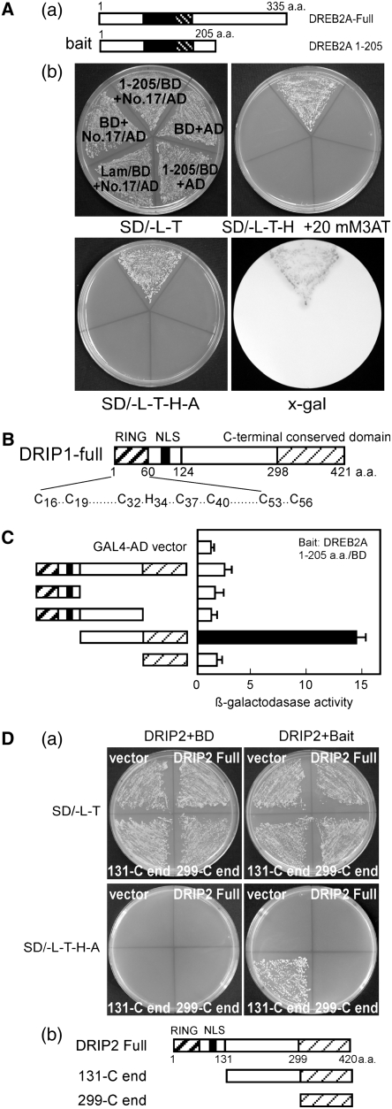
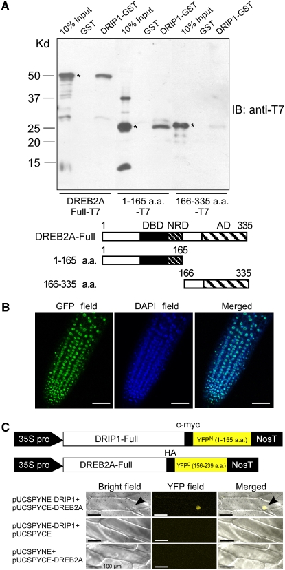
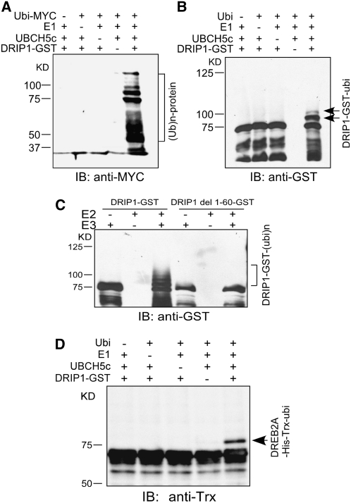
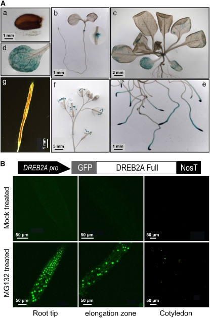
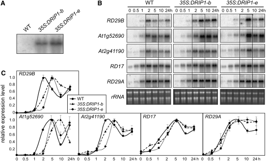
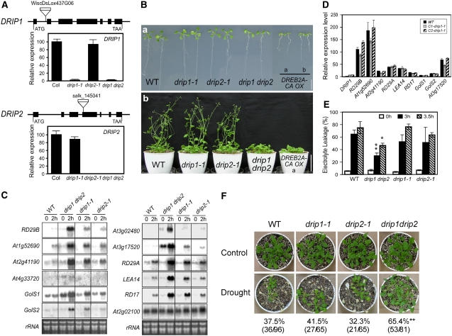
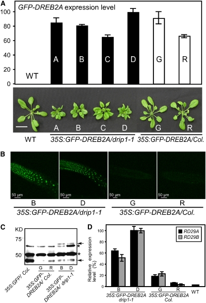
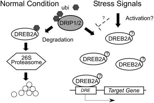
The DEHYDRATION-RESPONSIVE ELEMENT BINDING PROTEIN2A (DREB2A) transcription factor controls water deficit-inducible gene expression and requires posttranslational modification for its activation. The activation mechanism is not well understood; however, the stability of this protein in the nucleus was recently found to be important for its activation. Here, we report the isolation of Arabidopsis thaliana DREB2A-INTERACTING PROTEIN1 (DRIP1) and DRIP2, C3HC4 RING domain-containing proteins that interact with the DREB2A protein in the nucleus. An in vitro ubiquitination assay showed that they function as E3 ubiquitin ligases and are capable of mediating DREB2A ubiquitination. Overexpression of DRIP1 in Arabidopsis delayed the expression of DREB2A-regulated drought-responsive genes. Drought-inducible gene expression was slightly enhanced in the single T-DNA mutants of drip1-1 and drip2-1. By contrast, significantly enhanced gene expression was revealed in the drip1 drip2 double mutant under dehydration stress. Collectively, these data imply that DRIP1 and DRIP2 function negatively in the response of plants to drought stress. Moreover, overexpression of full-length DREB2A protein was more stable in drip1-1 than in the wild-type background. These results suggest that DRIP1 and DRIP2 act as novel negative regulators in drought-responsive gene expression by targeting DREB2A to 26S proteasome proteolysis.


Figures

Figure 1.
Figure 1.
Identification of DRIP1 and DRIP2 Interaction with DREB2A via Yeast Two-Hybrid Analysis. (A) Isolation of DRIP1 from yeast two-hybrid screening. (a) Diagram of the bait structure for DREB2A 1-205. Filled boxes indicate the AP2/ERF DNA binding domain; heavy striped boxes indicate the NRD domain. (b) The positive clone 17 (encoding DRIP1) growing on selective medium and exhibiting β-galactosidase activity. BD (for DNA binding domain) indicates pGBKT7 vector; AD indicates pGADT7 vector; Lam/BD indicates human lamin C fused to the pGBKT7 vector as a negative control. (B) Illustration of DRIP1 and DRIP2 protein domain organization. Heavy striped boxes indicate the RING domain; filled boxes indicate the nuclear localization domain (NLS); light striped boxes identify the conserved C-terminal region. (C) Localization of the DRIP1 interaction domain with yeast two-hybrid assays. β-Galactosidase activity was measured for each transformant. Error bars indicate se (n = 3). The enzyme activity of empty vector was defined as 1.0. The diagram for each construct is indicated (left) with domains indicated as in (B). (D) Identification of DRIP2–DREB2A protein interaction. (a) Yeast cells growing on SD/-L-T plates or selective medium SD/-L-T-H-A. (b) Illustration of each construct, with domains indicated as in (B).
Figure 2.
Figure 2.
In Vitro and in Vivo Interaction of the DRIP1 and DREB2A Proteins. (A) In vitro pull-down assays of full-length or truncated DREB2A protein with GST or DRIP1-GST fusion protein. DRIP1-GST fusion protein was used as a bait to pull down the full-length or truncated DREB2A-T7 protein from the induced cell extracts. GST protein was assayed as a negative control. Immunoblot detection of prey protein is with a T7 antibody. Asterisks indicate the corresponding target proteins. The bottom panel diagrams the three kinds of target proteins. (B) Nuclear localization of DRIP1-GFP fusion protein. Photomicrographs of transgenic Arabidopsis root harboring the 35S:DRIP1-GFP construct. GFP fluorescence (left), DAPI (for 4′,6-diamidine-2′-phenylindole dihydrochloride) staining (middle), and overlay images (right) are shown. Bars = 50 μm. (C) Verification of in vivo DRIP1–DREB2A interaction with the BiFC system. The top panel illustrates constructs of pUCSPYNE-DRIP1 and pUCSPYCE-DREB2A. Bright-field, YFP fluorescence, and merged images are shown for each kind of transformation combination. Arrows indicate nuclei. Bars = 100 μm.
Figure 3.
Figure 3.
DRIP1 Functions as an E3 Ubiquitin Ligase and Mediates DREB2A Protein Ubiquitination. (A) In the presence of the ubiquitin-myc, E1, and E2 enzymes, DRIP1-GST fusion proteins display ubiquitin E3 ligase activity. Protein bands with ubiquitin attached were detected by anti-myc immunoblot (IB) analysis (10% SDS-PAGE). (B) Detection of DRIP1-GST autoubiquitination. DRIP1-GST fusion proteins were detected with a GST antibody, and shifted bands indicate the attachment of one or two ubiquitin molecules (6% SDS-PAGE). (C) Assessment of E3 ubiquitin ligase activity. Wild-type and RING domain–deleted DRIP1-GST fusion proteins (DRIP1 del1-60-GST) were tested for E3 ubiquitin ligase activity in the presence of UBCH5c (E2), E1, and ubiquitin. A GST antibody was used to detect DRIP1-GST fusion protein (10% SDS-PAGE). (D) DRIP1 mediates the ubiquitination of DREB2A protein. The full-length DREB2A protein was fused with His and Trx tags (DREB2A-His-Trx) and used as the substrate for the assay. Anti-Trx was used in the immunoblot analysis for the detection of Trx-tagged substrate protein (6% SDS-PAGE).
Figure 4.
Figure 4.
Expression Profiling of DRIP1 in Different Tissues, and the Degradation of DREB2A-GFP Protein Is Inhibited by the MG132 Proteasome Inhibitor. (A) GUS staining of DRIP1pro:GUS transgenic plants from different growth stages. (a) Gene expression from mature seeds. (b) Ten-day-old seedlings growing on a GM agar plate. (c) Aerial portions of 3-week-old seedlings. (d) Higher magnification image of (c) for observing cotyledons. (e) Root tips and elongation zones from 3-week-old seedlings. (f) Flowers from 7-week-old plants. (g) Siliques from 7-week-old plants. Bars in each panel indicate actual lengths. (B) A native DREB2A promoter sequence was constructed to drive GFP-DREB2A gene expression. Three-week-old plants treated (or mock-treated) with MG132 under dim light overnight were observed with fluorescence microscopy.
Figure 5.
Figure 5.
Overexpression of DRIP1 in Plants Delays the Expression of DREB2A Target Genes in Response to Dehydration Stress. (A) RNA gel blot analysis of two transgenic lines overexpressing DRIP1 to show the levels of DRIP1 mRNA expression. Transgene expression levels of lines b and e are shown. (B) RNA gel blot analysis of some stress-responsive genes (indicated at left) under dehydration stress (times indicated above panels) in 3-week-old plants of the wild type and two overexpression lines (b and e). Changes in gene expression are highlighted by frames. Total RNA is shown to indicate equal loading. (C) Quantitative RT-PCR analysis of the gene expression level in (B). The highest expression level in each type of plant was defined as 1.0. Error bars indicate se (n = 2).
Figure 6.
Figure 6.
Phenotypic and Gene Expression Studies of drip1-1, drip2-1, and drip1 drip2 Mutants. (A) Relative DRIP1 and DRIP2 gene expression in drip1-1, drip2-1, and drip1 drip2 mutants as determined by quantitative RT-PCR. Error bars indicate se (n = 3). Expression of DRIP1 and DRIP2 in the wild type was determined as 100. The gene organizations of DRIP1 and DRIP2 and their T-DNA insertions are shown; exons are indicated as thick lines and introns as thin lines. (B) Phenotypes of drip1-1, drip2-1, and drip1 drip2 mutants. (a) Growth of 10-d-old seedlings on agar plates. Growth of DREB2A-CA overexpression line b is also shown for comparison (Sakuma et al., 2006a). (b) Three-week-old plants were transferred into soil and photographed at 5 weeks. Growth of DREB2A-CA overexpressor line b is also shown. (C) Stress-responsive gene expression analysis for the wild type and mutants in response to a 2-h dehydration stress. Total RNA is shown as a loading control. (D) Complementation of the drip1-1 mutant. The expression level of DRIP1 was determined in the wild type and two independent complementation lines (C1-drip1-1 and C2-drip1-1) under normal conditions by quantitative RT-PCR. Relative expression levels of stress-responsive genes under a 2-h dehydration stress were also compared. For each gene, the expression level in the wild type under nonstressed conditions was defined as 1.0. Error bars indicate se (n = 3). (E) Leaf electrolyte leakage of the wild type and drip1-1, drip2-1, and drip1 drip2 mutants before and after dehydration stress. Error bars indicate se of three replicates. * P < 0.05, ** P < 0.01, by t test. (F) Survival rates of wild-type, drip1-1, drip2-1, and drip1 drip2 plants after being exposed to drought stress for 14 to 16 d, which is when the most significant difference was observed. Photographs were taken after a 1-week recovery period subsequent to rewatering. ** P < 0.01, by t test.
Figure 7.
Figure 7.
The Phenotype and Expression of Downstream Genes in 35S:GFP-DREB2A/drip1-1 Transgenic Plants. (A) Relative transgene expression of GFP-DREB2A in 35S:GFP-DREB2A/drip1-1, 35S:GFP-DREB2A/Col, and wild-type plants as measured by quantitative RT-PCR. Error bars indicate se (n = 3). The highest gene expression level was designated as 100. The phenotypes for 4-week-old plants for all lines are shown at bottom. Bar = 2 cm. (B) Confocal microscopic observation of GFP fluorescence in the roots of 35S:GFP-DREB2A/drip1-1-B and -D and 35S:GFP-DREB2A/Col-G and -R. (C) Protein gel blot analysis of GFP or GFP-DREB2A protein levels in 35S:GFP/Col, 35S:GFP-DREB2A/drip1-1-B and -D, and 35S:GFP-DREB2A/Col-G and -R using anti-GFP antibody. The asterisks indicate unknown protein bands. The bottom arrow indicates the GFP protein band, and the top arrow indicates the GFP-DREB2A fusion protein band. (D) Relative gene expression of RD29A and RD29B in 35S:GFP-DREB2A/drip1-1-B and -D and 35S:GFP-DREB2A/Col-G and -R and wild-type plants as measured by quantitative RT-PCR. Error bars indicate se (n = 3).
Figure 8.
Figure 8.
A Model of DRIP1 and DRIP2 Functioning in Dehydration Stress Signaling by Targeting DREB2A Degradation. Under normal growth conditions, the DREB2A protein is expressed at low levels. To prevent the activation of a stress response in the absence of drought conditions, this protein is recognized and ubiquitinated by DRIP1 and DRIP2 protein and subjected to 26S proteasome proteolysis. Under stress, it is possible that the ubiquitination and proteolysis process is either blocked directly by stress-dependent signals or blocked via competition of a stress signal–dependent modification of DREB2A, indicated as question marks in circles. Thus, in drought conditions, plants are able to acquire a sufficient amount of effective DREB2A protein to activate downstream gene expression and produce a stress response.

Comment in

References

    1. Baker, S.S., Wilhelm, K.S., and Thomashow, M.F. (1994). The 5′-region of Arabidopsis thaliana cor15a has cis-acting elements that confer cold-, drought- and ABA-regulated gene expression. Plant Mol. Biol. 24 701–713. - PubMed
    1. Barlow, P.N., Luisi, B., Milner, A., Elliott, M., and Everett, R. (1994). Structure of the C3HC4 domain by 1H-nuclear magnetic resonance spectroscopy. A new structural class of zinc-finger. J. Mol. Biol. 237 201–211. - PubMed
    1. Bartels, D., and Sunkar, R. (2005). Drought and salt tolerance in plants. Crit. Rev. Plant Sci. 24 23–58.
    1. Borden, K.L. (2000). RING domains: Master builders of molecular scaffolds? J. Mol. Biol. 295 1103–1112. - PubMed
    1. Borden, K.L., and Freemont, P.S. (1996). The RING finger domain: A recent example of a sequence-structure family. Curr. Opin. Struct. Biol. 6 395–401. - PubMed

Publication types

MeSH terms