. 2008 Sep;84(3):769-79.
doi: 10.1189/jlb.1207817. Epub 2008 Jun 13.

Differential cell fates induced by all-trans retinoic acid-treated HL-60 human leukemia cells

Affiliations

Differential cell fates induced by all-trans retinoic acid-treated HL-60 human leukemia cells

Munetaka Ozeki et al. J Leukoc Biol. 2008 Sep.

Abstract

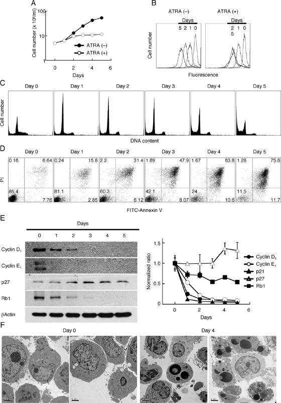
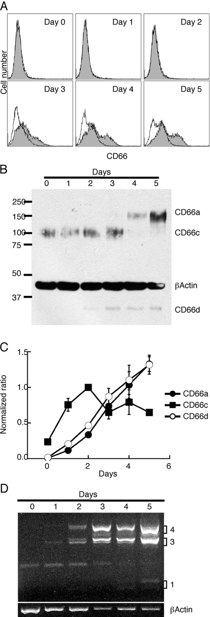
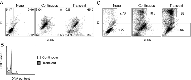
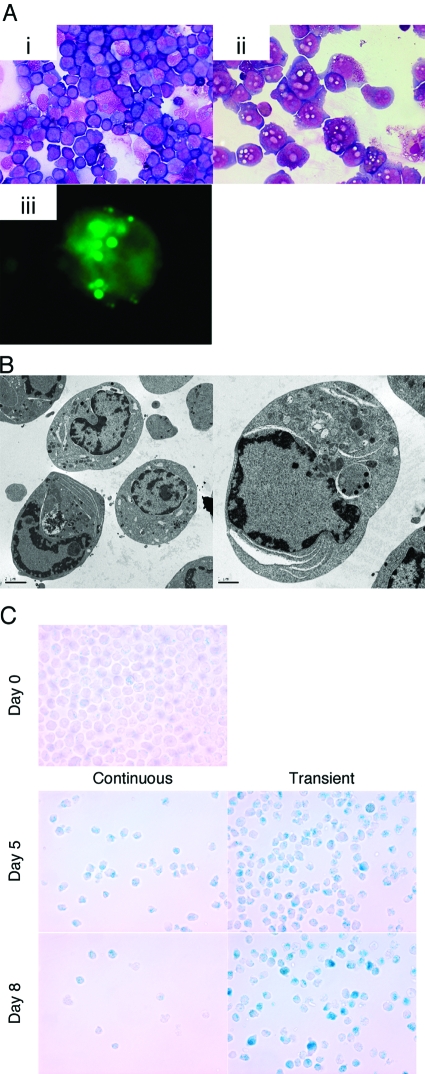
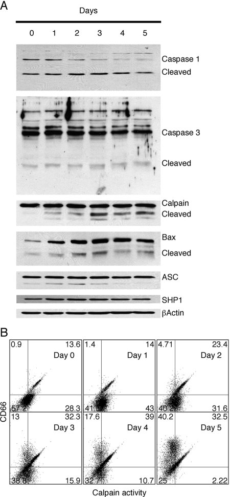
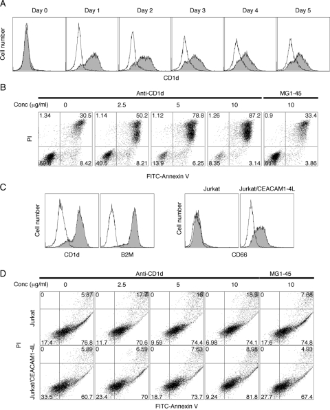
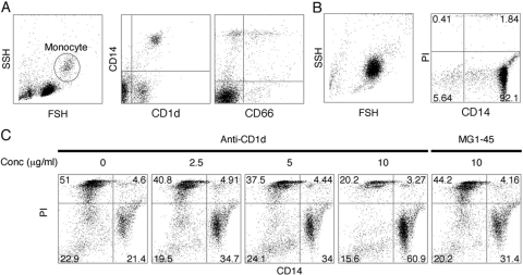
HL-60 human leukemia cells, differentiated into a neutrophil lineage by all-trans retinoic acid (ATRA) treatment, express three members of the carcinoembryonic antigen (CEA) gene family, CEA-related cell adhesion molecule 1 (CEACAM1; CD66a), CEACAM3 (CD66d), and CEACAM6 (CD66c). CD66d is a neutrophil lineage-specific marker, and CD66a and CD66c are found on epithelial and other cells. HL-60 cells continuously treated with ATRA underwent apoptosis, and cells transiently treated for 1 day underwent cell-cycle arrest, entered into senescence, and exhibited reduced apoptosis with CD66-positive cells accounting for the majority of live cells. CD66 antigens were also induced in NB4 leukemic cells upon continuous treatment with ATRA. NB4 cells underwent apoptosis with a higher frequency in transient versus continuous-treated cells (38% vs. 19% at Day 5), in contrast to HL-60 cells that underwent cell-cycle arrest and senescence when transiently treated with ATRA. CD66 antigens were not induced in transient, ATRA-treated NB4 cells compared with HL-60 cells. Cell-cycle arrest in HL-60 cells involved reduction in expression levels of p21, cyclins D and E, while Rb1 exhibited reduction in protein levels without changes in mRNA levels over the time course of ATRA treatment. Analysis of several proapoptotic proteins implicated the activation of calpain and cleavage of Bax in the intrinsic apoptotic pathway, similar to published studies about the apoptosis of neutrophils. CD1d expression was also induced by ATRA in HL-60 cells and ligation with anti-CD1d antibody-induced apoptosis. In contrast, CD1d-positive primary monocytes were protected from spontaneous apoptosis by CD1d ligation. These studies demonstrate distinct cell fates for ATRA-treated HL-60 cells that provide new insights into ATRA-induced cell differentiation.


Figures

Fig. 1.
Fig. 1.
Induction of apoptosis in HL-60 cells treated with ATRA. HL-60 cells (5×10⁵ cells/ml) were treated with 2 μM ATRA for up to 5 days and analyzed for (A) cell number by Coulter counter analysis, (B) cell proliferation by CFSE staining, (C) cell cycle by permeabilization and PI staining, (D) apoptosis by double-staining with FITC-annexin V and PI, and (E) expression of cell-cycle regulatory molecules by Western blotting and real-time RT-PCR. These experiments were repeated at least twice, and the representative results are shown. (F) EM (original magnification, ×1100) was performed on ATRA-treated HL-60 cells at Days 0 and 4. Note the presence of bilobed nuclei and abundant cytoplasmic granules in treated cells, as well as apoptotic cells with condensed nuclei with intact plasma membranes.
Fig. 2.
Fig. 2.
CD66 expression in ATRA-treated HL-60 cells. Time course of expression of (A) CD66a, -c, and -d by FACS analysis; (B) CD66a, -c, and -d by Western blot analysis; (C) CD66a, -c, and -d by qRT-PCR; and (D) CD66a isoforms by RT-PCR on ATRA-treated HL-60 cells. RT-PCR was normalized to β-actin expression. All experiments were repeated at least twice with similar results.
Fig. 3.
Fig. 3.
Comparison of continuous versus transient ATRA treatment of HL-60 cells, which (A) were untreated (None), continuously treated with ATRA (2 μM) for 9 days, or transiently treated with ATRA for 1 day, followed by culturing in ATRA-free medium for an additional 8 days. All cells were double-stained for CD66 and PI and analyzed by FACS at Day 9. (B) Cell-cycle analysis (permeabilized, PI) was performed at Day 11 on transiently (shaded) versus continuously ATRA-treated (open) HL-60 cells. (C) NB4 cells were untreated (None), continuously treated with ATRA (2 μM) for 5 days, or transiently treated with ATRA for 1 day, followed by culturing in ATRA-free medium for an additional 4 days. All cells were double-stained for CD66 and PI and analyzed by FACS at Day 5. All experiments were repeated at least twice to ensure the validity of results.
Fig. 4.
Fig. 4.
Microscopic analysis of ATRA-treated HL-60 cells, which (A) when treated transiently with ATRA, were stained with PI at Day 4. PI-negative cells were separated by Moflo and stained with Giemsa’s stain. (i) Untreated cells; (ii) PI-negative cells; (iii) lipid particles in cytosol were detected by BODIPY staining on transiently treated HL-60 cells at Day 4. (B) Further structure was analyzed by EM. (C) Untreated (Day 0), transiently treated, or continuously ATRA-treated HL-60 cells were stained for β-Gal using X-Gal at pH 6.0 for the presence of SA-β-Gal at Days 5 and 8. Note the decreased cell numbers in the continuously treated cells and the presence of blue-staining cells in the transiently treated cells. This experiment was repeated at least twice with similar results.
Fig. 5.
Fig. 5.
Analysis of ATRA-treated HL-60 cell for markers of apoptosis. (A) HL-60 cells were treated with 2 μM ATRA for 0–5 days and analyzed by Western blot analysis for expression of caspase-1, caspase-3, calpain, Bax, ASC, and SHP1. In each case, note the presence or absence of the cleaved, activated form of the proapoptotic protein. (B) FACS analysis of ATRA-treated cells was performed by double-staining with the calpain substrate rhodamine 110, bis-(t-BOC-L-leucyl-L-methionine amide), and CD66 antibody T84.1 All experiments were repeated at least twice, and similar results were obtained.
Fig. 6.
Fig. 6.
CD1d induction and apoptosis with its stimulation on leukemic cell lines. HL-60 cells were treated with 2 μM ATRA for 0–5 days, continuously or transiently. (A) CD1d expression (shaded) was determined by FACS. The isotype control is shown (open). (B) ATRA-treated HL-60 cells were stimulated with anti-CD1d antibody at Day 1 for an additional 2 days. Apoptosis was analyzed by FITC-annexin V/PI double-staining. MG1-45 isotype antibody was used as negative control. (C) CD1d, B2M, and CD66 expressions in Jurkat cells were measured by FACS. (D) Jurkat cells transfected with or without CEACAM1-4L were stimulated with anti-CD1d antibody for 2 days, and apoptosis was analyzed with FITC-annexin V/PI double-staining. All experiments were repeated at least twice with consistent results.
Fig. 7.
Fig. 7.
CD1d stimulation on human primary monocytes. (A) CD1d and CD66 expression on human primary monocytes, which (B) were isolated by immunomagnetic purification. Purity and viability were evaluated by CD14/PI staining. (C) Isolated monocytes were stimulated with anti-CD1d antibody overnight, and then cell viability was determined by CD14/PI staining. All experiments were repeated at least twice, and a typical result is shown here. SSH, side scatter height; FSH, forward scatter height.

Similar articles

Cited by

References

    1. Mehta K. Retinoids as regulators of gene transcription. J Biol Regul Homeost Agents. 2003;17:1–12. - PubMed
    1. Puccetti E, Ruthardt M. Acute promyelocytic leukemia: PML/RARα and the leukemic stem cell. Leukemia. 2004;18:1169–1175. - PubMed
    1. Tallman M S, Nabhan C, Feusner J H, Rowe J M. Acute promyelocytic leukemia: evolving therapeutic strategies. Blood. 2002;99:759–767. - PubMed
    1. Roninson I B, Dokmanovic M. Induction of senescence-associated growth inhibitors in the tumor-suppressive function of retinoids. J Cell Biochem. 2003;88:83–94. - PubMed
    1. Wright W E, Shay J W. Cellular senescence as a tumor-protection mechanism: the essential role of counting. Curr Opin Genet Dev. 2001;11:98–103. - PubMed

Publication types

MeSH terms