. 2008 Sep 1;112(5):2071-80.
doi: 10.1182/blood-2007-12-127480. Epub 2008 Jun 13.

Hematopoietic-specific Stat5-null mice display microcytic hypochromic anemia associated with reduced transferrin receptor gene expression

Affiliations

Hematopoietic-specific Stat5-null mice display microcytic hypochromic anemia associated with reduced transferrin receptor gene expression

Bing-Mei Zhu et al. Blood. .

Abstract

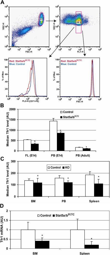
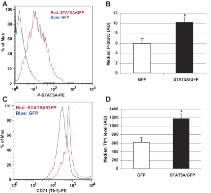
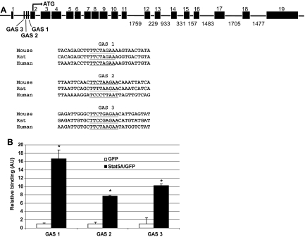
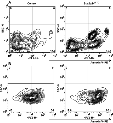
Iron is essential for all cells but is toxic in excess, so iron absorption and distribution are tightly regulated. Serum iron is bound to transferrin and enters erythroid cells primarily via receptor-mediated endocytosis of the transferrin receptor (Tfr1). Tfr1 is essential for developing erythrocytes and reduced Tfr1 expression is associated with anemia. The transcription factors STAT5A/B are activated by many cytokines, including erythropoietin. Stat5a/b(-/-) mice are severely anemic and die perinatally, but no link has been made to iron homeostasis. To study the function of STAT5A/B in vivo, we deleted the floxed Stat5a/b locus in hematopoietic cells with a Tie2-Cre transgene. These mice exhibited microcytic, hypochromic anemia, as did lethally irradiated mice that received a transplant of Stat5a/b(-/-) fetal liver cells. Flow cytometry and RNA analyses of erythroid cells from mutant mice revealed a 50% reduction in Tfr1 mRNA and protein. We detected STAT5A/B binding sites in the first intron of the Tfr1 gene and found that expression of constitutively active STAT5A in an erythroid cell line increased Tfr1 levels. Chromatin immunoprecipitation experiments confirmed the binding of STAT5A/B to these sites. We conclude that STAT5A/B is an important regulator of iron update in erythroid progenitor cells via its control of Tfr1 transcription.


Figures

Figure 1
Figure 1
Peripheral blood smears from control, Stat5a/bf/f; TC, and Stat5a/b−/− E14.5 embryos and neonates, subjected to Giemsa staining. Peripheral blood smears revealed microcytosis, anisocytosis, and hypochromia in Stat5a/b mutant mice, suggestive of iron deficiency. An increase in nucleated erythroblasts (long →) and reticulocytes (short →) further indicates stressed erythropoiesis in mutant mice. formula image 
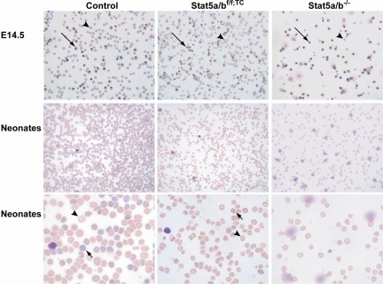
indicates mature red blood cells. Original magnifications: top and middle panels, ×200; bottom panel, ×600.
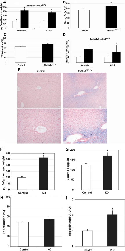
Figure 2
Figure 2
Iron overload in the absence of STAT5A/B. Iron content and hepcidin expression in control and Stat5a/bf/f; TC mice (A-E) and in mice that received a transplant of control or Stat5a/b−/− fetal liver cells (F-I). (A) Non–heme liver iron in adult and neonate female control and Stat5a/bf/f; TC mice. n = 5 neonates; n = 6 adults. (B,C) Serum iron (B) and transferrin saturation (C) in adult female control and Stat5a/bf/f; TC mice. n = 12 in each group. (D) Hepcidin mRNA expression in neonate and adult control and Stat5a/bf/f; TC mice (males and females). Hepcidin expression in control neonate and adult mice is set to 1. n = 9 in each group. (E) Perls Prussian blue iron staining of liver paraffin sections from adult female control and Stat5a/bf/f; TC mice. Original magnification, ×100 in top panel and ×200 in bottom panel. Non–heme iron is stained blue. (F) Non–heme liver iron. (G) Serum iron. (H) Serum transferrin saturation. (I) Hepcidin mRNA expression. Hepcidin expression in mice that received a transplant of control cells is set to 1. n = 5 in each group for all the measurements. *P < .05 compared with control samples. P values were determined by 2-tailed unpaired t test. Error bars represent SEM.
Figure 3
Figure 3
Decreased Tfr expression in the absence of STAT5A/B. Transferrin receptor expression in control and Stat5a/b mutant Ter119-positive cells and (A-C) transferrin receptor mRNA expression in Stat5a/bf/f; TC and control adult mice (D). (A,B) Gated Ter119-positive cells were analyzed for fluorescence of an α-CD71 (Tfr1) antibody in fetal liver and adult bone marrow and spleen cells. Peripheral blood data were generated by double staining with the α-CD71 antibody and thiazole orange dye, which stains RNA and can be used to isolate reticulocytes of a narrow age range as the amount of RNA changes significantly during maturation. (A) Representative data from 8 adult peripheral blood analyses. Top panels show the gate for thiazole orange–positive cells (right) in the whole red blood cell population (left panel). (B) Quantification of Tfr1 protein levels, expressed as the median measurements of α-CD71 fluorescence. E14 data were generated from Stat5a/b−/− embryos. n = 10 in each group. Adult data were generated from Stat5a/bf/f; TC mice. n = 6 for each group. (C) Quantification of Tfr1 protein levels in mice that received a transplant of control or Stat5a/b−/− fetal liver cells, expressed as the median measurements of α-CD71 fluorescence. n = 5 for each group. FL indicates fetal liver; PB, peripheral blood; and BM, bone marrow. *P < .05 compared with control mice. Error bars represent SEM. (D) Total RNA was extracted from Ter119-positive cells isolated from bone marrow and spleen. Tfr1 expression levels in control mice are set to 1. n = 3 in control group; n = 4 in Stat5a/bf/f; TC group. *P < .05 compared with control mice. Error bars represent SEM.
Figure 4
Figure 4
Transferrin receptor expression in murine erythroleukemia (MEL) cells transfected with a plasmid encoding constitutively active STAT5A. MEL cells were transfected with a plasmid encoding constitutively active STAT5A and GFP or GFP alone. (A,C) Histogram from FACS analyses of gated GFP-positive cells. (A) Phospho-STAT5A levels in MEL cells. (C) Transferrin receptor (CD71) expression in MEL cells. Quantification of phospho-STAT5A (B) and Tfr1 levels (D) in GFP- and STAT5A/GFP-transfected cells. Representative data are from 1 of 3 individual experiments. Error bars represent SEM. *P < .05 compared with GFP-transfected cells.
Figure 5
Figure 5
STAT5A/B binding sites in a putative Tfr1 enhancer. (A) Putative STAT5A/B binding (GAS) sites within intron 1 of the Tfr1 gene (underlined). Three conserved GAS sites were identified, with GAS 1 having the highest interspecies conservation. Sites are shown aligned with sequences from other species. (B) Chromatin immunoprecipitation (ChIP) analysis of binding of active STAT5A to the putative GAS sites within intron 1 of Tfr1. Biologic and technical triplicates were performed. Error bars represent SEM. *P < .05 compared with GFP-transfected cells.
Figure 6
Figure 6
Apoptosis in Stat5a/bf/f; TC and control adult splenic erythroid cells. (A) Freshly isolated splenic Ter119-positive cells were analyzed by FACS using an apoptosis detection kit and Ter119 staining. (B) After 24 hours in culture with Epo, cells were subjected to the same analysis. The left panels are from control mice; the right panels are from Stat5a/bf/f; TC mice. Populations shown were gated as Ter119 positive and 7AAD negative. Representative profile from 1 of 3 individual experiments.

Comment in

  • GASing up iron delivery via STAT5.
    Friedman JS. Friedman JS. Blood. 2008 Sep 1;112(5):1553-4. doi: 10.1182/blood-2008-07-167965. Blood. 2008. PMID: 18725572 No abstract available.

References

    1. Hentze MW, Muckenthaler MU, Andrews NC. Balancing acts: molecular control of mammalian iron metabolism. Cell. 2004;117:285–297. - PubMed
    1. Levy JE, Jin O, Fujiwara Y, Kuo F, Andrews NC. Transferrin receptor is necessary for development of erythrocytes and the nervous system. Nat Genet. 1999;21:396–399. - PubMed
    1. Andrews NC, Schmidt PJ. Iron homeostasis. Annu Rev Physiol. 2007;69:69–85. - PubMed
    1. Neubauer H, Cumano A, Muller M, Wu H, Huffstadt U, Pfeffer K. Jak2 deficiency defines an essential developmental checkpoint in definitive hematopoiesis. Cell. 1998;93:397–409. - PubMed
    1. Parganas E, Wang D, Stravopodis D, et al. Jak2 is essential for signaling through a variety of cytokine receptors. Cell. 1998;93:385–395. - PubMed

Publication types

MeSH terms