. 2008 Jul;9(7):676-82.
doi: 10.1038/embor.2008.79. Epub 2008 Jun 13.

Drosophila RalA is essential for the maintenance of Jak/Stat signalling in ovarian follicles

Affiliations

Drosophila RalA is essential for the maintenance of Jak/Stat signalling in ovarian follicles

Christian Ghiglione et al. EMBO Rep. 2008 Jul.

Abstract

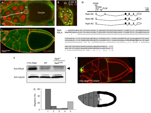
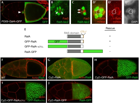
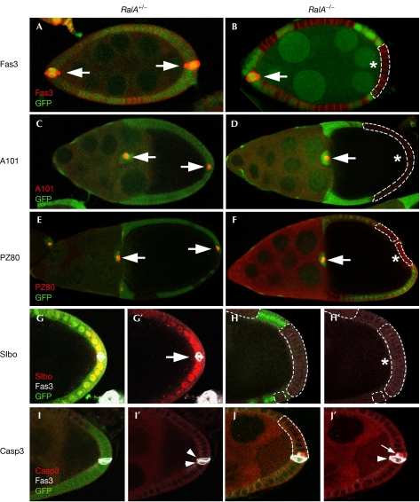
Small GTPases of the Ras-like (Ral) family are crucial for signalling functions in both normal and cancer cells; however, their role in a developing organism is poorly understood. Here, we identify the Drosophila Ral homologue RalA as a new key regulator of polar-cell differentiation during oogenesis. Polar cells have a crucial role in patterning the egg chamber and in recruiting border cells, which undergo collective and guided migration. We show that RalA function is essential for the maintenance of anterior and posterior polar-cell fate and survival. RalA is required cell autonomously to control the expression of polar-cell-specific markers, including the Jak/Stat ligand Unpaired. The loss of RalA also causes a cell non-autonomous phenotype owing to reduced Jak/Stat signalling in neighbouring follicle cells. As a result, border-cell assembly and migration as well as the polarization of the oocyte are defective. Thus, RalA is required in organizing centres to control proper patterning and migration in vivo.


Conflict of interest statement

The authors declare that they have no conflict of interest.

Figures

Figure 1
Figure 1
RalA controls border-cell migration in egg chambers. (A) Wild-type (WT) stage-10 egg chamber stained with phalloidin (green) and propidium iodide (red). Border cells (BCs; enclosed by dotted lines) have reached the anterior region of the oocyte, after delaminating from the anterior epithelium and migrating through the nurse-cell compartment (arrow). (B) BC cluster organization showing two centrally located polar cells (PCs; circles) surrounded by six outer BCs (oBCs; asterisks). (C) Mosaic stage-10 egg chamber containing homozygous RalAPG69 cells (GFP–negative). Note the absence of migration of BCs (enclosed by dotted lines). Actin is shown in red. (D) Schematic view of the genomic organization of the RalA locus and ClustalW alignment of RalA and human RALA (accession no. EAL23994) protein sequences. (E) Western blot of ovary extracts. RalA (black arrowhead) is detected using a polyclonal antibody directed against the human RALB protein. Tubulin antibodies were used to assess protein levels in each lane. (F) Expression of UAS–RalAS25N in BCs (enclosed by dotted lines) blocks their migration. (G) Stage-10 migration defects following expression of RalAS25N in BCs. The nurse-cell compartment (dark grey) has been divided into five regions (1–5) to determine the extent of migration. GFP, green fluorescent protein.
Figure 2
Figure 2
Expression of RalA and sequence determinants for RalA subcellular localization. (A) Expression of RalA in border cells (BCs) and follicle cells was detected using a RalA–Gal4 enhancer trap (RalAPG69) crossed to UAS–GFP. Expression of RalA–LacZ in BCs (B) during or (C) when migration has completed using the RalAPL56 enhancer-trap line. Expression of β-galactosidase is detected in outer BCs (oBCs) as well as in polar cells (PCs). Fas3 staining is used to mark PCs (red). (DD″) Fluorescent in situ hybridization of a stage-9 egg chamber using an antisense RalA probe, showing expression in PCs and oBCs. Red indicates RalA mRNA and green EYA (marks oBCs but not PCs, enclosed by dotted lines). (E) Schematic representation of RalA wild-type and GFP fusion proteins, showing the RAS domain and the CTLL motif at the C terminus. Each RalA construct was used in rescue experiments to assess their activity (see text for details). (F) The human RALB antibody (Fig 1E) does not detect endogenous levels of RalA expression but recognizes overexpressed wild-type RalA (using CY2–Gal4), which localizes to the plasma membrane (G). GFP–RalA expressed using CY2–Gal4 also localizes to the plasma membrane (H). (I) GFP–RalAΔCTLL, in which the last four C-terminal amino acids are deleted, is mislocalized and accumulated predominantly in the nucleus. (J) RalA–GFP fusion protein is also mislocalized in the nucleus. GFP, green fluorescent protein.
Figure 3
Figure 3
Drosophila RalA is essential in polar cells for border-cell recruitment and migration. (A) Mosaic border-cell (BC) cluster in which the two polar cells (PCs; arrows in B,B′) are wild type, whereas outer BCs (oBCs; enclosed by dotted lines) are mutant for RalAPG69. Mutant cells are negative for GFP (green). PCs are marked by Fas3 (red). Stage-10 egg chamber is shown. (B,B′) Enlargement of the boxed region shown in (A). (C) BC cluster in which only one PC is wild type (arrow; GFP and Fas3 positive) does not migrate. Early stage-10 egg chamber is shown. (D,D′) Enlargement of the boxed region shown in (C). (E) The two PCs are mutant in this egg chamber and BCs did not form. (F,F′) Enlargement of the boxed region shown in (E). Stage-10A egg chambers are shown in (A,C) and a stage-10B egg chamber is shown in (E). Green indicates GFP clonal marker and grey 4′,6-diamidino-2-phenylindole (DAPI) staining. The dotted lines indicate clonal limits. The table shows a summary of RalA-induced BC phenotypes shown in (AF′). Left column, number of wild-type PCs; middle column, number of recruited oBCs; right column, migration phenotype (+, wild type; −, no migration). GFP, green fluorescent protein.
Figure 4
Figure 4
Drosophila RalA controls polar-cell and terminal follicle cell differentiation. Specific markers for polar cells (PCs; Fas3, A101–lacZ, PZ80–lacZ) and terminal follicle cells (Slbo–lacZ) were used to analyse cell fate in RalA homozygous follicle cells (B,D,F,HH′,JJ′), as compared with heterozygous cells (A,C,E,GG′,I–I′). The expression of Fas3 (A,B), A101–lacZ (C,D) and PZ80–lacZ (E,F) is completely lost when PCs are mutant for RalA. The RalA mutations also lead to a complete loss of expression of the terminal follicle cell marker Slbo–lacZ (GG′,H–H′). PCs that are mutant for RalA show a specific activation of Casp3 (JJ′; in red; white arrow) and this is not observed in wild-type PCs at this late stage (II′; white arrowheads). Green indicates GFP clonal marker and grey Fas3 staining (GJ′). The dotted lines indicate clonal limits and the asterisks mutant PCs. GFP, green fluorescent protein.
Figure 5
Figure 5
RalA mutant polar cells block Jak/Stat signalling and oocyte anterior–posterior polarization. (A,A′) Mosaic egg chamber in which the two anterior polar cells (PCs) are wild type (enclosed by dotted lines). Domeless (Dome; in red) is found in intracellular vesicles (arrows). (B,B′) Mosaic egg chamber in which the two anterior PCs are mutant for RalA (asterisk). Note the absence of Dome endocytic vesicles. Mutant cells do not express the clonal marker GFP (green). PCs are marked with Fas3 (green). (C,C′) Graded nuclear expression of Stat in a heterozygous RalA+/− egg chamber. (D,D′) Expression of Stat is not affected in a mosaic egg chamber in which the two posterior PCs are wild type (GFP, clonal marker). (E) Nuclear Stat is completely lost when the two PCs are mutant for RalA. In (CE), red denotes Fas3 staining and grey Stat staining. (F,G) The Unpaired (Upd) ligand (in red) forms a gradient centred at the poles in heterozygous egg chambers (PCs are marked by Fas3 in green). By contrast, Upd expression is strongly reduced when the two PCs are mutant for RalA (G). (H,I) When posterior PCs are mutant, the oocyte shows anterior–posterior polarization defects, as evidenced by the mislocalization of the Staufen protein (in red). Green indicates GFP clonal marker. GFP, green fluorescent protein.

References

    1. Balakireva M et al. (2006) The Ral/exocyst effector complex counters c-Jun N-terminal kinase-dependent apoptosis in Drosophila melanogaster. Mol Cell Biol 26: 8953–8963 - PMC - PubMed
    1. Beccari S, Teixeira L, Rorth P (2002) The JAK/STAT pathway is required for border cell migration during Drosophila oogenesis. Mech Dev 111: 115–123 - PubMed
    1. Besse F, Pret AM (2003) Apoptosis-mediated cell death within the ovarian polar cell lineage of Drosophila melanogaster. Development 130: 1017–1027 - PubMed
    1. Bianco A, Poukkula M, Cliffe A, Mathieu J, Luque CM, Fulga TA, Rørth P (2007) Two distinct modes of guidance signalling during collective migration of border cells. Nature 448: 362–365 - PubMed
    1. Bourbon HM et al. (2002) A P-insertion screen identifying novel X-linked essential genes in Drosophila. Mech Dev 110: 71–83 - PubMed

Publication types