Nuclear to non-nuclear Pmel17/gp100 expression (HMB45 staining) as a discriminator between benign and malignant melanocytic lesions
- PMID: 18552823
- PMCID: PMC2570478
- DOI: 10.1038/modpathol.2008.100
Nuclear to non-nuclear Pmel17/gp100 expression (HMB45 staining) as a discriminator between benign and malignant melanocytic lesions
Abstract
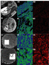
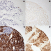
HMB45 is a mouse monoclonal antibody raised against Pmel17/gp100, a melanoma-specific marker, which is routinely used in the diagnosis of primary cutaneous malignant melanoma. The standard expression pattern for a positive HMB45 staining result on immunohistochemistry is based upon the results of chromogenic-based methods. We re-evaluated the patterns of HMB45 staining across the 480-core 'SPORE melanoma progression array' containing lesions representing the spectrum of melanocytic lesions ranging from thin nevus to visceral metastasis using the fluorescence-based staining technique and automated quantitative analysis (AQUA) of the obtained digital images. The methods validated the expected cytoplasmic HMB45 staining pattern in 70/108 malignant lesions and in the epithelial components of nevus specimens. However, the fluorescence-based approach revealed a nuclear gp100 localization present in the dermal component of all nevi that was not seen before. This nuclear localization could not be observed on routine chromogenic stains, because the standard hematoxylin nuclear counterstain overwhelms the weak nuclear HMB45 stain. The thin (0.450+/-0.253) and thick (0.513+/-0.227) nevi had strongly positive mean ln(nuclear/non-nuclear AQUA score ratios), which are significantly higher than those from the group of malignant lesions (P<0.0001). This finding was reproduced on a smaller but independent progression array composed of nevi and melanomas from the Yale Pathology archives (P<0.01). The odds ratio associated with a sample being a nevus was 2.24 (95% CI: 1.87-2.69, P<0.0001) for each 0.1 unit increase of the ln(nuclear/non-nuclear AQUA score ratio) to yield an ROC curve with 0.93 units of area and a simultaneously maximized sensitivity of 0.92 and specificity of 0.80 for distinguishing benign nevi from malignant melanomas. On the basis of this preliminary study, we propose that the ratio of nuclear to non-nuclear HMB45 staining may be useful for diagnostic challenges in melanocytic lesions.
Figures


References
-
- Hoashi T, Muller J, Vieira WD, Rouzaud F, Kikuchi K, Tamaki K, Hearing VJ. The repeat domain of the melanosomal matrix protein PMEL17/GP100 is required for the formation of organellar fibers. J Biol Chem. 2006;281:21198–208. - PubMed
-
- Valencia JC, Hoashi T, Pawelek JM, Solano F, Hearing VJ. Pmel17: controversial indeed but critical to melanocyte function. Pigment Cell Res. 2006;19:250–2. author reply 3-7. - PubMed
-
- Satzger I, Volker B, Meier A, Schenck F, Kapp A, Gutzmer R. Prognostic significance of isolated HMB45 or Melan A positive cells in Melanoma sentinel lymph nodes. Am J Surg Pathol. 2007;31:1175–80. - PubMed
-
- Yaziji H, Gown AM. Immunohistochemical markers of melanocytic tumors. Int J Surg Pathol. 2003;11:11–5. - PubMed
Publication types
MeSH terms
Substances
Grants and funding
LinkOut - more resources
Full Text Sources
Other Literature Sources
Medical

