

Imbalance between pSmad3 and Notch induces CDK inhibitors in old muscle stem cells
- PMID: 18552838
- PMCID: PMC7761661
- DOI: 10.1038/nature07034
Imbalance between pSmad3 and Notch induces CDK inhibitors in old muscle stem cells
Abstract
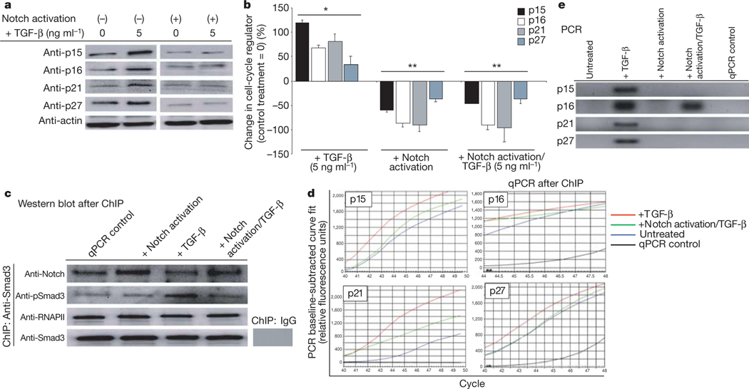
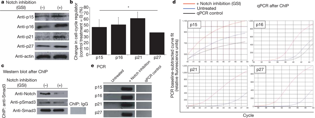
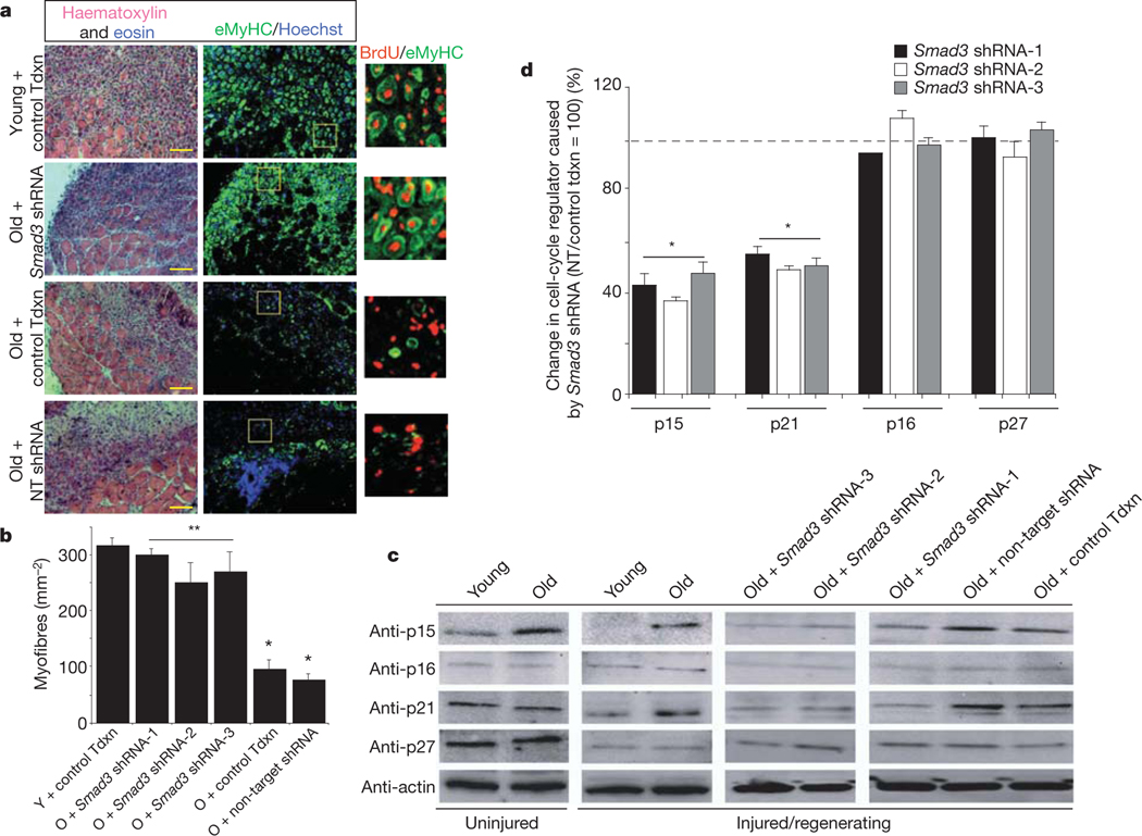
Adult skeletal muscle robustly regenerates throughout an organism's life, but as the muscle ages, its ability to repair diminishes and eventually fails. Previous work suggests that the regenerative potential of muscle stem cells (satellite cells) is not triggered in the old muscle because of a decline in Notch activation, and that it can be rejuvenated by forced local activation of Notch. Here we report that, in addition to the loss of Notch activation, old muscle produces excessive transforming growth factor (TGF)-beta (but not myostatin), which induces unusually high levels of TGF-beta pSmad3 in resident satellite cells and interferes with their regenerative capacity. Importantly, endogenous Notch and pSmad3 antagonize each other in the control of satellite-cell proliferation, such that activation of Notch blocks the TGF-beta-dependent upregulation of the cyclin-dependent kinase (CDK) inhibitors p15, p16, p21 and p27, whereas inhibition of Notch induces them. Furthermore, in muscle stem cells, Notch activity determines the binding of pSmad3 to the promoters of these negative regulators of cell-cycle progression. Attenuation of TGF-beta/pSmad3 in old, injured muscle restores regeneration to satellite cells in vivo. Thus a balance between endogenous pSmad3 and active Notch controls the regenerative competence of muscle stem cells, and deregulation of this balance in the old muscle microniche interferes with regeneration.
Figures

References
-
- Grounds MD Age-associated changes in the response of skeletal muscle cells to exercise and regeneration. Ann. NY Acad. Sci 854, 78–91 (1998). - PubMed
-
- Renault V, Thornell LE, Eriksson PO, Butler-Browne G & Mouly V. Regenerative potential of human skeletal muscle during aging. Aging Cell 1, 132–139 (2002). - PubMed
-
- Conboy IM, Conboy MJ, Smythe GM & Rando TA Notch-mediated restoration of regenerative potential to aged muscle. Science 302, 1575–1577 (2003). - PubMed
-
- Wagers AJ & Conboy IM Cellular and molecular signatures of muscle regeneration: current concepts and controversies in adult myogenesis. Cell 122, 659–667 (2005). - PubMed
-
- Collins CA & Partridge TA Self-renewal of the adult skeletal muscle satellite cell. Cell Cycle 4, 1338–1341 (2005). - PubMed
Publication types
MeSH terms
Substances
Grants and funding
LinkOut - more resources
Full Text Sources
Other Literature Sources
