. 2008 Jul;40(7):871-9.
doi: 10.1038/ng.104. Epub 2008 Jun 15.

Dishevelled controls apical docking and planar polarization of basal bodies in ciliated epithelial cells

Affiliations

Dishevelled controls apical docking and planar polarization of basal bodies in ciliated epithelial cells

Tae Joo Park et al. Nat Genet. 2008 Jul.

Abstract

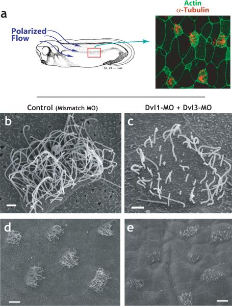
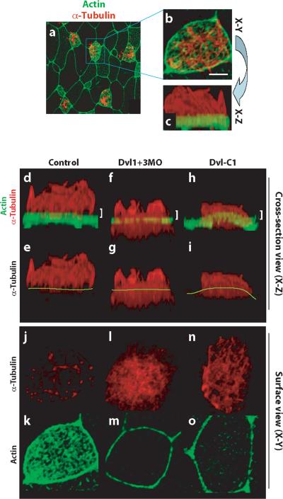
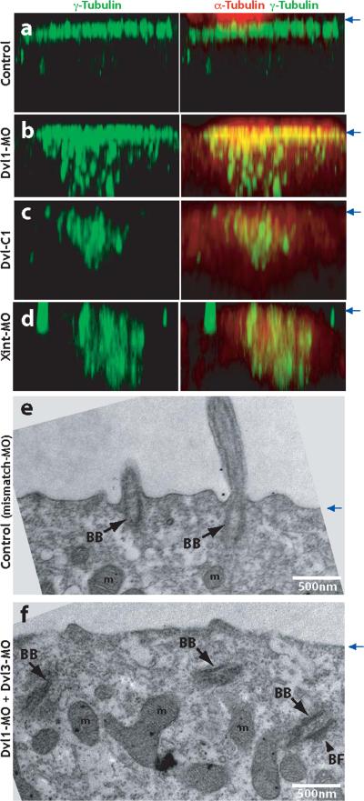
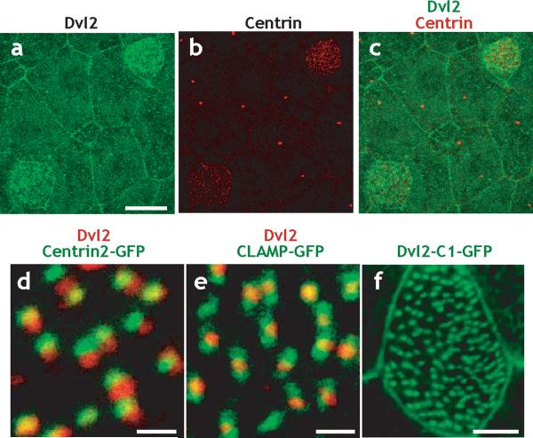
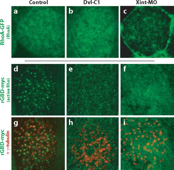
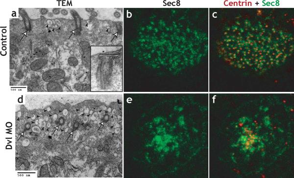
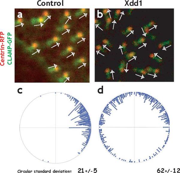
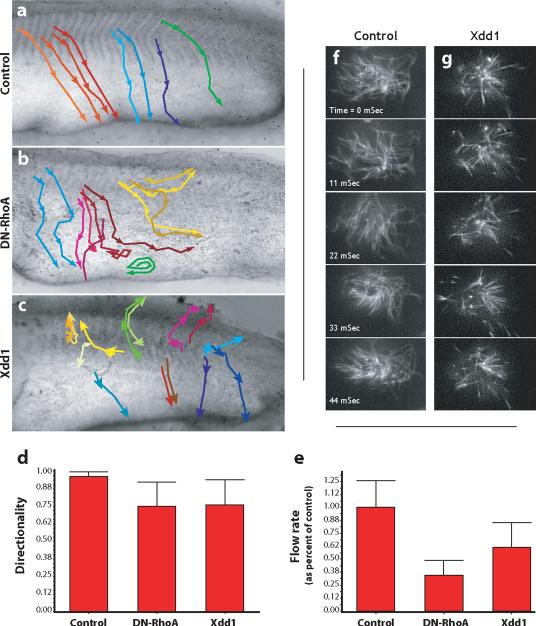
The planar cell polarity (PCP) signaling system governs many aspects of polarized cell behavior. Here, we use an in vivo model of vertebrate mucociliary epithelial development to show that Dishevelled (Dvl) is essential for the apical positioning of basal bodies. We find that Dvl and Inturned mediate the activation of the Rho GTPase specifically at basal bodies, and that these three proteins together mediate the docking of basal bodies to the apical plasma membrane. Moreover, we find that this docking involves a Dvl-dependent association of basal bodies with membrane-bound vesicles and the vesicle-trafficking protein, Sec8. Once docked, basal bodies again require Dvl and Rho for the planar polarization that underlies directional beating of cilia. These results demonstrate previously undescribed functions for PCP signaling components and suggest that a common signaling apparatus governs both apical docking and planar polarization of basal bodies.


Figures

Figure 1
Figure 1. Dishevelled is essential for ciliogenesis in multi-ciliated cells
A. The epidermis of the Xenopus embryo is composed of multi-ciliated cells and mucus secreting cells Ciliated cells, labelled by α-tubulin immunostaining (red), are evenly-spaced between mucus-secrteing cells. Polarized beating of cilia generates directional flow across the epithelium,. B. riposte2ormal ciliated cell from a control-injected embryo (injected with a mismatched morpholino-oligonucleotide). C. Defective cilia outgrowth in Dvl1/Dvl3 double morphants. D. Wider field view of control (mismatch-MO injected) embryo. E. Wider field view of Dvl1/Dvl3 double morphant embryo showing many affected cells.
Figure 2
Figure 2. Dishevelled is essential for normal cytoskeletal organization in multi-ciliated cells
A. Surface view of a 3D confocal dataset showing the ciliated epidermis of an intact Xenopus embryo. B. At higher magnification, cilia (red, α-tubulin) and an apical actin network (green, phalloidin) are visible. Scale bar = 5μm. C. Rotation of the 3D confocal dataset shown in B allows the same cell to be viewed in cross-section. D. In a control cell, axonemal microtubules are well organized and form atop dense apical actin filaments. d'. Same image as panel D, but without the green channel; yellow line indicates the apical cell surface. E and e'. Simultaneous knockdown of Dvl1 and Dvl3 disrupts ciliogenesis and results in microtubule accumulation below or at the apical surface. F and f'. Expression of Dvl-C1 disrupts ciliogenesis and results in microtubule accumulation below or at the apical surface. G. Thin confocal section at the apical surface (indicated by white bracket in D) reveals that ciliary microtubules project perpendicularly from the cell surface. g'. Actin filaments are highly enriched at the apical cell surface. H. Thin confocal section of a Dvl1/Dvl3 double morphant (indicated by white bracket in E) reveals that ciliary microtubules are interwoven parallel to the apical cell surface. h'. Apical actin filaments are lost at the surface of morphant cells, but remain at cell-cell boundaries. I. Thin confocal section of anembryo expressing Dvl-C1 (indicated by white bracket in F) reveals that ciliary microtubules are interwoven parallel to the apical cell surface. i'. Apical actin filaments are lost at the surface of DVl-C1 expressing cells, but remain at cell-cell boundaries.
Figure. 3
Figure. 3. Dishevelled and Inturned are essential for the apical positioning of basal bodies
A. Control embryos were stained with anti-γ-tubulin antibody to visualize basal bodies (green). Ciliary microtubules are visualized with anti-α-tubulin antibody (red). Serial confocal images are projected in X-Z plane, with the position of apical membrane indicated by blue arrows at right. B. Failure of apical basal body localization in Dvl1 morphants. C. Failure of apical basal body localization in Dvl-C1 expressing cells. D. Failure of apical basal body localization in Inturned morphants. E. TEM transverse section of control (Dvl1 mismatch MO injected) embryo reveals normal outgrowth of ciliary axonemes from basal bodies docked at the apical cell surface. Cilia can be observed in cross-section projecting above the apical surface. F. Basal bodies fail to dock at the apical membrane and remian in the cytoplasm in Dvl morphants. (BB: basal body, BF: basal foot, m: mitochondria).
Figure. 4
Figure. 4. Dishevelled localizes near the base of cilia in multi-ciliated cells
A. Dvl2 immunostaining reveals enrichment at cell membranes throughout the epidermis and enrichment at the apical surface of ciliated cells, as indicated by immunostaining for centrin (a', a”). Scale bar = 15μm. B. Signal for Dvl2 immunostaining (red) localizes immediately adjacent to the basal body, as indicated by Centrin2-GFP (green). Scale bar = 1μm. C. Signal for Dvl2 immunostaining (red) localizes to the center of the region marked by CLAMP-GFP (green); data in Supp. Fig. 4 indicate that CLAMP-GFP labels the ciliary rootlet. Scale bar = 1μm. D. The C-terminus of Dvl2 is sufficient to drive localization to basal body in multi-ciliated cells. Stage 27 embryos are shown throughout. Scale bar = 5μm.
Figure. 5
Figure. 5. Dvl and Inturned control the activation and localization of the Rho GTPase
A. RhoA-GFP is enriched apically in control ciliated cells. B. Apical enrichment of RhoA-GFP is normal in ciliated cells expressing Dvl-C1. C. Apical enrichment of RhoA-GFP is lost in Inturned morphant cells, while enrichment at cell-cell boundaries was not affected. D. In control cells, the active Rho sensor, rGBD-GFP, specifically localizes near the base of cilia, where α-tubulin (red in d') appears as puncta due to the perpendicular projection of ciliary axonemes. E. Disruption of Dvl function signficantly reduces the number of rGBD-GFP foci, but does not affect signal at cell-cell junctions. Loss of rGBD-GFP foci correlates with accumulation of interwoven microtubules (red in e') parallel to and below the apical cell surface. F. Disruption of Inturned function signficantly reduces the number of rGBD-GFP foci, but does not affect signal at cell-cell junctions. Loss of rGBD-GFP foci correlates with accumulation of interwoven microtubules (red in f') parallel to and below the apical cell surface.
Figure 6
Figure 6. Dishevelled links basal body docking to vesicle traffic and Sec8 localization
A. TEM of control embryo. Basal bodies are docked and apical cytoplasm contains very few vesicles (arrowheads). B. TEM of Dvl morphant; basal bodies have failed to dock apically, and the apical cytoplasm contains numerous vesicles (arrowheads). C. Immunostaining reveals an orderly distribution of Sec8 in the apical surface of control multi-ciliated cells. c'. The Sec8 signal (green) associates evenly with basal bodies as indicated by Centrin immunostaining (red). D. Sec8 immunostaining is disordered in Dvl morphants. d'. The Sec8 signal (green) fails to associate with basal bodies (red) in Dvl morphants.
Figure. 7
Figure. 7. Dishevelled governs planar polarization of basal bodies
A. Centrin-RFP and CLAMP-GFP localize to basal bodies and rootlets, respectively revealing the polarized morphology of the basal body complex. In control cells, basal body complexes align parallel to one another (white arrows). B. Polarization can be quantified by angular measurement of individual basal bodies. On the circular plot, each point preresents a single basal body. The circular standard deviation of these measurments is small (21), and is comparable to data obtained using TEM (See Supp. Fig. 4 and Ref. 16). C. Expression of Xdd1 severely disrupts the parallel alignment of basal bodies. D. A circular plot of the angles shows that basal bodies are pointing random directions in Xdd1 expressing cells; the circular standard deviation of these measurments is much larger than controls (62).
Figure. 8
Figure. 8. Dvl and Rho govern directional fluid flow across the ciliated epidermis
A. Control embryos were grown until St. 26 and latex bead were added to the media to visualize fluid flow over the epidermis. The movement of beads was recorded and representative beads were tracked. Representative bead movements were tracked from the horizontal myoseptum, as indicated by the colored lines. Beads move from dorso-anterior to ventral-posterior uniformly in control embryos. B. DN-RhoA expression severely disrupts directional flow; beads were turning and swirling frequently. C. Xdd1 expression severely disrupts directional flow; beads were turning and swirling frequently. D. The directionality of beads was quantified as the ratio of the linear distance between the first and last point of a track and the total diatance along the track. A straight line would be a value of 1.00. E. DN-RhoA or Xdd1 reduces the rate of bead movement across the epidermis. Finally, defects in polarized flow were evident in that even beads moving in comparatively straight paths moved at a greatly reduced rate across experimental embryos. F. Stills from a high-speed time lapse movie of beating cilia labelled with tau-GFP. G. Stills from a high-speed time lapse movie of beating cilia expressing Xdd1, frequency is comparable to control.

References

    1. Chilvers MA, O'Callaghan C. Local mucociliary defence mechanisms. Paediatr Respir Rev. 2000;1:27–34. - PubMed
    1. Hayes JM, et al. Identification of novel ciliogenesis factors using a new in vivo model for mucociliary epithelial development. Dev Biol. 2007;312:115–30. - PMC - PubMed
    1. Steinman RM. An electron microscopic study of ciliogenesis in developing epidermis and trachea in the embryo of Xenopus laevis. Am J Anat. 1968;122:19–55. - PubMed
    1. Raji AR, Naserpour M. Light and Electron Microscopic Studies of the Trachea in the One-Humped Camel (Camelus dromedarius) Anat Histol Embryol. 2007;36:10–3. - PubMed
    1. Dawe HR, Farr H, Gull K. Centriole/basal body morphogenesis and migration during ciliogenesis in animal cells. J Cell Sci. 2007;120:7–15. - PubMed

Publication types

MeSH terms