. 2008 Sep;155(2):253-64.
doi: 10.1038/bjp.2008.246. Epub 2008 Jun 16.

Interactions between CB(1) receptors and TRPV1 channels mediated by 12-HPETE are cytotoxic to mesencephalic dopaminergic neurons

Affiliations

Interactions between CB(1) receptors and TRPV1 channels mediated by 12-HPETE are cytotoxic to mesencephalic dopaminergic neurons

S R Kim et al. Br J Pharmacol. 2008 Sep.

Abstract

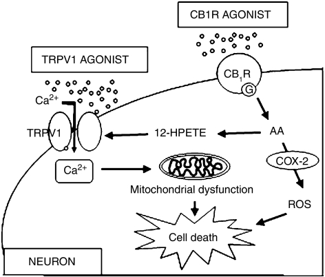
Background and purposes: We recently proposed the existence of neurotoxic interactions between the cannabinoid type 1 (CB(1)) receptor and transient receptor potential vanilloid 1 (TRPV1) channels in rat mesencephalic cultures. This study seeks evidence for the mediator(s) and mechanisms underlying the neurotoxic interactions between CB(1) receptors and TRPV1 in vitro and in vivo.

Experimental approach: The mediator(s) and mechanism(s) for the interactions between CB(1) receptors and TRPV1 were evaluated by cell viability assays, immunocytochemistry, Fura-2 calcium imaging, mitochondrial morphology assay, ELISA and Western blot assay in vitro in neuron-enriched mesencephalic cultures. Injections into the substantia nigra and subsequent cell counts were also used to confirm these interactions in vivo.

Key results: The neurotoxic interactions were mediated by 12(S)-hydroperoxyeicosatetraenoic acid (12(S)-HPETE), an endogenous TRPV1 agonist. CB(1) receptor agonists (HU210 and WIN55,212-2) increased the level of 12(S)-hydroxyeicosatetraenoic acid (12(S)-HETE), a downstream metabolite of 12(S)-HPETE, which stimulates TRPV1-mediated death of mesencephalic neurons, both in vitro and in vivo. The neurotoxicity was mediated by increased intracellular Ca(2+) concentration ([Ca(2+)](i)) through TRPV1, consequently leading to mitochondrial damage and was attenuated by baicalein, a 12-lipoxygenase inhibitor.

Conclusion and implications: Activation of CB(1) receptors in rat mesencephalic neurons was associated with biosynthesis of 12(S)-HPETE, which in turn stimulated TRPV1 activity, leading to increased [Ca(2+)](i), mitochondrial damage and neuronal death.


Figures

Figure 1
Figure 1
Neurotoxicity induced by WIN55,212-2 or HU210 in neuron-enriched mesencephalic cultures. Cultures were treated with 0.5% dimethyl sulphoxide (DMSO) as a control (ac), 6 μM WIN55,212-2 (WIN) (di) or 3 μM HU210 for 24 h. Where indicated, cells were pretreated with 5 μM transient receptor potential vanilloid subtype 1 (TRPV1) antagonist capsazepine (CZP; gi) or 5 μM CB1 receptor antagonist AM251 before 5 min, and stained with calcein-acetoxymethyl ester (calcein-AM, green for live; a, d, g) and ethidium homodimer-1 (Eth-1, red for dead; b, e, h), or immunostained with TH antibody (c, f, i), respectively. In all cultures, cell death was assessed by counting the number of Eth-1-positive dead cells or TH-immunopositive (ip) cells (j). All values represent the mean±s.e.mean of triplicate cultures in four separate platings. §P<0.001, significant from control; *P<0.01 and **P<0.05, significant from treatment with HU210 or WIN55,212-2. Scale bars: ab, de, gh, 40 μm; c, f, i, 100 μm. A colour version of this figure is available online.
Figure 2
Figure 2
Fluorescence in Fura-2 loaded cultured mesencephalic neurons. Intracellular Ca2+ concentration was measured in neuron-enriched mesencephalic cultures treated with 3 μM HU210 (left panel, arrowhead) or 6 μM WIN55,212-2 (right panel, arrowhead) in the presence (ab) or absence (cd) of 1.8 mM extracellular Ca2+. Response to 3 μM HU210 (e) or 6 μM WIN55,212-2 (f) of cultures pretreated with 5 μM capsazepine (CZP: arrow) in the presence of 1.8 mM extracellular Ca2+. Data were averaged from 20 to 25 randomly selected cells for each condition and the results shown are representative of four independent experiments. (gj) Mitochondrial disruption in neuron-enriched mesencephalic cultures treated with 3 μM HU210 (i, j) or 0.5% dimethyl sulphoxide (DMSO) as a control (g, h) in the presence of 1.8 mM extracellular Ca2+. (k, l) Pretreatment with 5 μM CZP inhibited HU210-induced mitochondrial disruption. In live cells, mitochondrial structure was stained with Mito-Tracker fluorescence dye (MT). Each coloured arrow indicates the same cells. (mo) Localization of cytochrome c (green) immunoreactivity and MT (red) in cells treated with vehicle (m), 3 μM HU210 (n) or 6 μM WIN55,212-2 (WIN; o) for 12 h. (p) Western blot analysis of cytochrome c levels after treatment of cells with 3 μM HU210 or 6 μM WIN55,212-2 for 12 h. The results are representative from three independent experiments. Cyt-c, cytochrome c; Con, control; Cyto, cytosolic fraction; Mito, mitochondrial fraction. Scale bars: gl, 20 μm; mo, 30 μm. A colour version of this figure is available online.
Figure 3
Figure 3
12(S)-hydroperoxyeicosatetraenoic acid (12(S)-HPETE) mediates the neurotoxic interactions between transient receptor potential vanilloid subtype 1 (TRPV1) and cannabinoid type 1 (CB1) receptors. (a) Number of dead cells and TH-immunopositive (ip) neurons in neuron-enriched mesencephalic cultures treated with 0.5% dimethyl sulphoxide (DMSO), 3 μM HU210, or 6 μM WIN55,212-2, or co-treated with 10 μM 12-lipoxygenase inhibitor baicalein (BCL) or 10 μM 5-lipoxygenase inhibitor REV-5901 (REV), and stained with calcein-acetoxymethyl ester and ethidium homodimer-1 (Eth-1), or immunostained with TH antibody, respectively, at 24 h later. All values represent the mean±s.e.mean of triplicate cultures in four separate platings. §P<0.001, significant from control; **P<0.05, significant from treatment with HU210 or WIN55,212-2. (be) Response to 3 μM HU210 or 6 μM WIN55,212-2 of cultures co-treated with 10 μM baicalein (bc, arrowhead) or REV (de, arrowhead) in the presence of 1.8 mM extracellular Ca2+. Data were averaged from 20 to 25 randomly selected cells for each condition and the results shown are representative of four independent experiments. (f) 12(S)-HETE-specific enzyme immunoassay showing the increase of 12(S)-HETE at 30 min after treatment with 3 μM HU210 or 6 μM WIN55,212-2 (WIN) in cultured mesencephalic neurons. CON, non-treated control. Veh, 0.5% DMSO. The statistical significance of differences was assessed using one-way ANOVA, followed by Student–Newman–Keuls analyses. All values represent the mean±s.e.mean of triplicate cultures in four separate platings. *P<0.001, significant from control (CON or Veh). A colour version of this figure is available online.
Figure 4
Figure 4
Neurotoxicity induced by 12(S)-hydroperoxyeicosatetraenoic acid (12(S)-HPETE) in neuron-enriched mesencephalic cultures. (a) Cultures were treated with 6 μM 5(S)-, 12(S)-, or 15(S)- HPETE for 24 h. Where indicated, cells were pretreated with 5 μM capsazepine (CZP) 5 min earlier than treatment with HPETEs and immunostained with TH antibody. Death of TH-immunopositive (ip) neurons was assessed by counting the number of TH-ip cells. All values represent the mean±s.e.mean of triplicate cultures in four separate platings. §P<0.001 and **P<0.01, significant from control; *P<0.01, significant from treatment with 12-HPETE. (bc) Changes of fluorescence were measured in cultures treated with 6 μM 12-HPETE (b, arrowhead), or pretreated with 5 μM CZP (arrow) before treatment with 12-HPETE (c, arrowhead) in the presence of 1.8 mM extracellular Ca2+. Data were averaged from 20 to 25 randomly selected cells for each condition and the results are representative from three independent experiments. (d, e) Mitochondrial disruption in neuron-enriched mesencephalic cultures treated with 6 μM 12-HPETE in the presence of 1.8 mM extracellular calcium. (f, g) Inhibition of 12-HPETE-induced mitochondrial disruption by pretreatment with 5 μM CPZ. Scale bars: dg, 20 μm. A colour version of this figure is available online.
Figure 5
Figure 5
Neurotoxicity induced by 12(S)-hydroperoxyeicosatetraenoic acid (12(S)-HPETE) in the substantia nigra (SN) in vivo. (al) 12-HPETE-induced neurotoxicity in the substantia nigra of rat brains. Animals were administered a unilateral injection of 200 pmol 12-HPETE in 3 μL of Phosphate-buffered saline containing 14% ethanol (c-d, gh, kl) or vehicle (ab, ef, ij) into the substantia nigra and killed 7 days later. Brain tissues were stained with cresyl violet (ad) or immunostained with antibodies to neuron-specific nuclear protein (NeuN) (eh) or TH (il). Dotted lines indicate substantia nigra pars compacta (where dopaminergic neurons were degenerated). Scale bars: a, c, e, g, i, k, 300 μm; b, d, f, h, j, l, 50 μm. SNpc, substantia nigra pars compacta; VTA, ventral tegmental area. (m) Number of TH-immunopositive (ip) neurons in the SN treated with 12-HPETE in the absence or presence of capsazepine. Animals receiving intranigral 12-HPETE (200 pmol) with or without administration of capsazepine (500 pmol) were killed 7 days after injection. Brain tissues were cut and immunostained with antibody to TH. TH-ip neurons were counted using a stereological technique in the whole SN. Six to eight animals were used for each experimental group. **P<0.001, significant from vehicle; *P<0.05, significant from 12-HPETE (12-H).
Figure 6
Figure 6
COX-2 mediates CB1 receptor-induced neurotoxicity in neuron-enriched mesencephalic cultures. (a) Western blot analysis showing levels of COX-1 and COX-2 expression at 5 h after treatment with 3 μM HU210 or 6 μM WIN55,212-2 (WIN) in neuron-enriched mesencephalic cultures. The results are representative from three independent experiments. Non-treated- (CON) or 0.5% dimethyl sulphoxide (DMSO)- (Veh) treated cultures were used as controls. (b) Mean±s.e.mean of four pooled samples per each treatment. §P<0.001, significant from control. (c) Number of TH-immunopositive (ip) cells in neuron-enriched mesencephalic cultures treated with 0.5% DMSO, 3 μM HU210, or 6 μM WIN55,212-2, or co-treated with 5 μM indomethacin (IM, COX inhibitor), 5 μM NS398 or 5 μM DuP-697 (COX-2 inhibitors), and immunostained with TH antibody at 24 h later. In all cultures, cell death was assessed by counting the number of TH-ip cells. All values represent the mean±s.e.mean of triplicate cultures in four separate platings. §P<0.001, significant from control; *P<0.001 and **P<0.01, significant from HU210 or WIN55,212-2. (de) Changes of fluorescence were measured in cultures co-treated with 5 μM IM and 3 μM HU210 (d), or 6 μM WIN55,212-2 (e) in the presence of 1.8 mM extracellular Ca2+. Data were averaged from 20 to 25 randomly selected cells for each condition and the results are representative from three independent experiments.
Figure 7
Figure 7
Diagram showing that 12(S)-hydroperoxyeicosatetraenoic acid (12(S)-HPETE) mediates functional interactions between cannabinoid type 1 (CB1) receptors and transient receptor potential vanilloid subtype 1 (TRPV1). The CB1 receptor couples to a G-protein activating arachidonic acid (AA), which can be metabolized by 12-lipoxygenase to (12(S)-HPETE). 12(S)-HPETE is an endogenous agonist of TRPV1 and activates TRPV1, leading to Ca2+ influx by TRPV1, mitochondrial disruption and eventual cell death. In addition, activation of CB1 receptors is also able to enhance COX-2 expression, which can induce neuronal cell death regardless of Ca2+ influx by TRPV1. ROS, reactive oxygen species.

Similar articles

Cited by

References

    1. Campbell VA. Tetrahydrocannabinol-induced apoptosis of cultured cortical neurones is associated with cytochrome c release and caspase-3 activation. Neuropharmacology. 2001;40:702–709. - PubMed
    1. Canals S, Casarejos MJ, de Bernardo S, Rodriguez-Martin E, Mena MA. Nitric oxide triggers the toxicity due to glutathione depletion in midbrain cultures through 12-lipoxygenase. J Biol Chem. 2003;278:21542–21549. - PubMed
    1. Cannizzaro C, D'Amic OM, Preziosi P, Martire M. Presynaptic effects of anandamide and WIN55,212-2 on glutamatergic nerve endings isolated from rat hippocampus. Neurochem Int. 2006;48:159–165. - PubMed
    1. Carrasco E, Casper D, Werner P. Dopaminergic neurotoxicity by 6-OHDA and MPP+: differential requirement for neuronal cyclooxygenase activity. J Neurosci Res. 2005;81:121–131. - PubMed
    1. Caterina MJ, Julius D. The vanilloid receptor: a molecular gateway to the pain pathway. Annu Rev Neurosci. 2001;24:487–517. - PubMed

Publication types