

Streamlining the evaluation of low back pain in children
- PMID: 18553213
- PMCID: PMC2584263
- DOI: 10.1007/s11999-008-0296-2
Streamlining the evaluation of low back pain in children
Abstract
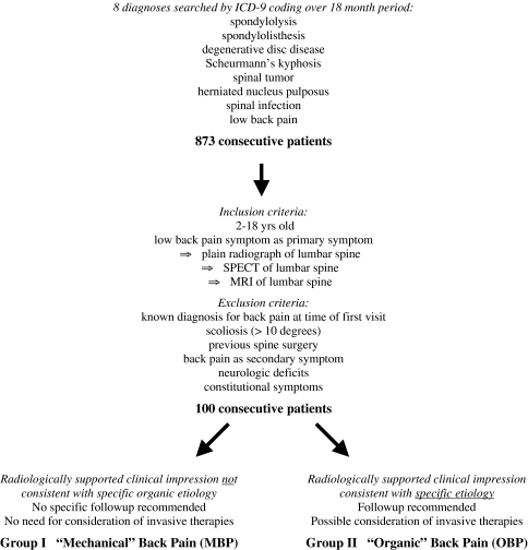
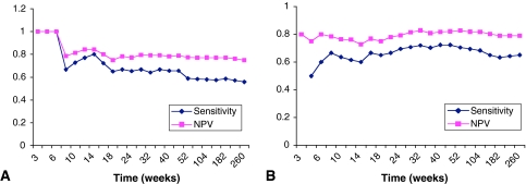
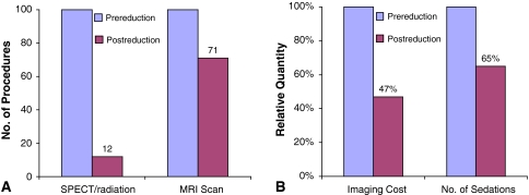
The workup of low back pain in children often results in overimaging so as not to miss organic back pain. The primary goal of this study was to identify which combination of imaging modalities provides the most sensitive and specific screening protocol for children with low back pain. Medical records from 100 consecutive patients between 2 and 18 years of age presenting with low back pain, without night pain or constitutional symptoms, were evaluated. A hyperextension test combined with a radiograph showed a negative predictive value of 0.81 and sensitivity of 0.90. The addition of a bone scan was highly effective in achieving good negative predictive value and sensitivity. Bone scans had perfect negative predictive value and sensitivity when symptom duration was less than 6 weeks. We identified a set of factors that is highly predictive for distinguishing organic back pain from mechanical back pain. Painless hyperextension combined with negative anteroposterior, lateral, and oblique lumbar radiographs and magnetic resonance images predicts mechanical back pain. For patients with nonneurologic back pain of less than 6 weeks duration, bone scan is the most useful screening test because it is accurate, accessible, inexpensive, and unlikely to require sedation.
Level of evidence: Level III, diagnostic study.
Figures
References
-
- {'text': '', 'ref_index': 1, 'ids': [{'type': 'PubMed', 'value': '2976526', 'is_inner': True, 'url': 'https://pubmed.ncbi.nlm.nih.gov/2976526/'}]}
- Balague F, Dutoit G, Waldburger M. Low back pain in schoolchildren: an epidemiological study. Scand J Rehabil Med. 1988;20:175–179. - PubMed
-
- {'text': '', 'ref_index': 1, 'ids': [{'type': 'PubMed', 'value': '1829845', 'is_inner': True, 'url': 'https://pubmed.ncbi.nlm.nih.gov/1829845/'}]}
- Bellah RD, Summerville DA, Treves ST, Micheli LJ. Low-back pain in adolescent athletes: detection of stress injury to the pars interarticularis with SPECT. Radiology. 1991;180:509–512. - PubMed
-
- {'text': '', 'ref_index': 1, 'ids': [{'type': 'DOI', 'value': '10.1097/00007632-198810000-00018', 'is_inner': False, 'url': 'https://doi.org/10.1097/00007632-198810000-00018'}, {'type': 'PubMed', 'value': '2974625', 'is_inner': True, 'url': 'https://pubmed.ncbi.nlm.nih.gov/2974625/'}]}
- Bodner RJ, Heyman S, Drummond DS, Gregg JR. The use of single photon emission computed tomography (SPECT) in the diagnosis of low-back pain in young patients. Spine. 1988;13:1155–1160. - PubMed
-
- {'text': '', 'ref_index': 1, 'ids': [{'type': 'PubMed', 'value': '14960688', 'is_inner': True, 'url': 'https://pubmed.ncbi.nlm.nih.gov/14960688/'}]}
- Bono CM. Low-back pain in athletes. J Bone Joint Surg Am. 2004;86:382–396. - PubMed
-
- {'text': '', 'ref_index': 1, 'ids': [{'type': 'DOI', 'value': '10.1007/s00256-004-0878-3', 'is_inner': False, 'url': 'https://doi.org/10.1007/s00256-004-0878-3'}, {'type': 'PubMed', 'value': '15668821', 'is_inner': True, 'url': 'https://pubmed.ncbi.nlm.nih.gov/15668821/'}]}
- Campbell RS, Grainger AJ, Hide IG, Papastefanou S, Greenough CG. Juvenile spondylolysis: a comparative analysis of CT, SPECT and MRI. Skeletal Radiol. 2005;34:63–73. - PubMed
MeSH terms
LinkOut - more resources
Full Text Sources
Medical
Research Materials
