. 2008 Jul 8;47(27):7097-107.
doi: 10.1021/bi8005835. Epub 2008 Jun 14.

Characterization of a streptococcal cholesterol-dependent cytolysin with a lewis y and b specific lectin domain

Affiliations

Characterization of a streptococcal cholesterol-dependent cytolysin with a lewis y and b specific lectin domain

Stephen Farrand et al. Biochemistry. .

Abstract

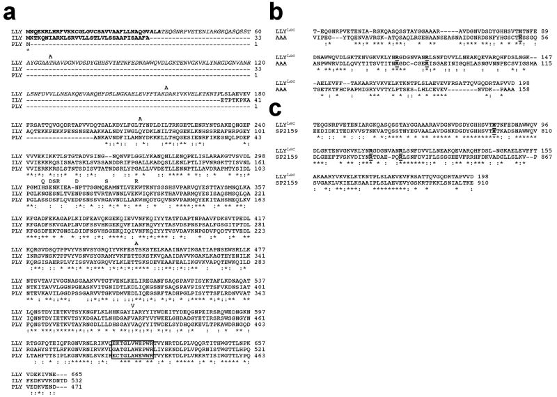
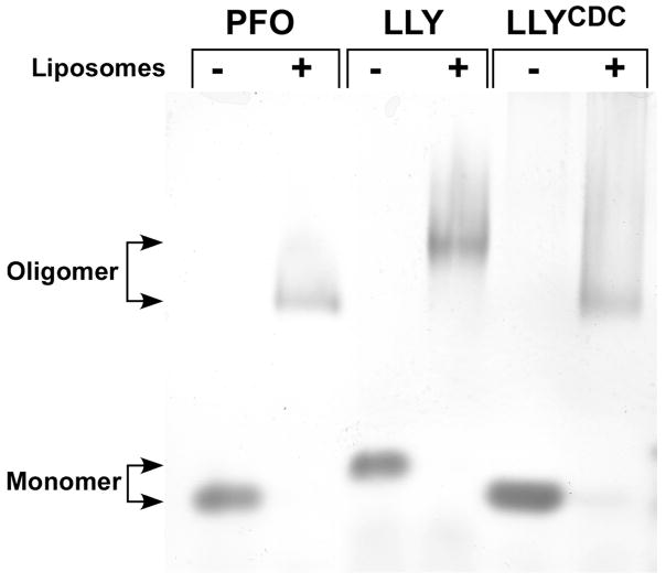
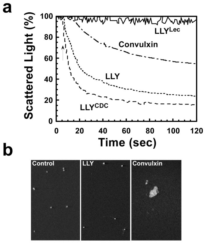
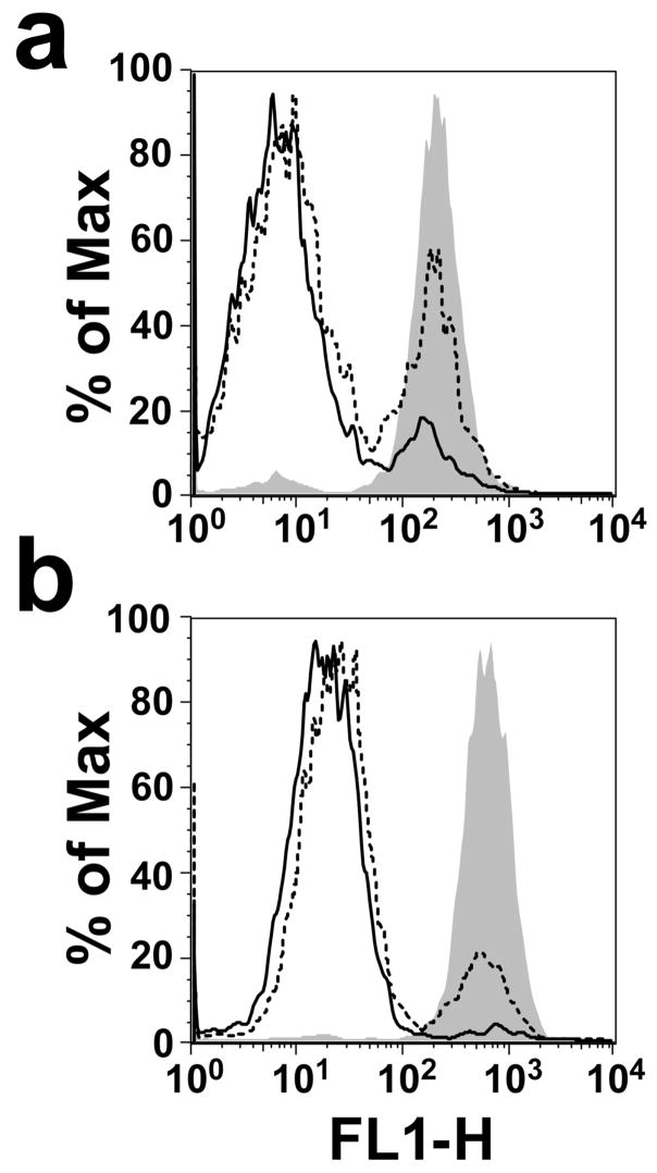
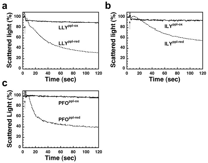
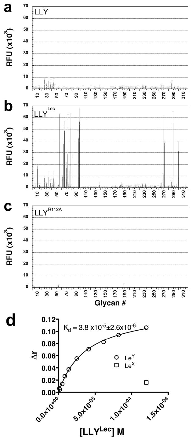
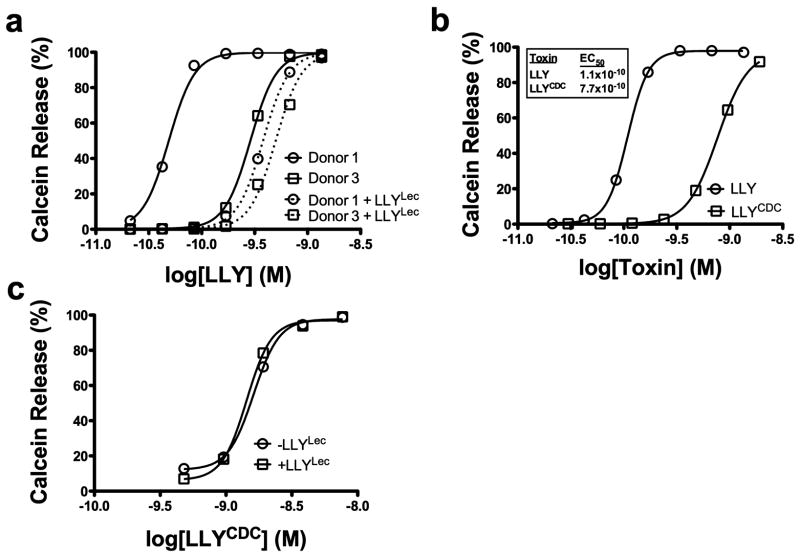
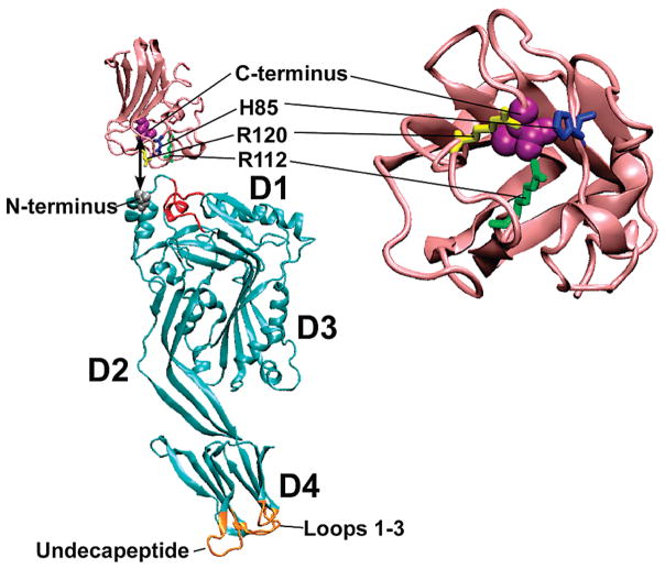
The cholesterol-dependent cytolysins (CDCs) are a large family of pore-forming toxins that often exhibit distinct structural changes that modify their pore-forming activity. A soluble platelet aggregation factor from Streptococcus mitis (Sm-hPAF) was characterized and shown to be a functional CDC with an amino-terminal fucose-binding lectin domain. Sm-hPAF, or lectinolysin (LLY) as renamed herein, is most closely related to CDCs from Streptococcus intermedius (ILY) and Streptococcus pneumoniae (pneumolysin or PLY). The LLY gene was identified in strains of S. mitis, S. pneumoniae, and Streptococcus pseudopneumoniae. LLY induces pore-dependent changes in the light scattering properties of the platelets that mimic those induced by platelet aggregation but does not induce platelet aggregation. LLY monomers form the typical large homooligomeric membrane pore complex observed for the CDCs. The pore-forming activity of LLY on platelets is modulated by the amino-terminal lectin domain, a structure that is not present in other CDCs. Glycan microarray analysis showed the lectin domain is specific for difucosylated glycans within Lewis b (Le (b)) and Lewis y (Le (y)) antigens. The glycan-binding site is occluded in the soluble monomer of LLY but is apparently exposed after cell binding, since it significantly increases LLY pore-forming activity in a glycan-dependent manner. Hence, LLY represents a new class of CDC whose pore-forming mechanism is modulated by a glycan-binding domain.


Figures

Figure 1
Figure 1
Primary structure of LLY. (a) The primary structure of LLY and its comparison with the primary structures of ILY and PLY. Comparison of the primary structure of LLYLec with that of the (b) A. anguilla agglutinin (AAA) (10) and (c) the glycan binding domain of the family 98 glycoside hydrolases from S. pneumoniae (SP2159) (19). The letters above the LLY sequence in panel a represent the amino acid differences with the GenBank sequence of Sm-hPAF (accession number AB051299.1). Homology comparisons were carried out using CLC Free Workbench version 4.6 (CLCBio). The conserved undecapeptide sequence of the CDCs is boxed in panel a. The conserved active site residues of AAA are bolded and underlined in panels b and c. Conserved residues (*), conservative substitutions (:).
Figure 2
Figure 2
Oligomer formation by LLY. Purified PFO, LLY, and LLYCDC were incubated in the absence and presence cholesterol-rich liposomes, and the monomer and oligomer species were separated by SDS–AGE. Shown is the Coomassie stained gel. The smearing of the oligomer bands results from the presence of the liposome-derived lipids in the oligomer samples.
Figure 3
Figure 3
Platelet aggregation by LLY. Shown in (a) are the aggregometer recordings for platelets treated with LLY (71 nM), LLYCDC (95 nM), and LLYLec (280 nM) and the positive control convulxin (500 ng/mL). Shown in (b) are confocal microscopic images of untreated, LLY-treated, and convulxin-treated platelets. These fields are representative of 12 separate fields.
Figure 4
Figure 4
Calcein release from LLY-treated platelets. Calcein-loaded platelets were assayed by flow cytometry before (shaded peak) and after treatment with 0.7 nM (dashed line) or 1.4 nM (solid line) LLY (a) or LLYCDC (b).
Figure 5
Figure 5
Platelet lysis requires pore formation. Shown are aggregometer readings for platelets treated with prepore locked mutants of LLY (a), ILY (b), and PFO (c) in their oxidized (LLYppl-ox, ILYppl-ox, and PFOppl-ox) and reduced forms (LLYppl-red, ILYppl-red, and PFOppl-red). Conditions were similar to those in Figure 3a; all toxins were added at approximately 70 nM. The reduction of the disulfide allows each prepore-locked mutant to convert to the pore complex.
Figure 6
Figure 6
Glycan binding by LLY, LLYLec, and LLYLec-R112A. Version 3.0 of the printed glycan microarray, containing 320 eukaryotic-derived glycans (see Methods for a link to the full list of glycans on this array), was probed with fluorescently tagged (Alexa-488) versions of (a) LLY (670 nM), (b) LLYLec (560 nM), and (c) LLYLec-R112A (560 nM) that had been labeled at the cysteine-substituted Gln-190. Standard error of the mean is shown as gray error bars. Note: The microarray analyses herein were carried out using the updated version 3.0 of the glycan array whereas the results in Table 2 were obtained using version 2.1. This was a result of a change in the microarray version during the course of these studies. There are only five differences in fucose-containing glycans in the two versions. Only two of these glycans, numbers 290 and 301 on the version 3.0 array, contain the H-antigen structure and were shown to be bound by LLYLec whereas the other three glycans were not (data not shown). In panel d the Kd of the Ley–LLYLec interaction was determined by measuring changes in the anisotropy (Δr) of Ley (kept constant at 167 nm) in the presence of LLYLec that was varied from 300 nM to 120 μM. Lex incubated with the highest concentration of LLY (120 μM) exhibited a Δr of ≈10% of that observed for Ley.
Figure 7
Figure 7
The glycan-binding domain of LLY modulates pore-forming activity. LLY-dependent calcein release from calcein-loaded PRP was measured by flow cytometry. (a) LLY-dependent calcein release from platelets of high (donor 1) and low responding donors (donor 3) which were preincubated without (solid lines) or with (dashed lines) LLYLec (56 nM). No change is observed with LLYLec alone (not shown). (b) Calcein release from platelets treated with LLY or LLYCDC. (c) Same as the experiments shown in (a) with donor 1 platelets except that LLYCDC was substituted for LLY.
Figure 8
Figure 8
Molecular models of LLYCDC and LLYLec. Shown are the ribbon representations of the LLYCDC (cyan) and LLYLec (pink) molecular models generated by Swiss Model (swissmodel.expasy-.org). The LLYCDC model was generated using the ILY crystal structure (47) as the template whereas the LLYlec was modeled based on the structure of the Ley-binding module of a S. pneumoniae virulence factor of the family 98 glycoside hydrolases (19). The locations are shown for the fucose-binding residues [His-85 (blue), Arg-112 (yellow), and Arg-120 (green)] and the C-terminal residue (space filled, magenta) of LLYLec and the amino-terminal residue of LLYCDC (space filled, gray). The L1-L3 loops and undecapeptide at the tip of domain 4 are shown in gold. The red-colored regions of the LLYCDC structural model exhibited a nonfavorable energy environment when analyzed with ANOLEA (29). Molecular structures were drawn with VMD (48).

References

    1. Kennedy MJ, Jackson MA, Kearns GL. Delayed diagnosis of penicillin-resistant Streptococcus mitis endocarditis following single-dose amoxicillin prophylaxis in a child. Clin Pediatr (Philadelphia) 2004;43:773–776. - PubMed
    1. Gowda RM, Ansari AW, Khan IA. Complete endocardial cushion defect (complete atrioventricular canal) manifested in adult life by Streptococcus mitis endocarditis of the common atrioventricular valve. Int J Cardiol. 2003;89:109–110. - PubMed
    1. Hall GE, Baddour LM. Apparent failure of endocarditis prophylaxis caused by penicillin-resistant Streptococcus mitis. Am J Med Sci. 2002;324:51–53. - PubMed
    1. Huang IF, Chiou CC, Liu YC, Hsieh KS. Endocarditis caused by penicillin-resistant Streptococcus mitis in a 12-year-old boy, J. Microbiol. Immunol, Infect (China) 2002;35:129–132. - PubMed
    1. Lu HZ, Weng XH, Zhu B, Li H, Yin YK, Zhang YX, Haas DW, Tang YW. Major outbreak of toxic shock-like syndrome caused by Streptococcus mitis. J Clin Microbiol. 2003;41:3051–3055. - PMC - PubMed

Publication types

Substances