Review
. 2008 Oct;12(5B):1830-47.
doi: 10.1111/j.1582-4934.2008.00387.x. Epub 2008 Jun 28.

Endogenous anti-inflammatory neuropeptides and pro-resolving lipid mediators: a new therapeutic approach for immune disorders

Affiliations
Review

Endogenous anti-inflammatory neuropeptides and pro-resolving lipid mediators: a new therapeutic approach for immune disorders

Per Anderson et al. J Cell Mol Med. 2008 Oct.

Abstract

Identification of the factors that regulate the immune tolerance and control the appearance of exacerbated inflammatory conditions is crucial for the development of new therapies of inflammatory and autoimmune diseases. Although much is known about the molecular basis of initiating signals and pro-inflammatory chemical mediators in inflammation, it has only recently become apparent that endogenous stop signals are critical at early checkpoints within the temporal events of inflammation. Some neuropeptides and lipid mediators that are produced during the ongoing inflammatory response have emerged as endogenous anti-inflammatory agents that participate in the regulation of the processes that ensure self-tolerance and/or inflammation resolution. Here we examine the latest research findings, which indicate that neuropeptides participate in maintaining immune tolerance in two distinct ways: by regulating the balance between pro-inflammatory and anti-inflammatory factors, and by inducing the emergence of regulatory T cells with suppressive activity against autoreactive T-cell effectors. On the other hand, we also focus on lipid mediators biosynthesized from omega-3 and omega-6 polyunsaturated fatty-acids in inflammatory exudates that promote the resolution phase of acute inflammation by regulating leucocyte influx to and efflux from local inflamed sites. Both anti-inflammatory neuropeptides and pro-resolving lipid mediators have shown therapeutic potential for a variety of inflammatory and autoimmune disorders and could be used as biotemplates for the development of novel pharmacologic agents.


Figures
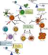
Fig 1
Fig 1
Loss of immune tolerance compromises immune homeostasis and results in the onset of autoimmune disorders. Host invasion by pathogens or trauma initially stimulate prostaglandins (PGs) and initiate early events in acute inflammation. Inflammatory leucocytes migrate to the inflamed site or area of tissue damage, with neutrophils being the first cell types at the scene (A). Secretion of leucotrienes (LTB4) and chemokines promotes the recruitment of more phagocytes. Once pathogens are killed or phagocytosed, neutrophils are cleared from the inflamed site by returning to the circulation or suffering apoptosis and subsequent phagocytosis by newly migrated macrophages, following an active program of resolution, where lipoxins (LXA4) and other lipid mediators play a major role (B). If inflammation is not maintained under control or complete resolution fails, acute inflammation can lead to chronic inflammation, scarring and eventual loss of tissue function. A further damage arises from potential autoimmune responses occurring during the chronic inflammatory response, in which the antigen presenting cells (APC) and molecules that respond to pathogen-derived antigens can also cross-react to self-antigens (C). Progression of the autoimmune response (multiple sclerosis is shown as example) involves the development of self-reactive T helper 1 (Th1) cells (D), their entry into the central nervous system (or target tissue), release of proinflammatory cytokines (tumour-necrosis factor-α (TNFα) and interferon-γ (IFNγ/)) and chemokines and subsequent recruitment and activation of inflammatory cells (macrophages and neutrophils), which produce cytotoxic factors, such as cytokines, nitric oxide and free radicals (E). Finally, regulatory T (Treg) cells are key players in maintaining tolerance by their suppression of self-reactive Th1 cells (F). Unbalance of Treg versus Th1 cells, and/or of anti-inflammatory cytokines versus proinflammatory factors, is the cause of autoimmune disorders. Therapeutic opportunities to manage inflammatory and autoimmune disorders should be found in agents that regulate inflammation resolution, control Th1 expansion, inhibit inflammatory mediators and/or induced Treg cell generation. Interleukin-10, IL-10; transforming growth factor-β, TGFβ; cytotoxic T lymphocyte associated antigen 4, CTLA4; T cell receptor, TCR.
Fig 2
Fig 2
Control of immune tolerance by anti-inflammatory neuropeptides. Vasoactive intestinal peptide (VIP), α-melanocyte-stimulating hormone (αMSH), urocortin (UCN), adrenomedullin (AM), ghrelin (GHR) and cortistatin (CST) are produced by T cells or macrophages in response to antigenic and inflammatory stimulation. These neuropeptides induce immune tolerance and inhibit the autoimmune response through different non-excluding mechanisms. (A) They decrease TH1-cell functions through direct actions on differentiating T cells, or indirectly by regulating dendritic cell (DC) functions. As a consequence, the inflammatory and autoimmune responses are impaired because the infiltration and activation of neu-trophils and macrophages by interferon-γ (IFNγ) and the production of complement-fixing IgG2a antibodies are avoided. (B) Neuropeptides inhibit the production of inflammatory cytokines, chemokines, free radicals (i.e. nitric oxide) and high-mobility group box 1 (HMGB1) by macrophages and microglia. In addition, they impair the costimulatory activity of macrophages on effector T cells, inhibiting the subsequent clonal expansion. This avoids the infiltration of leucocytes and the inflammatory response and the subsequent cytotoxicity against the target tissue. (C) Neuropeptides induce the new generation of regulatory T cells (Treg) that suppress activation of autoreactive T cells through a mechanism that involves production of interleukin-10 (IL-10) and transforming growth factor-β (TGFβ), and/or expression of the cytotoxic T lymphocyte-associated protein 4 (CTLA4). In addition, neuropeptides indirectly generate Treg through the differentiation of tolerogenic DCs. This effect contributes to the maintenance of an anti-inflammatory state and restores the immune tolerance. Black arrows indicate a stimulatory effect. Red crosses indicate an inhibitory effect.
Fig 3
Fig 3
Role of new PUFA-derived lipid mediators in the progression and resolution of acute inflammation. A schematic of the biosynthetic pathways for lipid mediators derived from ω-6 (AA, arachidonic acid) and ω-3 (EPA, eicosapentanoic acid; DHA, docosahexaenoic acid) with key enzymes is shown. AA is metabolized by cycloxygenase 1 (COX1) or COX2 to prostaglandin (PG) G2 and then PGH2, which in turn serves as a substrate for a series of downstream synthases to give rise to the PGs. PGs (such as PGE2) and leucotrienes (LTB4) participate in the initiation of the inflammatory response. PGH2 is also metabolized to PGD2 and then broken down to PGJ2 and the cyclopentenone PGs (cyPGs), which act as anti-inflammatory factors. After the initiation of acute inflammation by PGs and LTs, a class switching occurs with time towards pro-resolving lipid mediators that start with the generation of lipoxins (LXs) from AA through three distinct biosynthetic routes. First, AA is sequentially metabolized in a transcellular manner by 5-lipoxygenase (5-LOX) in polymorphonuclear cells (PMNs) and platelet 12-LOX to lypoxin A4 (LXA4) and LXB4. Second, AA can be transformed via sequential actions of the 5-LOX in monocytes or epithelial cells and the 5-LOX in PMNs yielding an epoxide intermediate that is converted to LXA4 and LXB4 by leucocyte epoxide hydrolases. Third, aspirin acetylates the active site of COX2 (Asp-COX2) that now is able to metabolize AA to 15(R)-hydroxyeicosatetraenoic (15R-HETE), which when released from endothelial and epithelial cells is converted by leucocyte 5-LOX to the called aspirin-triggered LXs (ATLs), 15-epi-LXA4 and 15-epi-LXB4. Once AA is metabolized to ATLs, it is substituted by the ω-3 EPA and DHA as substrates of the E-series and D-series resolvins and protectins. Vascular endothelial Asp-COX2 converts EPA to 18R-hydroxyperoxy-EPE (18R-H(p)EPE), which is further sequentially metabolized by leucocyte 5-LOX to lead the formation of resolving E1 (RvE1). Microbial P-450s may also convert EPA in RvE1. 5-LOX can also generate RvE2 from 18R-H(p)EPE and further reduction. DHA is transformed by the leucocyte LOX to 17S-H(p)DHA, which is rapidly converted by PMN LOX into two epoxide intermediates that finally lead the formation of the bioactive products 17S-resolvin D series (RvD1 to RvD4). Alternatively, Asp-COX2 can metabolize DHA to a 17R-H(p)DHA, which in turn generate the 17R-resolvin D series (AT-RvDs) by action of the leucocyte 5-LOX. Finally, by action of the 15-LOX in microglia, brain leucocytes, retinal cells or Th2 cells, DHA is converted to 17S-H(p)DHA, which following further enzymatic epoxidation and hydrolysis form protectin 1 (PD1), or neuroprotectin 1 (NPD1) if formed in the brain. LXA4, ATLs, resolvins and PD1 share some anti-inflammatory and pro-resolving actions, although they have distinct roles within the induction of resolution. dendritic cells, DC; inducible nitric oxide synthase, iNOS; peroxisome proliferator-activating receptor γ, PPARγ; transforming growth factor, TGF.

References

    1. Firestein GS. Evolving concepts of rheumatoid arthritis. Nature. 2003;423:356–61. - PubMed
    1. Goodnow CC. Multistep pathogenesis of autoimmune diseases. Cell. 2007;130:25–35. - PubMed
    1. Nathan C. Points of control in inflammation. Nature. 2002;420:846–52. - PubMed
    1. Mills KHG. Regulatory T cells: friend or foe in immunity to infection? Nat Rev Immunol. 2004;4:841–55. - PubMed
    1. Bluestone JA. Regulatory T-cell therapy: is it ready for the clinic? Nat Rev Immunol. 2005;5:343–9. - PubMed

Publication types

MeSH terms