. 2008 Aug;69(3):736-46.
doi: 10.1111/j.1365-2958.2008.06324.x. Epub 2008 Jun 28.

EsaC substrate for the ESAT-6 secretion pathway and its role in persistent infections of Staphylococcus aureus

Affiliations

EsaC substrate for the ESAT-6 secretion pathway and its role in persistent infections of Staphylococcus aureus

Monica L Burts et al. Mol Microbiol. 2008 Aug.

Abstract

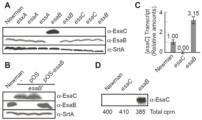
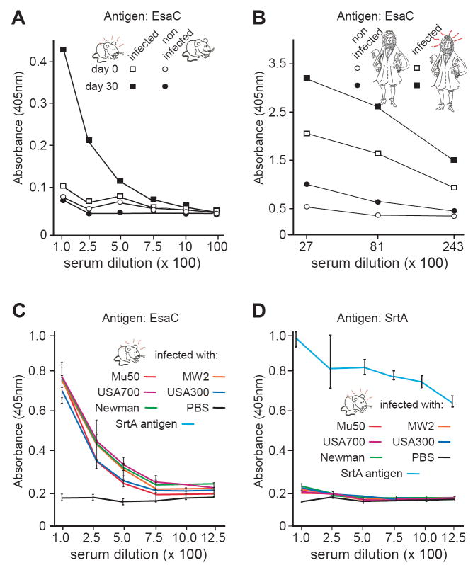
Staphylococcus aureus encodes the specialized secretion system Ess (ESAT-6 secretion system). The ess locus is a cluster of eight genes (esxAB, essABC, esaABC) of which esxA and esxB display homology to secreted ESAT-6 proteins of Mycobacterium tuberculosis. EsxA and EsxB require EssA, EssB and EssC for transport across the staphylococcal envelope. Herein, we examine the role of EsaB and EsaC and show that EsaB is a negative regulator of EsaC. Further, EsaC production is repressed when staphylococci are grown in broth and increased when staphylococci replicate in serum or infected hosts. EsaB is constitutively produced and remains in the cytoplasm whereas EsaC is secreted. This secretion requires an intact Ess pathway. Mutants lacking esaB or esaC display only a small defect in acute infection, but remarkably are unable to promote persistent abscesses during animal infection. Together, the data suggest a model whereby EsaB controls the production of effector molecules that are important for host pathogen interaction. One such effector, EsaC, is a secretion substrate of the Ess pathway and implements its pathogenic function during infection.


Figures

Fig. 1
Fig. 1
Schematic drawing of the ess cluster found in various Gram-positive bacteria as well as M. tuberculosis. Color of genes and proteins indicates: FtsK-SpoIIIE ATPases (FSD factors), yellow color; ESAT-6 like protein, red color; conserved proteins, grey shades. Genes located at the position of esaC are depicted in various colors.
Fig. 2
Fig. 2
EsaB regulates EsaC production. (A-B) Total cell cultures of strain Newman and variants were examined for production of EsaC. Staphylococci were grown in tryptic soy broth. Proteins in whole culture lysates were precipitated with TCA, separated by SDS-PAGE and detected by immunoblotting with specific antibodies [α-EsaC, α-EsaB and α-SrtA as a loading control]. Panel A shows extracts of wild type Newman and isogenic mutants as indicated. Complementation analysis of esaB mutant is shown in panel B. Immunoblot analysis of total cell extracts of Newman, esaB- with no vector (-), vector alone (pOS), vector carrying esaB (pOS-esaB). (C) Quantitative RT-PCR analysis of esaC transcripts was performed by isolating RNA from S. aureus isogenic strains Newman, esaC, and esaB. Reverse transcriptional polymerase chain reaction (RT-PCR) was carried out using oligos specific for sdrE and esaC transcripts. sdrE transcript levels did not change in all three backgrounds (not shown). The ratio of sdrE/esaC transcripts in Newman was 3/1. (D) Cultures of wild type (Newman) and esaB or esaC mutant cells were radiolabeled with [35S]-methionine for 2 min. Labeling was quenched by addition of trichloroacetic acid, staphylococci were lysed with lysostaphin and extracts solubilized in hot SDS. Total radioactive counts were measured using 5 μl of each sample in a scintillation counter. Total cell extracts were subjected to immunoprecipitation with anti-EsaC antibodies. Samples were separated on SDS-PAGE and analyzed by autoradiography using a PhosphorImager.
Fig. 3
Fig. 3
Staphylococci grown in serum produce EsaC. Staphylococci, Newman, esaC mutant with no vector (-), vector alone (pOS), vector carrying esaC (pOS-esaC), were grown in TSB or serum to the same density, washed and lysed with lysostaphin. Proteins in these extracts were precipitated with TCA, separated by SDS-PAGE and detected by immunoblotting with specific antibodies [α-EsaC, and α-SrtA as a loading control].
Fig. 4
Fig. 4
EsaC is a ubiquitous secreted antigen of the S. aureus Ess pathway. (A) S. aureus USA300 and USA700 secrete EsaC into the extracellular medium (MD). As control, regulation of EsaC expression in S. aureus Newman as well as USA300 is dependent on esaB as measured in whole culture lysates (WC). Antibodies against ribosomal protein L6 were used as a control for proper fractionation. (B) EssC is required for secretion of EsaC. Immunoblot analysis of total cell extracts of Newman or isogenic essC mutant, with vector alone (pOS) or vector carrying esaC (pOS-esaC). Production and secretion of EsaC was measured in whole culture lysates (WC) and culture supernatants (MD). Antibodies against ribosomal protein L6 were used as a control for loading and fractionation. (C) Subcellular location of EsaC. S. aureus cultures of strains Newman, Newman esaB, USA300 and USA300 esxB were grown to OD660nm 0.8. Equal volumes of cultures were removed for preparation of whole cell lysates (WC) and fractionation of staphylococci into cytoplasm (C), membrane (M), cell wall (W) and medium (MD) fractions. Hence each cellular compartment is kept equimolar to the WC fraction. Proteins were precipitated with TCA, separated on SDS/PAGE, and detected by immunoblotting with specific antibodies [α-EsaC, α-ribosomal protein L6, α-SrtA, α-Spa (protein A)].
Fig. 5
Fig. 5
Mice and humans infected with S. aureus generate EsaC IgG specific antibodies. (A) Three-week-old BALB/c mice were injected retro-orbitally with ~ 106 CFU of strain Newman. Sera were collected on day 0 and 30 days post infection and analyzed for the presence of EsaC reactive antibodies. (B) Quantification of EsaC IgG levels in human sera obtained from patients infected or not with S. aureus (two sera each, respectively). (C-D) Three-week-old BALB/c mice were injected as in panel A with clinical strains as indicated on the figure. Sera were collected 0 and 30 days post infection (the 30-day data set is shown). IgG titers to EsaC and Sortase A are shown in panels C and D, respectively. In panel D, a rabbit polyclonal antibody raised against recombinant SrtA was used as a control. All IgG titers were determined in triplicate by ELISA and reported as an absorbance at 405 nm.
Fig. 6
Fig. 6
Virulence of S. aureus esaB and esaC mutants. BALB/c mice were infected retro-orbitally with ~ 106 CFU for each strain. Both kidneys were harvested from mock (PBS) infected animals or mice infected with Newman, esaB or esaC isogenic variants, for 5 and 36 days and the right kidney for each animal was homogenized. Viable bacteria were counted after dilution and colony formation on tryptic soy agar. Statistical significance was examined with Student’s t test, and averages and P values are indicated. The limit of detection was determined to be 10 CFU.
Fig. 7
Fig. 7
Pathological substrate of infection caused by S. aureus wild type and esaB or esaC mutants. Kidneys of mice infected as described in figure 6 were removed 5 and 36 days post infection. The right kidney was used for CFU counts and the left was fixed with formalin. Formalin-fixed tissues were embedded, sectioned, and stained with hematoxylin/eosin. Microscopic images of whole kidneys (x10, top panels) or organ tissue at higher magnification (x100, lower panels) revealed fewer and less persistent abscesses in esaB or esaC infected animals. White arrows point to abscesses with a central concentration of staphylococci and PMN infiltrate. Numbers under each panel indicate the average number of abscesses per kidney with standard deviation, between 8 and 12 kidneys were examined per group. Statistical significance was examined with the Student t test, and P values were recorded.

References

    1. Abdallah AM, Verboom T, Hannes F, Safi M, Strong M, Eisenberg D, Musters RJ, Vandenbroucke-Grauls CM, Appelmelk BJ, Luirink J, Bitter W. A specific secretion system mediates PPE41 transport in pathogenic mycobacteria. Mol Microbiol. 2006;62:667–679. - PubMed
    1. Abdallah AM, Gey van Pittius NC, Champion PA, Cox J, Luirink J, Vandenbroucke-Grauls CM, Appelmelk BJ, Bitter W. Type VII secretion--mycobacteria show the way. Nat Rev Microbiol. 2007;5:883–891. - PubMed
    1. Andersen P, Andersen AB, Sorensen AL, Nagai S. Recall of long-lived immunity to Mycobacterium tuberculosis infection in mice. J Immunol. 1995;154:3359–3372. - PubMed
    1. Archer GL. Staphylococcus aureus: a well-armed pathogen. Clin Infect Dis. 1998;26:1179–1181. - PubMed
    1. Baba T, Bae T, Schneewind O, Takeuchi F, Hiramatsu K. Genome sequence of Staphylococcus aureus strain Newman and comparative analysis of staphylococcal genomes: polymorphism and evolution of two major pathogenicity islands. J Bacteriol. 2008;190:300–310. - PMC - PubMed

Publication types

MeSH terms