. 2008 Aug;69(4):794-808.
doi: 10.1111/j.1365-2958.2008.06299.x. Epub 2008 Jun 28.

The specialized secretory apparatus ESX-1 is essential for DNA transfer in Mycobacterium smegmatis

Affiliations

The specialized secretory apparatus ESX-1 is essential for DNA transfer in Mycobacterium smegmatis

Abbie Coros et al. Mol Microbiol. 2008 Aug.

Abstract

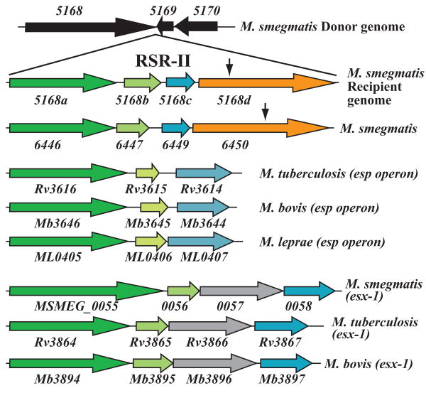
Conjugal DNA transfer in Mycobacterium smegmatis occurs by a mechanism distinct from plasmid-mediated DNA transfer. Previously, we had shown that the secretory apparatus, ESX-1, negatively regulated DNA transfer from the donor strain; ESX-1 donor mutants are hyper-conjugative. Here, we describe a genome-wide transposon mutagenesis screen to isolate recipient mutants. Surprisingly, we find that a majority of insertions map within the esx-1 locus, which encodes the secretory apparatus. Thus, in contrast to its role in donor function, ESX-1 is essential for recipient function; recipient ESX-1 mutants are hypo-conjugative. In addition to esx-1 genes, our screen identifies novel non-esx-1 loci in the M. smegmatis genome that are required for both DNA transfer and ESX-1 activity. DNA transfer therefore provides a simple molecular genetic assay to characterize ESX-1, which, in Mycobacterium tuberculosis, is necessary for full virulence. These findings reinforce the functional intertwining of DNA transfer and ESX-1 secretion, first described in the M. smegmatis donor. Moreover, our observation that ESX-1 has such diametrically opposed effects on transfer in the donor and recipient, forces us to consider how proteins secreted by the ESX-1 apparatus can function so as to modulate two seemingly disparate processes, M. smegmatis DNA transfer and M. tuberculosis virulence.


Figures

Fig. 1
Fig. 1
A genetic map comparing the esx-1 loci of M. smegmatis and M. tuberculosis. The two maps are aligned, with colour-coding used to indicate homologous gene pairs in the two species. The M. tuberculosis esp operon is shown above the M. tuberculosis esx-1 locus, and is aligned with gene homologues found in the first segment of esx-1. The map of the M. smegmatis recipient esx-1 locus is based on the donor DNA sequence (TIGR), but it is modified based on our sequencing analyses. Note that donor genes Msmeg_0072-0075 are deleted. The 3′-end of the M. smegmatis locus is predicted to encode additional genes (Msmeg_0077-0080) that are not present in M. tuberculosis; these genes are related to M. smegmatis genes Msmeg_0055-0058 as indicated by their colours. Above the M. smegmatis map, the +/- symbols indicate the sites of transfer-defective insertions. A ‘+’ indicates that the insertion disrupts EsxB secretion and, therefore, the gene is required for both transfer and ESX-1 activities. A ‘-’ indicates the insertion has no effect on EsxB secretion, but does disrupt DNA transfer.
Fig. 2
Fig. 2
Clustered insertions identify loci required for DNA transfer. Genes with defined function are named and shaded in light grey, while genes that have a putative function or that are classified as hypothetical orfs are shaded in dark grey. A. Multiple insertions (indicated by vertical arrows) mapped in Msmeg_0044-0045 disrupted both DNA transfer and EsxB secretion. These two genes are part of a three-gene operon found upstream of the esx-1 locus. The esx-1 locus begins at Msmeg_0055 (in charcoal). B. Three transfer-defective insertions map to two operons transcribed from overlapping promoter regions. One insertion was mapped in Msmeg_1955 (conserved hypothetical) and two insertions were located in Msmeg_1959 (encoding a putative membrane protein). Msmeg_1954 encodes a putative ABC1 transporter, ATP-binding protein. Msmeg_1958 encodes a PDZ domain family protein. C. Insertions in Msmeg_3888 and 3889 disrupt recipient function. These two genes are both predicted to encode DNA binding proteins.
Fig. 3
Fig. 3
A recipient-specific region is located within esx-1 and encodes genes required for DNA transfer, but not for EsxB secretion. The esx-1 loci from donor and recipient are aligned to highlight the significant differences between them. Note that not only are genes Msmeg_0072-0075 deleted in the recipient, but genes Msmeg_0069R-0071R are only weakly related to their donor homologues. This heterogeneity ends within 0071R; over the last 812 bp of the gene, 0071R has 88% nucleotide identity with the donor copy. The junction between esx-1 and the 5′ end of RSR-I has not yet been mapped. Current sequencing information indicates that two insertion sequences (indicated by yellow boxes) flank 0069R (Coros et al., 2008).
Fig. 4
Fig. 4
Recipient-specific locus II and a second operon, Msmeg_6446-6450, encode homologues of genes found in the esp locus of M. tuberculosis. RSR-II is inserted between genes Msmeg_5168 and 5169, with respect to the mc2155 donor genome sequence. An insertion (vertical arrow) in Msmeg_5168d abolishes DNA transfer. 5168d is significantly similar to Msmeg_6450 (36% aa identity, 53% aa similarity; Table 3); 6450 is also required for DNA transfer (vertical arrow indicates the site of transposon insertion). Importantly, 5168a-d and 6446-6450 form two syntenous operons that are similar to the M. tuberculosis esp operon (middle panel) and the first segment of the highly conserved esx-1 operon (bottom three alignments).
Fig. 5
Fig. 5
A majority of transfer-defective insertions map to the esx-1 locus and disrupt ESX-1 activity. A. A map of the M. smegmatis chromosome showing sites of transfer-defective insertions; charcoal circles indicate insertions that disrupt EsxAB secretion; open circles exhibit wild-type EsxAB secretion. B. Representative Western analyses showing the genetic requirements for ESX-1 activity, as monitored by EsxB secretion. Top panel: A majority of insertions in esx-1 disrupt secretion into the culture supernatant (e.g. those insertions in Msmeg_0059, 0062), as compared to the wild-type control (MKD8). However, insertions in Msmeg_0071 and 0076 result in wild-type levels of EsxB secretion but are defective in recipient function. In addition, insertions in Msmeg_0044, 0045 and 5447, which are not esx-1 genes, disrupt secretion, indicating that genes other than those associated with esx-1 mediate ESX-1 activity. Lower panel; The presence of EsxB in total cell lysates indicates that EsxB is stably expressed in the mutants and that its absence from the culture filtrate is due to disruption of ESX-1 activity.

References

    1. Abdallah AM, Gey van Pittius NC, Champion PA, Cox J, Luirink J, Vandenbroucke-Grauls CM, Appelmelk BJ, Bitter W. Type VII secretion--mycobacteria show the way. Nat Rev Microbiol. 2007;5:883–891. - PubMed
    1. Alteri CJ, Xicohtencatl-Cortes J, Hess S, Caballero-Olin G, Giron JA, Friedman RL. Mycobacterium tuberculosis produces pili during human infection. Proc Natl Acad Sci U S A. 2007;104:5145–5150. - PMC - PubMed
    1. Bardarov S, Kriakov J, Carriere C, Yu S, Vaamonde C, McAdam RA, Bloom BR, Hatfull GF, Jacobs WRJ. Conditionally replicating mycobacteriophages: A system for transposon delivery to Mycobacterium tuberculosis. Proceedings of the National Academy of Sciences of the United States of America; 1997. pp. 10961–10966. - PMC - PubMed
    1. Bardarov S, Bardarov S, Jr, Pavelka MS, Jr, Sambandamurthy V, Larsen M, Tufariello J, Chan J, Hatfull G, Jacobs WR., Jr Specialized transduction: an efficient method for generating marked and unmarked targeted gene disruptions in Mycobacterium tuberculosis, M. bovis BCG and M. smegmatis. Microbiology. 2002;148:3007–3017. - PubMed
    1. Behr MA, Wilson MA, Gill WP, Salamon H, Schoolnik GK, Rane S, Small PM. Comparative genomics of BCG vaccines by whole-genome DNA microarray. Science. 1999;284:1520–1523. - PubMed

Publication types