

Non-invasive screening method for simultaneous evaluation of in vivo growth factor release profiles from multiple ectopic bone tissue engineering implants
- PMID: 18554743
- PMCID: PMC2601638
- DOI: 10.1016/j.jconrel.2008.05.004
Non-invasive screening method for simultaneous evaluation of in vivo growth factor release profiles from multiple ectopic bone tissue engineering implants
Abstract
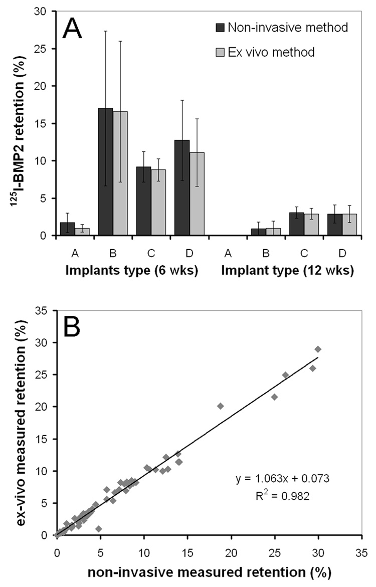
The purpose of this study was to develop and validate a screening method based on scintillation probes for the simultaneous evaluation of in vivo growth factor release profiles of multiple implants in the same animal. First, we characterized the scintillation probes in a series of in vitro experiments to optimize the accuracy of the measurement setup. The scintillation probes were found to have a strong geometric dependence and experience saturation effects at high activities. In vitro simulation of 4 subcutaneous limb implants in a rat showed minimal interference of surrounding implants on local measurements at close to parallel positioning of the probes. These characteristics were taken into consideration for the design of the probe setup and in vivo experiment. The measurement setup was then validated in a rat subcutaneous implantation model using 4 different sustained release carriers loaded with (125)I-BMP-2 per animal. The implants were removed after 42 or 84 days of implantation, for comparison of the non-invasive method to ex vivo radioisotope counting. The non-invasive method demonstrated a good correlation with the ex vivo counting method at both time-points of all 4 carriers. Overall, this study showed that scintillation probes could be successfully used for paired measurement of 4 release profiles with minimal interference of the surrounding implants, and may find use as non-invasive screening tools for various drug delivery applications.
Figures


Similar articles
-
Non-invasive monitoring of BMP-2 retention and bone formation in composites for bone tissue engineering using SPECT/CT and scintillation probes.J Control Release. 2009 Mar 19;134(3):169-76. doi: 10.1016/j.jconrel.2008.11.023. Epub 2008 Dec 3. J Control Release. 2009. PMID: 19105972 Free PMC article.
-
Enhancement of bone growth by sustained delivery of recombinant human bone morphogenetic protein-2 in a polymeric matrix.Pharm Res. 2001 Dec;18(12):1747-53. doi: 10.1023/a:1013382832091. Pharm Res. 2001. PMID: 11785696
-
Characterization of absorbable collagen sponges as rhBMP-2 carriers.Int J Pharm. 1999 Sep 30;187(1):91-9. doi: 10.1016/s0378-5173(99)00174-x. Int J Pharm. 1999. PMID: 10502616
-
Ectopic bone formation after implantation of a slow release system of polylactic acid and rhBMP-2.Clin Oral Implants Res. 2009 Jan;20(1):24-30. doi: 10.1111/j.1600-0501.2008.01613.x. Clin Oral Implants Res. 2009. PMID: 19126104
-
Collective review: bioactive implants coated with poly(D,L-lactide) and growth factors IGF-I, TGF-beta1, or BMP-2 for stimulation of fracture healing.J Long Term Eff Med Implants. 2006;16(1):61-9. doi: 10.1615/jlongtermeffmedimplants.v16.i1.70. J Long Term Eff Med Implants. 2006. PMID: 16566746 Review.
Cited by
-
Effect of Biomaterial Electrical Charge on Bone Morphogenetic Protein-2-Induced In Vivo Bone Formation.Tissue Eng Part A. 2019 Jul;25(13-14):1037-1052. doi: 10.1089/ten.TEA.2018.0140. Epub 2019 Jun 14. Tissue Eng Part A. 2019. PMID: 30612538 Free PMC article.
-
In Vitro and In Vivo Correlation of Bone Morphogenetic Protein-2 Release Profiles from Complex Delivery Vehicles.Tissue Eng Part C Methods. 2018 Jul;24(7):379-390. doi: 10.1089/ten.TEC.2018.0024. Tissue Eng Part C Methods. 2018. PMID: 29756545 Free PMC article.
-
Pre-clinical characterization of tissue engineering constructs for bone and cartilage regeneration.Ann Biomed Eng. 2015 Mar;43(3):681-96. doi: 10.1007/s10439-014-1151-0. Epub 2014 Oct 16. Ann Biomed Eng. 2015. PMID: 25319726 Free PMC article. Review.
-
Phosphate Functional Groups Improve Oligo[(Polyethylene Glycol) Fumarate] Osteoconduction and BMP-2 Osteoinductive Efficacy.Tissue Eng Part A. 2018 May;24(9-10):819-829. doi: 10.1089/ten.TEA.2017.0229. Epub 2018 Apr 23. Tissue Eng Part A. 2018. PMID: 29065776 Free PMC article.
-
Effect of autologous bone marrow stromal cell seeding and bone morphogenetic protein-2 delivery on ectopic bone formation in a microsphere/poly(propylene fumarate) composite.Tissue Eng Part A. 2009 Mar;15(3):587-94. doi: 10.1089/ten.tea.2007.0376. Tissue Eng Part A. 2009. PMID: 18925831 Free PMC article.
References
-
- Hori K, Sotozono C, Hamuro J, Yamasaki K, Kimura Y, Ozeki M, Tabata Y, Kinoshita S. Controlled-release of epidermal growth factor from cationized gelatin hydrogel enhances corneal epithelial wound healing. J Control Release. 2006 - PubMed
-
- Ruhe PQ, Boerman OC, Russel FG, Mikos AG, Spauwen PH, Jansen JA. In vivo release of rhBMP-2 loaded porous calcium phosphate cement pretreated with albumin. J Mater Sci Mater Med. 2006;17(10):919–927. - PubMed
-
- Woo BH, Fink BF, Page R, Schrier JA, Jo YW, Jiang G, DeLuca M, Vasconez HC, DeLuca PP. Enhancement of bone growth by sustained delivery of recombinant human bone morphogenetic protein-2 in a polymeric matrix. Pharm Res. 2001;18(12):1747–1753. - PubMed
-
- Li RH, Bouxsein ML, Blake CA, D'Augusta D, Kim H, Li XJ, Wozney JM, Seeherman HJ. rhBMP-2 injected in a calcium phosphate paste (alpha-BSM) accelerates healing in the rabbit ulnar osteotomy model. J Orthop Res. 2003;21(6):997–1004. - PubMed
-
- Abdiu A, Walz TM, Wasteson A. Uptake of 125I-PDGF-AB to the blood after extravascular administration in mice. Life Sci. 1998;62(21):1911–1918. - PubMed
Publication types
MeSH terms
Substances
Grants and funding
LinkOut - more resources
Full Text Sources
