. 2008 Aug 1;440(2):113-8.
doi: 10.1016/j.neulet.2008.05.031. Epub 2008 May 15.

Herpes simplex virus type 1 induces filopodia in differentiated P19 neural cells to facilitate viral spread

Affiliations

Herpes simplex virus type 1 induces filopodia in differentiated P19 neural cells to facilitate viral spread

Rohan Dixit et al. Neurosci Lett. .

Abstract

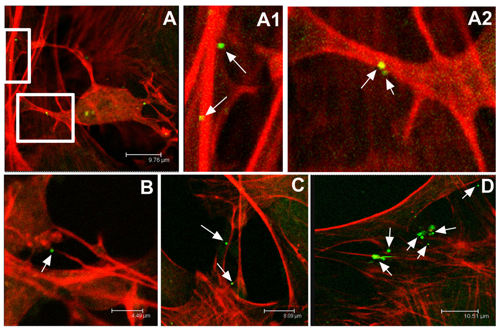
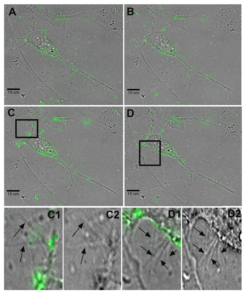
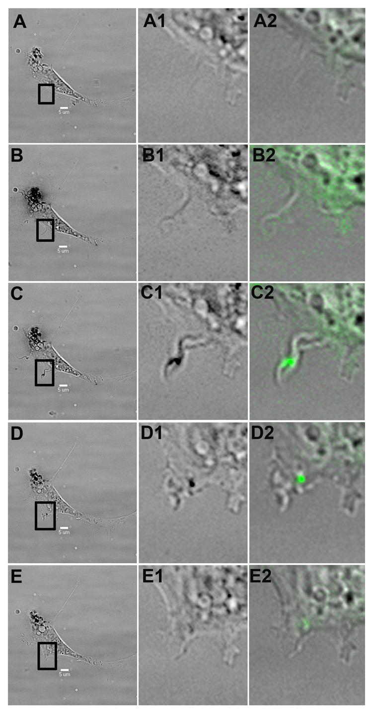
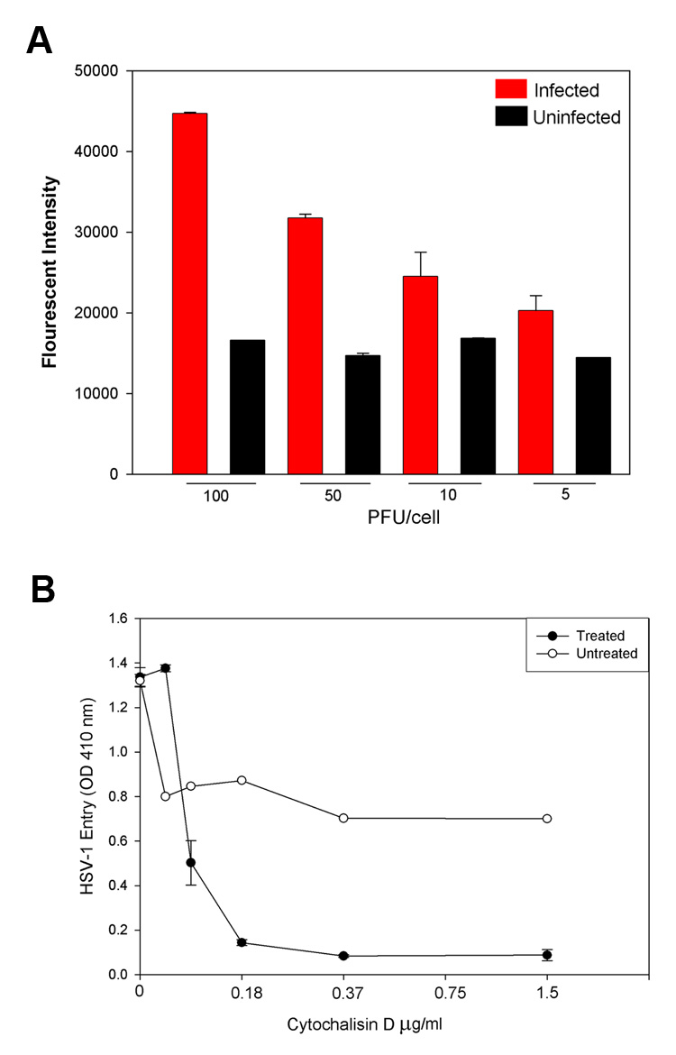
Herpes simplex virus type-1 (HSV-1) is a neurotropic virus with significant potential as a viral vector for central nervous system (CNS) gene therapy. This study provides visual evidence that recombinant green fluorescent protein (GFP)-expressing HSV-1 travel down dendrites in differentiated P19 neuronal-like cells to efficiently reach the soma. The virus also promotes cytoskeletal rearrangements which facilitate viral spread in vitro, including often dramatic increases in dendritic filopodia. Viral movements, cell infection and filopodia induction were each reduced with the actin polymerization inhibitor cytochalasin D, suggesting the involvement of the actin cortex in these processes. The observation of neural cytoskeletal reorganization in response to HSV-1 may shed light on the mechanisms by which acute viral infection associated with herpes encephalitis produces cognitive deficits in patients.


Figures

Figure 1
Figure 1
Confocal microscope images of neurons infected with the neurotropic herpes simplex type 1 virus. Actin networks in P19 cell cultures are visualized in red through cell staining with rhodamine-conjugated phalloidin. Individual viral particles express green fluorescent protein. The virus preferentially associates with long, actin-rich dendritic processes. A, An infected P19 neuron and A1, A higher magnification of a dendrite revealing two green virions attached. A2, A higher magnification of the neuron pictured in A again showing virus attached to red actin-rich dendrites. B, Virus attached to an actin-rich process. C, Multiple HSV-1 virions adjacent to dendritic networks in P19 culture. D, The virus continues to appear bound to actin-rich processes even within complex dendritic networks.
Figure 2
Figure 2
Surfing of HSV-1 and the induction of dendritic filopodia formation. Still frames taken from a live-cell recording of multiple HSV-1 virions surfing on dendrites towards the soma of a P19 neuron (Supplementary video 1). A, At 5 minutes post-infection, the virus particles have begun to attach to the neuron and its dendrites. B, At 16 minutes post-infection, multiple virions begin surfing along dendrites towards the neuron’s cell body. C, At 25 minutes post-infection, viral particles continue to surf towards the cell body, while significant new dendritic filopodial growth emerges from a previously stable dendrite highlighted in C1, a FITC and brightfield composite image, and C2, a brightfield image alone. D, At 32 minutes post-infection, many viral particles originally found on the neuron’s dendrites have now reached the soma. Another burst of new filopodia appears near the axon hillock and the region highlighted in D1, a FITC and brightfield composite image, and D2, a brightfield image alone.
Figure 3
Figure 3
Virus intake via surfing. A P19 cell exposed to HSV-1 begins to express filopodia, which are then used by HSV-1 virions (green) to surf into the cell. The first column shows a brightfield, low magnification cell image, while the second and third column represent closeups of the highlighted region in brightfield and FITC-brightfield composite, respectively. A, At 17 minutes post-infection, no filopodia are observed in the region of interest. B, At 20 minutes post-infection, a new filopodia emerges from the cell. C, At 22 minutes post-infection, a viral particle binds to the new filopodia. D, At 34 minutes, the virus is using the filopodia to surf towards the cell body. E, At 42 minutes post-infection, the virus appears to have reached the cell by surfing along the newly emerged filopodia.
Figure 4
Figure 4
Actin dynamics and HSV-1 viral spread. A, The F-actin fluorescence increases dramatically as a function of viral dose relative to uninfected controls. Viral dose was estimated in plaque-forming units (PFU) per P19 cell. A higher expression of F-actin implies filopodia formation and actin cortex reorganization in response to increasing HSV-1 concentrations. B, The actin polymerization inhibitor cytochalasin D greatly reduces viral entry, implicating actin dynamics in viral infection strategies in P19 neural cell cultures. Cytochalasin D prevented both surfing behavior and filopodia formation after HSV-1 application.

References

    1. Abraham VC, Krishnamurthi V, Taylor DL, Lanni F. The actin-based nanomachine at the leading edge of migrating cells. Biophysical Journal. 1999;77:1721–1732. - PMC - PubMed
    1. Bacon C, Lakics V, Machesky L, Rumsby M. N-WASP regulates extension of filopodia and processes by oligodendrocyte progenitors, oligodendrocytes, and Schwann cells - implications for axon ensheathment at myelination. Glia. 2007;55:844–858. - PubMed
    1. Bentley D, Toroian-Raymond A. Disoriented pathfinding by pioneer neurone growth cones deprived of filopodia by cytochalasin treatment. Nature. 1996;323:712–715. - PubMed
    1. Berges BK, Wolfe JH, Fraser NW. Transduction of brain by herpes simplex virus vectors. Molecular Therapy. 2007;15:20–29. - PubMed
    1. Billig I, Foris JM, Enquist LW, Card JP, Yates BJ. Definition of neuronal circuitry controlling activity of phrenic and abdominal motoneurons in the ferret using recombinant strains of pseudorabies virus. J. Neurosci. 2000;20:7446–7454. - PMC - PubMed

Publication types

MeSH terms

LinkOut - more resources