

Dense feature deformation morphometry: Incorporating DTI data into conventional MRI morphometry
- PMID: 18555734
- PMCID: PMC2702325
- DOI: 10.1016/j.media.2008.03.010
Dense feature deformation morphometry: Incorporating DTI data into conventional MRI morphometry
Abstract
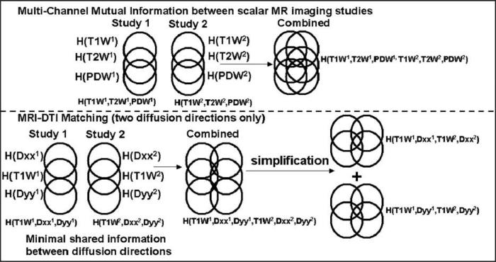
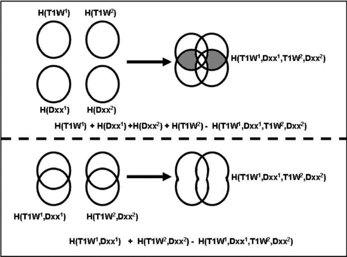
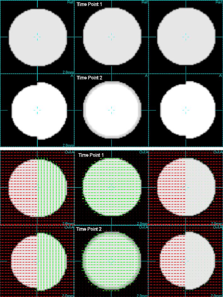
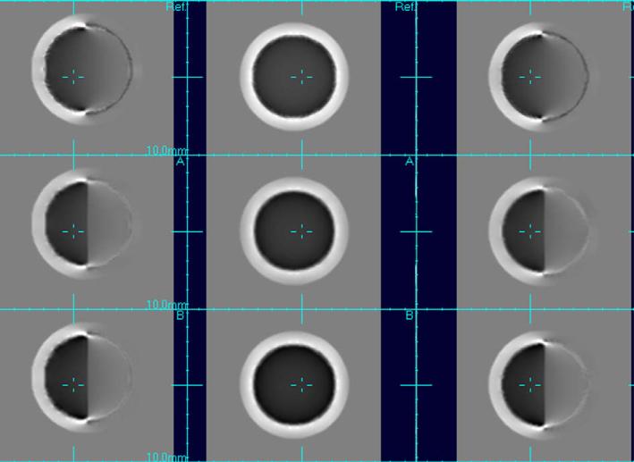
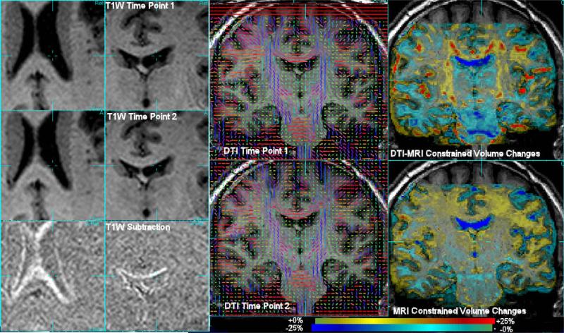
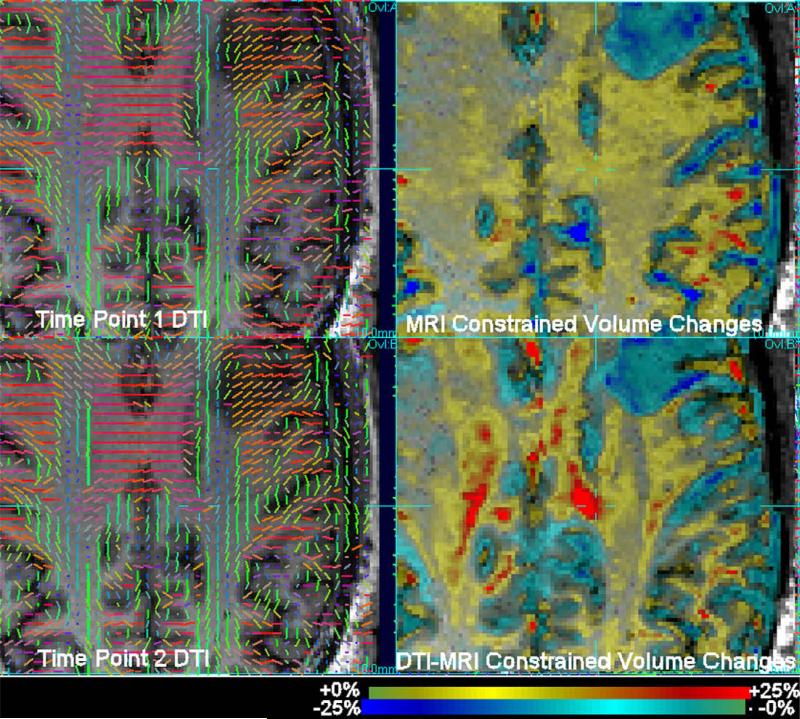
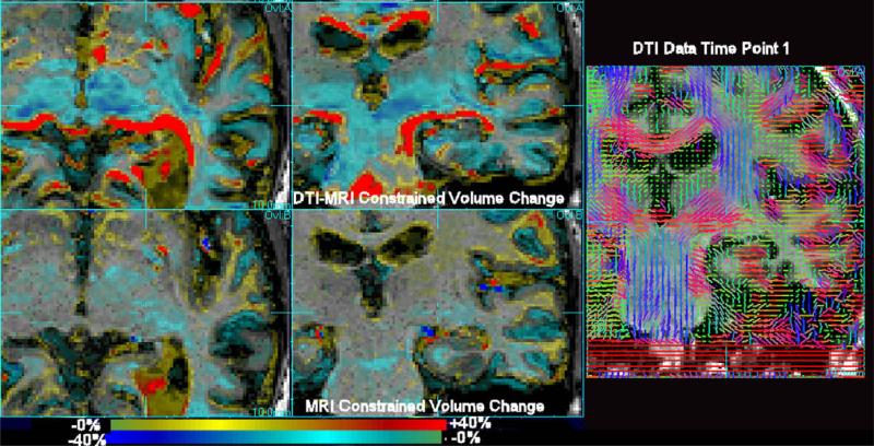
Registration based mapping of geometric differences in MRI anatomy allows the detection of subtle and complex changes in brain anatomy over time that provides an important quantitative window on the process of both brain development and degeneration. However, methods developed for this have so far been aimed at using conventional structural MRI data (T1W imaging) and the resulting maps are limited in their ability to localize patterns of change within sub-regions of uniform tissue. Alternative MRI contrast mechanisms, in particular Diffusion Tensor Imaging (DTI) data are now more commonly being used in serial studies and provide valuable complementary microstructural information within white matter. This paper describes a new approach which incorporates information from DTI data into deformation tensor morphometry of conventional MRI. The key problem of using the additional information provided by DTI data is addressed by proposing a novel mutual information (MI) derived criterion termed diffusion paired MI. This combines conventional and diffusion data in a single registration measure. We compare different formulations of this measure when used in a diffeomorphic fluid registration scheme to map local volume changes. Results on synthetic data and example images from clinical studies of neurodegenerative conditions illustrate the improved localization of tissue volume changes provided by the incorporation of DTI data into the morphometric registration.
Figures
Similar articles
-
Incorporating DTI data as a constraint in deformation tensor morphometry between T1 MR images.Inf Process Med Imaging. 2007;20:223-32. doi: 10.1007/978-3-540-73273-0_19. Inf Process Med Imaging. 2007. PMID: 17633702 Free PMC article.
-
Simultaneous consideration of spatial deformation and tensor orientation in diffusion tensor image registration using local fast marching patterns.Inf Process Med Imaging. 2009;21:63-75. doi: 10.1007/978-3-642-02498-6_6. Inf Process Med Imaging. 2009. PMID: 19694253
-
Brain tissue segmentation based on DTI data.Neuroimage. 2007 Oct 15;38(1):114-23. doi: 10.1016/j.neuroimage.2007.07.002. Epub 2007 Jul 13. Neuroimage. 2007. PMID: 17804258 Free PMC article.
-
Population based analysis of directional information in serial deformation tensor morphometry.Med Image Comput Comput Assist Interv. 2007;10(Pt 2):311-8. doi: 10.1007/978-3-540-75759-7_38. Med Image Comput Comput Assist Interv. 2007. PMID: 18044583 Free PMC article.
-
Contributions to magnetic susceptibility of brain tissue.NMR Biomed. 2017 Apr;30(4):10.1002/nbm.3546. doi: 10.1002/nbm.3546. Epub 2016 May 30. NMR Biomed. 2017. PMID: 27240118 Free PMC article. Review.
Cited by
-
Genetic influences on brain asymmetry: a DTI study of 374 twins and siblings.Neuroimage. 2010 Aug 15;52(2):455-69. doi: 10.1016/j.neuroimage.2010.04.236. Epub 2010 Apr 27. Neuroimage. 2010. PMID: 20430102 Free PMC article.
-
White matter damage in frontotemporal dementia and Alzheimer's disease measured by diffusion MRI.Brain. 2009 Sep;132(Pt 9):2579-92. doi: 10.1093/brain/awp071. Epub 2009 May 12. Brain. 2009. PMID: 19439421 Free PMC article.
-
Tensor-based morphometry using scalar and directional information of diffusion tensor MRI data (DTBM): Application to hereditary spastic paraplegia.Hum Brain Mapp. 2018 Dec;39(12):4643-4651. doi: 10.1002/hbm.24278. Epub 2018 Sep 25. Hum Brain Mapp. 2018. PMID: 30253021 Free PMC article.
-
Mapping directionality specific volume changes using tensor based morphometry: an application to the study of gyrogenesis and lateralization of the human fetal brain.Neuroimage. 2012 Nov 1;63(2):947-58. doi: 10.1016/j.neuroimage.2012.03.092. Epub 2012 Apr 6. Neuroimage. 2012. PMID: 22503938 Free PMC article.
-
Reprint of "Quantitative evaluation of brain development using anatomical MRI and diffusion tensor imaging".Int J Dev Neurosci. 2014 Feb;32:28-40. doi: 10.1016/j.ijdevneu.2013.11.006. Epub 2013 Dec 2. Int J Dev Neurosci. 2014. PMID: 24295553 Free PMC article.
References
-
- Alexander DC, Gee JC, Bajcsy RK. Strategies for data reorientation during nonrigid warps of diffusion tensor images. Proceedings of MICCAI. 1999;99:463–472.
-
- Basser P, Mattiello J, le Bihan D. Estimation of the effective self-diffusion tensor from the NMR spin echo. Journal of Magnetic Resonance. 1994;103:247–254. - PubMed
-
- Cao Y, Miller M, Winslow R, Younes L. Large deformation diffeomorphic metric mapping of vector fields. IEEE Transactions on Medical Imaging. 2005;24(9):1216–1230. - PubMed
Publication types
MeSH terms
Grants and funding
LinkOut - more resources
Full Text Sources
Other Literature Sources
Medical
