. 2008 Jun 13;133(6):963-77.
doi: 10.1016/j.cell.2008.04.039.

The amyotrophic lateral sclerosis 8 protein VAPB is cleaved, secreted, and acts as a ligand for Eph receptors

Affiliations

The amyotrophic lateral sclerosis 8 protein VAPB is cleaved, secreted, and acts as a ligand for Eph receptors

Hiroshi Tsuda et al. Cell. .

Abstract

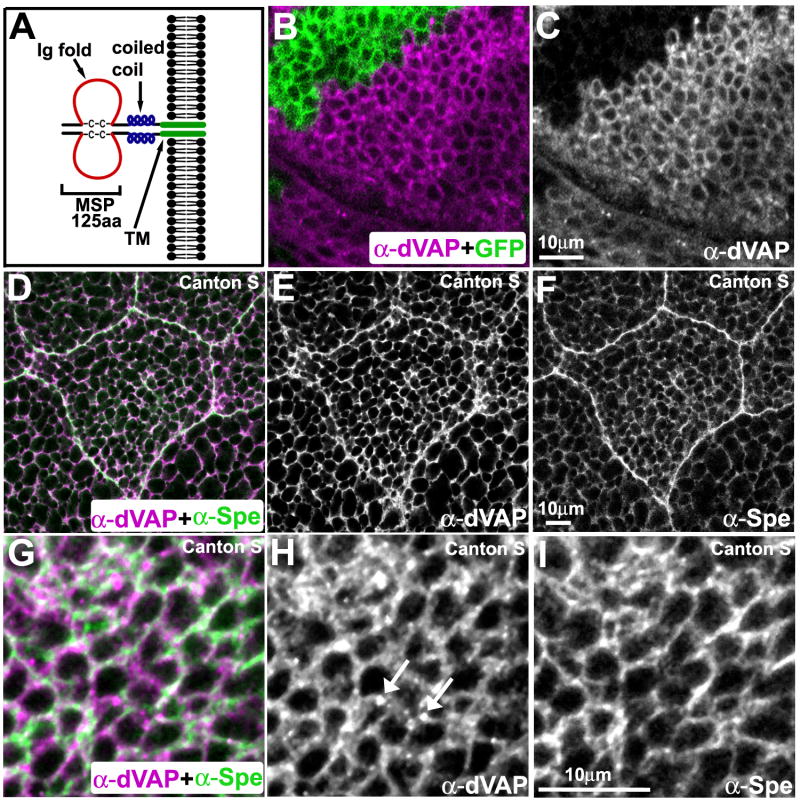
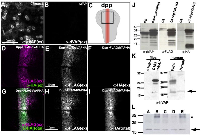
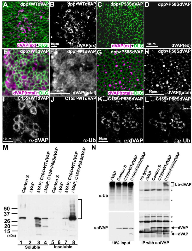
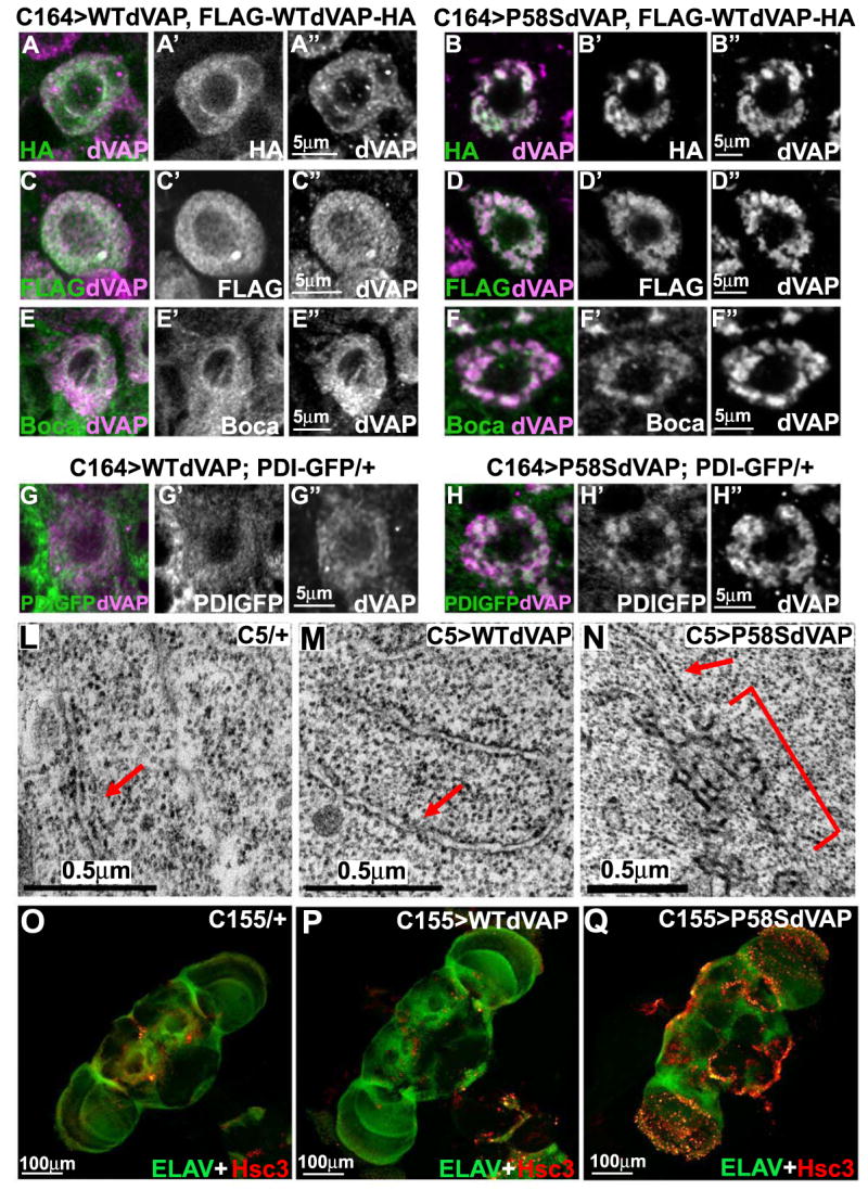
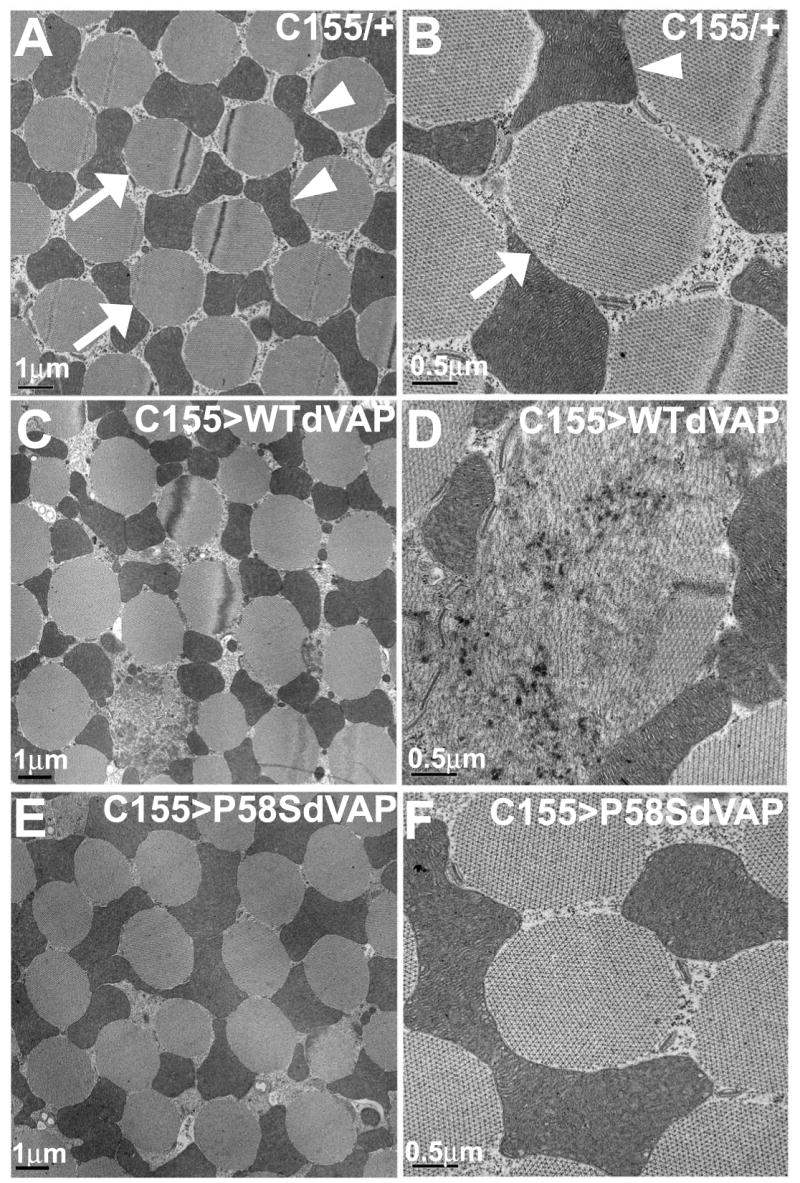
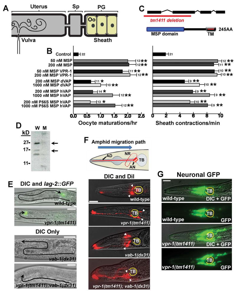
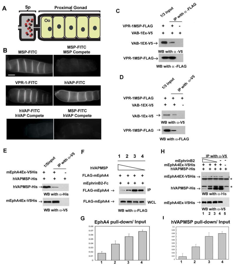
VAP proteins (human VAPB/ALS8, Drosophila VAP33, and C. elegans VPR-1) are homologous proteins with an amino-terminal major sperm protein (MSP) domain and a transmembrane domain. The MSP domain is named for its similarity to the C. elegans MSP protein, a sperm-derived hormone that binds to the Eph receptor and induces oocyte maturation. A point mutation (P56S) in the MSP domain of human VAPB is associated with Amyotrophic lateral sclerosis (ALS), but the mechanisms underlying the pathogenesis are poorly understood. Here we show that the MSP domains of VAP proteins are cleaved and secreted ligands for Eph receptors. The P58S mutation in VAP33 leads to a failure to secrete the MSP domain as well as ubiquitination, accumulation of inclusions in the endoplasmic reticulum, and an unfolded protein response. We propose that VAP MSP domains are secreted and act as diffusible hormones for Eph receptors. This work provides insight into mechanisms that may impact the pathogenesis of ALS.


Figures

Figure 1
Figure 1. VAP localization
(A) The structure of VAPs. Note that VAPs form dimers. (B-C) MARCM analysis showing the specificity of rabbit anti-dVAP antibody. A portion of the wing imaginal disc stained with anti-dVAP antibody (Rb92). GFP marks the mutant region (B). Single channel view of (B) showing only anti-dVAP (C). (D-I) dVAP partially colocalizes with Spectrin. Salivary gland (D-F) and wing imaginal disc (G-I) of Canton S flies.
Figure 2
Figure 2. A portion of VAP is secreted
(A-B) dVAP in the wing disc is localized extracellularly. Staining of a wing pouch of Canton S (A) and dVAP null mutant (ΔVAP) (B) with anti-dVAP antibody (Rb92) without membrane permeabilization. Note that not all cells are decorated by dVAP. (C) Pattern of expression of dpp-GAL4 in the wing imaginal disc. Anterior is to the left, ventral is to the top. The square in the center corresponds to Figure 2D-I. (D-F) The N-terminal but not the C-terminal of dVAP is localized extracellularly. The wing disc expressing FLAG-dVAP-HA was co-stained with anti-FLAG and anti-HA without permeabilization. (G-I) The N-terminal portion of dVAP is distributed more widely than the C-terminal portion. The wing disc expressing FLAG-dVAP-HA was stained with anti-FLAG extracellularly, and subsequently stained with anti-HA intracellularly (G). (total) intra- and extracellular staining. (ex) extracellular staining only. (J) The N-terminal region of dVAP is cleaved from the full length protein. da-GAL4 was used to express FLAG-dVAP-HA ubiquitously. Extracts from Canton S (CS) and and flies expressing FLAG-dVAP-HA were immunoblotted with anti-dVAP (left panel), anti-HA (middle panel) and anti-FLAG (right panel). (K) Truncated hVAP is expressed in human serum. Protein of wild type flies and those expressing hVAP were extracted and immunoblotted with anti-hVAP antibody (left panel). The extracts of human white blood cells and serum are immunoblotted with anti-hVAP antibody (right panel). Note that only the truncated hVAP protein is expressed in the serum (arrow in right panel). (L) hVAP is expressed in human serum of five different individuals. The extracts of human serum were immunoblotted with anti-hVAP antibody. * indicates non-specific band (see Figure S1D).
Figure 3
Figure 3. The P58S mutation leads to a failure to secrete dVAP and forms ubiquitinated inclusions
(A-D) The P58S mutation leads to a loss of the extracellular localization of dVAP. The wing discs expressing WT dVAP (A, B) and P58S dVAP (C, D) were stained using the anti-dVAP (magenta) extracellular staining protocol, and subsequently stained with anti-DLG (green) upon permeabilization. (E-H) The P58S mutation causes dVAP protein to be localized as intracellular inclusions. Wing discs expressing WT dVAP (E, F) and P58S dVAP (G, H). (I-L) Expression of P58S dVAP causes ubiquitin-containing inclusions. Neurons expressing WT dVAP (I, J) or P58S dVAP (K, L) were stained with anti-dVAP (I, K) and anti-Ubiquitin (J, L). The laser intensity was adjusted for the signals of overexpressed proteins. Hence, endogenous expression of dVAP cannot be observed (Figure 3A-L). (M) The P58S mutation causes detergent insoluble aggregates. Extracts of Canton S, null mutant (ΔVAP), and null mutant flies overexpressing WT or P58S dVAP were immunoblotted with anti-dVAP. Detergent soluble fractions (lane1-4); insoluble fractions (lane 5-8). (N) P58S dVAP is ubiquitinated. Anti-dVAP (GP33) was used to immunoprecipitate dVAP from extracts of ΔVAP and flies expressing WT (C155>WTdVAP) or P58S dVAP (C155>P58SdVAP). Immunoblots of anti-Ubiqutin (right top panel) and anti-dVAP (right bottom panel). (bracket) ubiquitin positive high molecular weight products * non-specific signals.
Figure 4
Figure 4. P58S dVAP protein accumulates in the ER, causes morphological changes in the ER, and induces an UPR
(A-D′′) Single motorneurons expressing WT FLAG-dVAP-HA probed with HA or FLAG antibodies. WT dVAP (A-A′′, C-C′′) or P58S dVAP (B-B′′, D-D′′) and FLAG-WTdVAP-HA are expressed in motor neurons with the C164-GAL4 driver. Note that all motor neurons expressing P58S dVAP have protein aggregates. (E-F′′) Boca is localized diffusely in the cytoplasm in flies expressing WT dVAP. In contrast, Boca is localized in inclusions in flies expressing P58S dVAP. (G-H′′) PDI-GFP is localized diffusely in the cytoplasm when WT dVAP is co-expressed in PDI-GFP transgenic flies. In contrast, PDI-GFP is present in inclusions when P58S dVAP is expressed. (L-N) TEM analysis of wing disc cells expressing WT or P58S dVAP with C5-GAL4, a wing pouch driver. Note that flies expressing P58S dVAP (N) contain clusters of aberrant electron-dense material (bracket) that is continuous with the rough ER (arrow). These clusters were commonly observed in C5>P58SdVAP (n=3 flies) and were never observed in wild type control cells (n=3 flies). (O-Q) Overexpression of P58S dVAP causes an UPR. Anti-Hsc3 and anti-Elav costaining of adult brains of control flies (O), flies expressing WT dVAP (P) or P58S dVAP (Q).
Figure 5
Figure 5. Overexpression of P58S dVAP does not phenocopy WT dVAP overexpression
TEM analysis of the dorsal ventral muscle (DVM) of flies overexpressing various dVAPs in neurons. Control animals (A-B), flies overexpressing neuronal WT dVAP (C-D) and P58S dVAP (E-F). Five animals, >30 muscles/animal were examined of each genotype.
Figure 6
Figure 6. VAPs have extracellular signaling activity and act in common genetic pathways with Eph receptors
(A-B) VAP MSP domains have extracellular signaling activity. (A) Microinjection is used to test molecules for the ability to induce oocyte (Oo) maturation and sheath contraction in unmated fog-2(q71) females, which lack sperm. Injected solutions diffuse into the spermatheca (Sp) and proximal gonad (PG). (B) Microinjections of purified, recombinant MSP domains are compared to a buffer-alone control injection. Additional negative controls are described in Miller et al. 2001, Miller et al. 2003, and Corrigan et al. 2005. Oocyte maturations per hour and basal sheath contractions per minute were scored. Number of gonads is to the right of SEM bars. *, P < 0.01; **, P < 0.001. (C) VPR-1 encodes the sole VAP homolog in the C. elegans genome. The tm1411 allele deletes the translational start site and the MSP domain. (D) Western analysis of mixed stage wild type (W) and vpr-1(tm1411) mutant (M) worms using antibodies against Drosophila VAP (Rb 92). (E) Distal Tip Cell (DTC) migration paths (black lines) in wild type and mutant hermaphrodites. vpr-1(tm1411) and vab-1(dx31) null mutants have incompletely penetrant and variably expressed defects in DTC migration (also see Fig. S7A). vpr-1(tm1411); vab-1(dx31) double mutants exhibit the same defects as vpr-1(tm1411) single mutants. (F) Amphid neuron positions in wild type and mutant hermaphrodites. Amphid neurons (AN, diagram) migrate in an anterior to posterior direction during embryogenesis. Amphid cell body positions are shown by arrowheads. Loss of vab-1 partially suppresses the posterior positioning defect of vpr-1(tm1411) mutants, although the double mutants have positioning and nerve ring (NR) defects not observed in either single mutant. The terminal bulb (TB) is outlined in yellow. Bars, 20 μm. (G) Head neuron positions in wild type and vpr-1(tm1411) hermaphrodites. A pan-neuronal transgenic reporter shows that many head neurons are positioned too far posteriorly in vpr-1(tm1411) mutants.
Figure 7
Figure 7. VAP MSP domains bind to Eph receptor extracellular domains
(A) Diagram of the C. elegans proximal gonad. MSP binds to receptors on the oocyte (Oo) and sheath cell surfaces in the proximal gonad (Miller et al. 2003; Corrigan et al. 2005). (B) Receptor binding sites are visualized using FITC-conjugated MSP domains. FITC-conjugates are biological active in promoting oocyte maturation and sheath contraction (Miller et al. 2003; data not shown). Compete includes a 25-fold molar excess of unlabelled protein. Bar, 20 μm. (C, D) VPR-1 MSP domain can bind to VAB-1 Eph receptor. Purified FLAG-tagged VPR-1 MSP (VPR-1MSP-FLAG) and V5 tagged VAB-1 ECT domain (VAB-1 Ex-V5) were co-immunoprecipitated using FLAG antibody (C) and V5 antibody (D). IP, immunoprecipitation. (E) His- and V5- tagged mouse EphA4 ectodomain (mEphA4Ex-V5His) and a His tagged hVAPB MSP (hVAPMSP-His) co-immunoprecipitate using anti-V5 antibody. (F) Conditioned medium containing hVAPMSP disrupts the interaction between mouse EphrinB2 (mEphrinB2) and EphA4. WCL: whole cell lysates. (G) Quantification of the fraction of IP of (F). Lanes 1 to 4 correspond to the lanes in Figure 7F. (H) Ephrin B2 competes with hVAPB MSP for binding of EphA4 in a dose dependent manner. As decreasing amounts of EphrinB2 are added, increasing amounts of hVAP MSP are pulled down by mEphA4Ex-V5 His. Note that EphrinB2-His cannot be detected in the top panel because it has a similar molecular weight to the IgG light chain. * shows non-specific heavy and light chains of IgG. (I) The quantification of the fraction of IP of (H). The lanes correspond to the lanes 1 to 4 in Figure 7H.

Comment in

References

    1. Amarilio R, Ramachandran S, Sabanay H, Lev S. Differential regulation of endoplasmic reticulum structure through VAP-Nir protein interaction. The Journal of biological chemistry. 2005;280:5934–5944. - PubMed
    1. Atkin JD, Farg MA, Turner BJ, Tomas D, Lysaght JA, Nunan J, Rembach A, Nagley P, Beart PM, Cheema SS, et al. Induction of the unfolded protein response in familial amyotrophic lateral sclerosis and association of protein-disulfide isomerase with superoxide dismutase 1. The Journal of biological chemistry. 2006;281:30152–30165. - PubMed
    1. Baker AM, Roberts TM, Stewart M. 2.6 A resolution crystal structure of helices of the motile major sperm protein (MSP) of Caenorhabditis elegans. J Mol Biol. 2002;319:491–499. - PubMed
    1. Basso M, Massignan T, Samengo G, Cheroni C, De Biasi S, Salmona M, Bendotti C, Bonetto V. Insoluble mutant SOD1 is partly oligoubiquitinated in amyotrophic lateral sclerosis mice. The Journal of biological chemistry. 2006;281:33325–33335. - PubMed
    1. Blelloch R, Anna-Arriola SS, Gao D, Li Y, Hodgkin J, Kimble J. The gon-1 gene is required for gonadal morphogenesis in Caenorhabditis elegans. Dev Biol. 1999;216:382–393. - PubMed

Publication types

MeSH terms