. 2008 Jun 13;133(6):1043-54.
doi: 10.1016/j.cell.2008.04.040.

Rewiring the specificity of two-component signal transduction systems

Affiliations

Rewiring the specificity of two-component signal transduction systems

Jeffrey M Skerker et al. Cell. .

Abstract

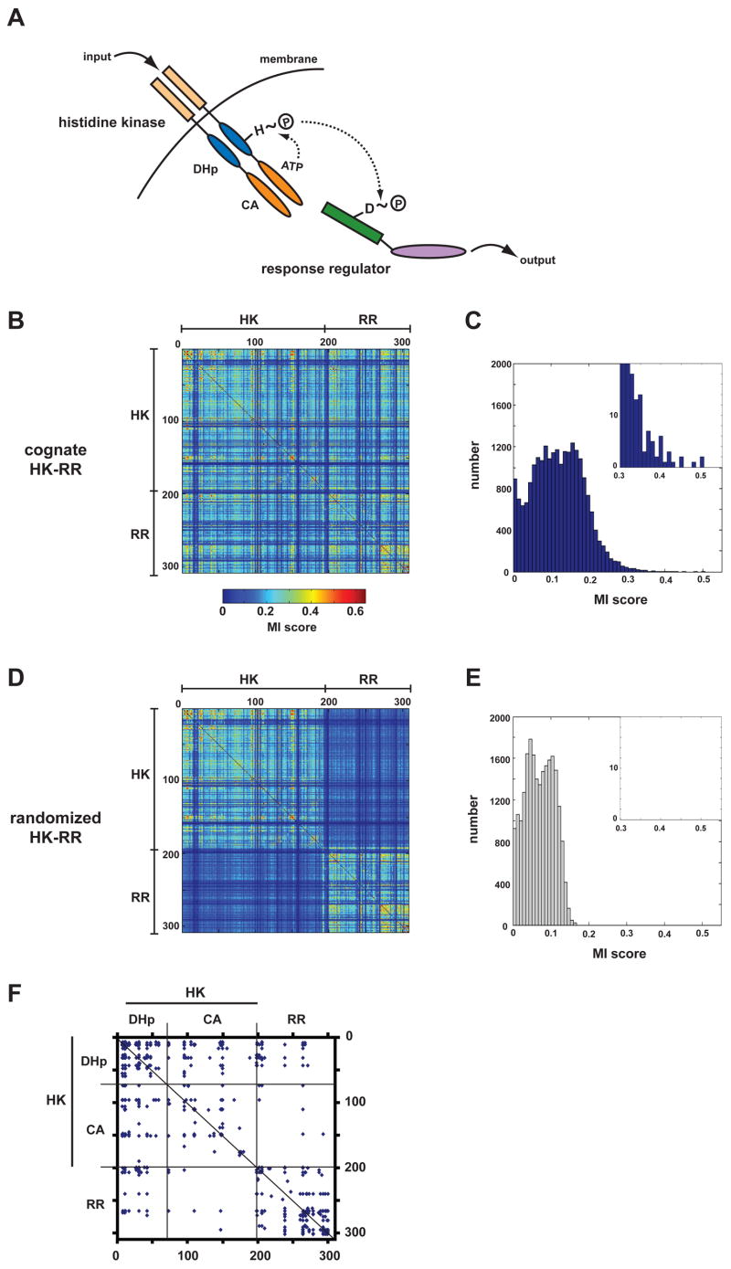
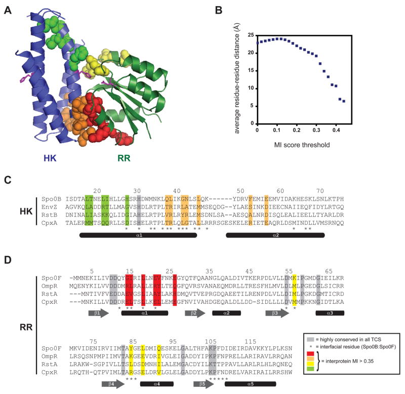
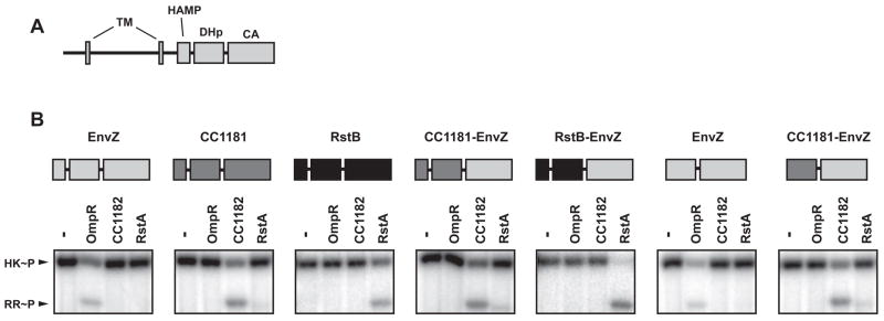
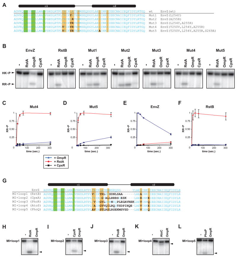
Two-component signal transduction systems are the predominant means by which bacteria sense and respond to environmental stimuli. Bacteria often employ tens or hundreds of these paralogous signaling systems, comprised of histidine kinases (HKs) and their cognate response regulators (RRs). Faithful transmission of information through these signaling pathways and avoidance of detrimental crosstalk demand exquisite specificity of HK-RR interactions. To identify the determinants of two-component signaling specificity, we examined patterns of amino acid coevolution in large, multiple sequence alignments of cognate kinase-regulator pairs. Guided by these results, we demonstrate that a subset of the coevolving residues is sufficient, when mutated, to completely switch the substrate specificity of the kinase EnvZ. Our results shed light on the basis of molecular discrimination in two-component signaling pathways, provide a general approach for the rational rewiring of these pathways, and suggest that analyses of coevolution may facilitate the reprogramming of other signaling systems and protein-protein interactions.


Figures

Figure 1
Figure 1. Mutual information analysis of two-component signal transduction proteins
(A) Schematic of the two-component signaling paradigm. Receipt of a signal stimulates autophosphorylation of a histidine kinase (HK). The phosphoryl group is subsequently passed to a cognate response regulator (RR), which can trigger changes in gene expression or other physiological processes. (B) Identification of covarying residues in histidine kinases and response regulators. Cognate, operon pairs of histidine kinases and response regulators were concatenated and aligned. Using this multiple sequence alignment, every pair of positions was examined for covariation using an analysis of mutual information. The matrix of scores for each pair of positions in the alignment is plotted as a heatmap, according to the color legend shown. Positions within the alignment corresponding to the histidine kinase and the response regulator are indicated. (C) Histogram of interprotein (HK-RR) mutual information scores from panel B. (D) The pairings of histidine kinases and response regulators from panel B were randomized and the mutual information analysis repeated. The heatmap of scores shows that the randomization process reduces interprotein, but not intraprotein (HK-HK, RR-RR), covariation. (E) Histogram of interprotein mutual information scores for the alignment in which HK-RR pairings were scrambled. (F) For the multiple sequence alignment containing cognate HK-RR pairs, the graph indicates the pairs of amino acids with scores >0.35. Regions of the alignment corresponding to the HK and RR are indicated as well as the two HK domains, the dimerization and histidine phosphotransfer (DHp) domain and the catalytic and ATP-binding (CA) domain (see panel A).
Figure 2
Figure 2. Specificity determining residues map to the interface of a two-component signal transduction complex
(A) Structure of the histidine phosphotransferase Spo0B in complex with the response regulator Spo0F (PDB: 1F51). No HK-RR cocrystal structure has yet been solved. However, the Spo0B:Spo0F serves as a suitable proxy because the four-helix bundle of Spo0B is similar to that formed by the DHp domain of histidine kinases. Residues with the highest intermolecular mutual information scores (> 0.35) are shown by spacefilling. Spo0B is a dimer, but for clarity, only one set of residues is shown. Each molecule contains two regions of high-scoring residues; those in the histidine kinase are colored green and orange and those in the response regulator are colored red and yellow. Additional views are shown in Figure S1. (B) Plot of average residue-residue distances as a function of covariation score for all pairs of positions from the multiple sequence alignment (see Figure 1B and Supplemental File 1) that include one site within the DHp domains of the kinase and one site within the response regulators. (C) Primary sequence alignment of the histidine kinases EnvZ, RstB, and CpxA with the histidine phosphotransferase Spo0B. Residues showing strongest covariation with residues in the response regulator are highlighted in green and orange, as in panel A. (D) Primary sequence alignment of the response regulators Spo0F, OmpR, RstA, and CpxR. Residues showing strongest covariation with residues in the histidine kinase are highlighted in red and yellow, as in panel A. Highly conserved residues are shaded in grey. Interprotein contact residues are marked by asterisks below the sequence alignments along with approximate locations of secondary structure elements.
Figure 3
Figure 3. Specificity of phosphotransfer resides in the DHp domain of a histidine kinase
(A) Schematic of a prototypical histidine kinase. Two transmembrane (TM) regions flank a periplasmic sensor domain followed by the cytoplasmic portion containing HAMP, DHp, and CA domains. (B) Phosphotransfer specificity of domain-swapped chimeric histidine kinases. Each chimera was autophosphorylated and incubated alone or tested for phosphotransfer to the response regulators OmpR, CC1182 and RstA at a 10 second time-point. Schematics above each gel indicate the identity of each domain in the chimera tested. For complete protein sequences, see Supplemental File 2.
Figure 4
Figure 4. The base of the DHp domain dictates phosphotransfer specificity of histidine kinases
(A) Sequences of the DHp domain in the sub-domain chimeras. A small region of the EnvZ DHp domain was replaced with the corresponding portion of five other kinases indicated in parentheses. Amino acids mutated, relative to wild-type EnvZ, are indicated in bold. Colored bars indicate residues with high interprotein (HK-RR) covariation scores. For complete protein sequences, see Supplemental File 2. A schematic of the histidine kinase domain structure and the secondary structure of the DHp domain are shown above the sequences. (B) Rewiring the phosphotransfer specificity of EnvZ to match that of RstB. Autophosphorylated histidine kinases were tested for phosphotransfer to RstA or OmpR and as a control, were incubated without regulator (indicated by a minus sign). The three gel images show the phosphotransfer behavior of (in order from top to bottom): EnvZ, RstB, and the sub-domain chimera Chim1. Similar sets of gel images are shown for rewiring of EnvZ to match the specificity of CpxA (C), PhoR (D), AtoS (E), and PhoQ (F). Phosphotransfer reactions were incubated for 10 seconds (B, C) or 5 minutes (D-F). Black arrowheads indicate the position of the band corresponding to OmpR (top gel in each panel) or the alternative response regulator (bottom two gels in each panel). Kinetic analysis of phosphotransfer specificity of EnvZ (G), RstB (H), CpxA (I), Chim1 (J), and Chim2 (K). In each case an autophosphorylated kinase, indicated above each graph, was examined for phosphotransfer to OmpR, RstA, and CpxR at 0, 10, 30, 60, and 300 seconds. Normalized levels of phosphorylated RR are plotted as fraction maximal versus reaction time in seconds. Data points and error bars represent the mean and standard deviation (n=3). Representative gel images are shown in Figure S4. The slow decrease in phosphorylated RR after reaching a maximal value is due to hydrolysis and phosphatase activity of the kinase. (L) Phosphotransfer profiles for Chim1 and Chim2 versus all 32 E. coli response regulators. Both chimeras phosphorylate only the intended, target response regulator, indicated by black arrowheads.
Figure 5
Figure 5. Rewiring two-component signal transduction pathways
(A) Sequences of point mutants constructed in which amino acids with high interprotein mutual information scores were mutated in EnvZ to match the corresponding amino acid in RstB. Sequences shown correspond only to the DHp domain. Sites differing from the wild-type EnvZ are in bold. Colored bars indicate residues with high interprotein mutual information scores. (B) Phosphotransfer specificity of EnvZ, RstB, and Mut1-Mut5. In each case the kinase was autophosphorylated and then incubated alone or examined for phosphotransfer to RstA, OmpR, and CpxR after 10 second incubations. (C,D) Kinetics of phosphotransfer for Mut4 and Mut5. Normalized RR~P levels are plotted as fraction maximal versus reaction time in seconds. Data points and error bars represent the mean and standard deviation (n=3). Representative gel images are shown in Figure S4. (E, F) Graphs showing the kinetic preference of wild-type EnvZ and RstB (see Figures 4G-4H) are shown for comparison to Mut4 and Mut5. (G) Sequences of the MI+loop (mutual information+loop) mutants. Amino acids that were mutated, relative to wild-type EnvZ, are indicated in bold. Colored bars indicate residues with high interprotein (HK-RR) covariation scores. MI+loop mutant kinases (MI+loop1-5) were tested for phosphotransfer to OmpR and the new intended target: RstA (H), CpxR (I), PhoB (J), AtoC (K), or PhoP (L). Black arrowheads indicate position of the target response regulator band within each gel. Experiments were performed exactly as in Figures 4B-4F and can be compared directly.
Figure 6
Figure 6. Rewiring histidine kinase specificity in vivo
(A) Schematic of the reporter construct used to assess CpxR phosphorylation in vivo. The gfp gene is driven by a cpxP promoter, which depends on CpxR~P for activation. (B) GFP fluorescence normalized by OD600 for the cpxP transcriptional reporter strain AFS161 (envZ cpxA PcpxP-gfp) containing the indicated plasmids. (C) Schematic of the reporter construct used to assess PhoP phosphorylation in vivo. The yfp gene is driven by a mgrB promoter, which depends on PhoP~P for activation. The cfp gene is driven by a constitutively active promoter and is used for normalization. (D) YFP/CFP fluorescence ratio of the mgrB transcriptional reporter strain AFS237 (envZ phoQ PmgrB-yfp PtetA-cfp) containing the indicated plasmids. In each case, the average of measurements from three independent cultures is shown. Error bars indicate standard deviations. The pEnvZ and p(MI+loop) plasmids express full-length versions of EnvZ and the chimeras (see Figure 5), respectively.

Comment in

References

    1. Atchley WR, Wollenberg KR, Fitch WM, Terhalle W, Dress AW. Correlations among amino acid sites in bHLH protein domains: an information theoretic analysis. Mol Biol Evol. 2000;17:164–178. - PubMed
    1. Batchelor E, Goulian M. Robustness and the cycle of phosphorylation and dephosphorylation in a two-component regulatory system. Proc Natl Acad Sci USA. 2003;100:691–696. - PMC - PubMed
    1. Batchelor E, Walthers D, Kenney LJ, Goulian M. The Escherichia coli CpxA-CpxR envelope stress response system regulates expression of the porins ompF and ompC. J Bacteriol. 2005;187:5723–5731. - PMC - PubMed
    1. Bloom JD, Meyer MM, Meinhold P, Otey CR, MacMillan D, Arnold FH. Evolving strategies for enzyme engineering. Curr Opin Struct Biol. 2005;15:447–452. - PubMed
    1. Buck MJ, Atchley WR. Networks of coevolving sites in structural and functional domains of serpin proteins. Mol Biol Evol. 2005;22:1627–1634. - PubMed

Publication types

MeSH terms