. 2008 Jun 13;133(6):1093-105.
doi: 10.1016/j.cell.2008.04.048.

Asymmetric homotypic interactions of the atypical cadherin flamingo mediate intercellular polarity signaling

Affiliations

Asymmetric homotypic interactions of the atypical cadherin flamingo mediate intercellular polarity signaling

Wei-Shen Chen et al. Cell. .

Abstract

Acquisition of planar cell polarity (PCP) in epithelia involves intercellular communication, during which cells align their polarity with that of their neighbors. The transmembrane proteins Frizzled (Fz) and Van Gogh (Vang) are essential components of the intercellular communication mechanism, as loss of either strongly perturbs the polarity of neighboring cells. How Fz and Vang communicate polarity information between neighboring cells is poorly understood. The atypical cadherin, Flamingo (Fmi), is implicated in this process, yet whether Fmi acts permissively as a scaffold or instructively as a signal is unclear. Here, we provide evidence that Fmi functions instructively to mediate Fz-Vang intercellular signal relay, recruiting Fz and Vang to opposite sides of cell boundaries. We propose that two functional forms of Fmi, one of which is induced by and physically interacts with Fz, bind each other to create cadherin homodimers that signal bidirectionally and asymmetrically, instructing unequal responses in adjacent cell membranes to establish molecular asymmetry.


Figures

Figure 1
Figure 1. Mutual recruitment of Fz and Vang
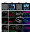
(A) Schematic of core PCP protein localization; early (<6 hrs APF), and later stages (> 24 hrs APF). (B–B″) Prehairs (phalloidin; B′; red in B″) initiate at the distal vertex where Fz is enriched (Fz::GFP; B; green in B″). Distal to the right in this and all subsequent figures. (C–F) fz, vang, fmi, and vang, fz double mutant clones. Using MARCM, clones are marked by DsRed (blue in [C″–F″]) and Fz::GFP is excluded from the clone ([C–F]; green in [C″–F″]). Prehairs stained with phalloidin ([C′–F′]; red in [C″–F″]). Scale bars in all figures indicate 15 microns. (C–C″) fzR52 clone showing distal non-autonomy ([C′]; red in [C″]). FzGFP is recruited to the clone border in wildtype neighbors ([C]; green in [C″]; yellow arrowheads). (D–D″) vangA3clone showing proximal non-autonomy ([D′]; red in [D″]). FzGFP is excluded from the clone border in wildtype neighbors ([D]; green in [D″]; yellow arrowheads). (E–E″) fmiE45 clone showing cell-autonomous polarity ([E′]; red in [E″]). FzGFP in wildtype neighbors is absent but not repelled from the clone border; note the characteristic accumulation at cell borders perpendicular to the clone border, and contrast with the vangA3clone ([E]; green in [E″]; compare to [D]; yellow arrowheads). (F–F″) vangA3; fzR52 double mutant clone shows a cell-autonomous phenotype ([F′]; red in [F″]). FzGFP is neither recruited to nor repelled from the clone border ([F]; green in [F″]; yellow arrowheads). (G–J) Schematics of hair polarity and Fz protein localization (green) in wildtype cells neighboring fzR52 (G), vangA3 (H), fmiE45 (I) and vangA3; fzR52 clones (J). (K–N) FzΔCRD rescues the fzK21/fzD21 phenotype. fz mutant showing disarrayed polarity in the wing (B) and thorax (D). (A, C) Rescue by the FzΔCRD transgene. (O) VangΔECDYFP shows asymmetric localization in pupal wing at 30hrs APF. (P) Schematics of VangΔECDand FzΔCRD. Extracellular loops of Vang are replaced by HA and FLAG tags in VangΔECD, and the CRD domain of Fz is deleted in FzΔCRD. (Q) Absence of polarity defect in wing clones simultaneously mutant for the five Drosophila Wnts expressed in the wing: Wnt2, Wg, Wnt4, Wnt6 and Wnt10.
Figure 2
Figure 2. Flamingo is required for Fz-Vang intercellular communication
(A–B) Paradigm to test functional requirements for individual PCP components in Fz signaling cells. A clone that simultaneously overexpresses Fz while mutant for a PCP gene of interest is generated using forward MARCM (positively labeled by DsRed [red dots]). Flip-on induced expression of VangYFP (green) in neighboring wildtype cells assays recruitment of VangYFP. (C–D) Paradigm to test functional requirements for individual PCP components in responding cells. Reverse MARCM generates a clone of cells mutant for PCP gene of interest (marked by loss of LacZ [blue; yellow dots]) and a twin spot that overexpresses Fz (bright blue; marked by DsRed [red dots]). Simultaneous flip-on induced expression of VangYFP (green) in the mutant cells assays recruitment of VangYFP. (E–F) Forward MARCM control. VangYFP from wildtype cells is recruited to the border of a Fz overexpressing LacZ clone (arrowhead in [E]; green in [F]). Fz overexpressing clone is positively marked by co-expression of DsRed (red). (G–H) Reverse MARCM control. VangYFP inside a LacZ clone (yellow dots [G]; absence of blue [H]) is recruited toward the Fz overexpressing clone (arrowhead in [G]; green in [H]). (I–J) VangYFP in cells neighboring a Fz overexpressing clone that is mutant for dgo380. VangYFP is recruited to the clone border (arrowhead in [I]; green in [J]). (K–L) VangYFP inside a dgo380 clone (yellow dots and absence of blue) is recruited toward the Fz overexpressing clone (arrowhead in [K]; green in [L]). (M–N) VangYFP in cells neighboring a Fz overexpressing clone that is mutant for fmiE45. VangYFP is not recruited to the clone border (arrowhead in [M]; green in [N]). (O–P) VangYFP inside a fmiE45 (yellow dots and absence of blue) clone is not recruited toward the Fz overexpressing clone (yellow arrow in [O]; green in [P]). Recruitment is seen near non-mutant cells (blue arrow).
Figure 3
Figure 3. Fmi generates an instructive signal
(A–D) Fmi overexpression in LacZ (A), fzR52 (B), vangA3 (C) or vangA3; fzR52 (D) clones non-autonomously recruits Fz from neighboring wildtype cells to the clone border (arrowheads). Clones overexpressing Fmi are positively marked by DsRed (blue in [A″–D″]) and exclude FzGFP expression ([A–D]; green in [A″–D″]). Prehairs stained with phalloidin ([A′–D′]; red in [A″–D″]). Hairs are repolarized to point toward the clone.
Figure 4
Figure 4. Flamingo acts homophilically
(A–B) A Fmi overexpression clone and its twin-spot that is mutant for fmiE45 (MARCM). Fmi overexpressing clone positively marked by DsRed (red in [B]) and excluding FzGFP expression. FzGFP in the fmiE45 clone ([A]; green in [B]; yellow dots outline the fmi clone) fails to localize to the cell membrane and remains largely intracellular (yellow arrowheads), whereas it is recruited from wildtype neighbors (blue arrowhead). (C–D) FmiΔC overexpression clone, positively marked by DsRed (blue in [D]), and excluding FzGFP expression ([C]; green in [D]), repolarizes neighboring wildtype cells and recruits FzGFP to the clone border (arrowhead). Prehairs stained with phalloidin (red in [D]). (E–F) FmiΔC overexpression in fmiE45 mutant clone, positively marked by DsRed (blue in [F]), and excluding FzGFP expression ([E]; green in [F]), repolarizes neighboring wildtype cells (arrowhead in [E]). Prehairs stained with phalloidin (red in [F]). Cells homozygous for fmiE45 fail to recruit Fz from neighbors (asterisk in [E]). (G–H) Schematic of FmiΔC overexpression clone in wildtype (G) and in fmi mutant cells (H).
Figure 5
Figure 5. Fmi exists in two functional forms
(A–C) vangA3 mutant clone overexpressing Fmi, positively labeled by DsRed (blue), recruits Fz but not Vang across cell boundaries. In the neighboring cells, FzGFP (green in [A]; [B]) is recruited to the Fmi overexpression clone border, but Vang (red in [A]; [C]) is excluded. Arrowhead marks a portion of the clone border. (D) Vang (visualized with anti-Vang antibody) is recruited to vangA3 mutant clone borders (arrowheads). (E–G) Negatively marked (red) fmi clones unrescued (E), rescued with FmiYFP (F), or rescued with FmiΔC (G), all stained with anti-Fmi (green) at 28 hrs APF. Note the zigzag pattern of Fmi within the clones. (E′, F′) fmi clones unrescued (E′) or rescued with FmiYFP (F′), showing phalloidin stained actin at 35 hrs APF. (H, I, L–N) Simultaneous overexpression of Fz and Fmi in a clone of wildtype cells (H, I) and a clone of vangA3 cells (L–N) can modify the direction of Fmi-Fmi signal relay. Overexpression clones are marked by loss of FzGFP expression ([H, I, M]; green in [L]) and endogenous Vang expression ([N]; red in [L]; MARCM). Though some clone borders still recruit FzGFP ([H, I, M]; green in [L]), many borders lose FzGFP expression (yellow dots in [H, I]) and recruit Vang instead ([N]; red in [L]). Cell by cell variation likely results from the variable over-expression levels and thus varying ratio of Fmi and Fz known to be achieved using the GAL4 system. (J–K) Schematics showing recruitment of Fz across cell boundaries by Fmi overexpression (J), and Vang recruitment in cells neighboring a Fmi + Fz co-overexpression clone (K). F-Fmi (blue) and V-Fmi (orange). Blue cells overexpress Fmi or Fmi + Fz and lack Vang. (O, P) While overexpression of FmiΔC recruits Fz to neighboring cell boundaries (O; blue dots mark wildtype neighbors), simultaneous overexpression of FmiΔC and Fz reverses this recruitment (P; yellow dots mark wildtype neighbors with decreased Fz recruitment), as was seen for wildtype Fmi (H, I, L, M).
Figure 6
Figure 6. Fz and Fmi physically interact
(A) Fmi co-immunoprecipitates Fz from inducible S2 cells. Fmi or FmiΔC transfected into S2 cells with or without CuSO4 induction of Fz expression. Immunoblots of total extract (left). Fz is co-immunoprecipitated by Fmi (center) or FmiΔC (right) only when Fmi or FmiΔC is transfected and Fz expression is induced. Upper panels probed for Fmi and lower panels probed for Fz. Arrowhead marks the dye front. (B) FmiΔNYFP co-immunoprecipitates Fz. FmiYFP or FmiΔNYFP transfected into S2 cells with Fz expression induced. Immunoblots of total extract (left). Fz is co-immunoprecipitated by FmiYFP or FmiΔNYFP. Anti-Fmi detects FmiYFP but not FmiΔNYFP (center), while anti-GFP detects both (right). Some degradation of Fmi is observed. (C) Fmi co-immunoprecipitates Fz from pupae. Total extracts from wildtype (OreR) probed for Fmi and Fz (left). Fmi co-immunoprecipitates Fz from OreR but only trace amounts from fzR52 pupae. Control anti-Flag does not precipitate either Fmi or Fz. Note that Fz runs at approximately 65 Kd in these gels, whereas in the S2 cell experiments, the majority runs at approximately 50 Kd, with a minor band at 65 Kd (not shown). This difference likely depends on different denaturing conditions, as samples cannot be boiled without losing all Fz signal.
Figure 7
Figure 7. Model
(A) Fmi exists in two functional forms: a Fz-associated form (F-Fmi; dark blue) that interacts with and is induced by Fz in the same cell, and a Vang-associated form (V-Fmi, orange) that interacts with Vang in the same cell. The two functional forms preferentially interact with each other, rather than with their like forms. (B) The Fz scalar slope and the PCP protein asymmetry models in clones surrounded by fmi mutant cells. Shading represents Fz activity and/or localization. Cells in the Fz scalar model cannot be arranged to allow a polarity swirl with hairs pointing from high toward low Fz activity levels. (C–E) Wildtype cells (positively marked by DsRed; green in [D]) surrounded by fmiE45 mutant cells shows a swirling polarity defect. Prehairs stained with phalloidin ([C]; red in [D]). (E) Schematic of polarity pattern. (F–H) Wildtype cells (positively marked by DsRed; yellow in [G]) surrounded by fmiE45 mutant cells shows another swirl. Prehairs stained with phalloidin (magenta in [G]). FzGFP ([F]; green in [G]).

Similar articles

Cited by

References

    1. Adler PN, Krasnow RE, Liu J. Tissue polarity points from cells that have higher Frizzled levels towards cells that have lower Frizzled levels. Curr Biol. 1997;7:940–949. - PubMed
    1. Adler PN, Taylor J, Charlton J. The domineering non-autonomy of frizzled and van Gogh clones in the Drosophila wing is a consequence of a disruption in local signaling. Mech Dev. 2000;96:197–207. - PubMed
    1. Amonlirdviman K, Khare NA, Tree DR, Chen WS, Axelrod JD, Tomlin CJ. Mathematical modeling of planar cell polarity to understand domineering nonautonomy. Science. 2005;307:423–426. - PubMed
    1. Axelrod JD. Unipolar membrane association of Dishevelled mediates Frizzled planar cell polarity signaling. Genes Dev. 2001;15:1182–1187. - PMC - PubMed
    1. Bastock R, Strutt H, Strutt D. Strabismus is asymmetrically localised and binds to Prickle and Dishevelled during Drosophila planar polarity patterning. Development. 2003;130:3007–3014. - PubMed

Publication types