. 2008 Jul;8(6):389-396.
doi: 10.1016/j.gep.2008.05.001. Epub 2008 May 10.

Expression of EGFL7 in primordial germ cells and in adult ovaries and testes

Affiliations

Expression of EGFL7 in primordial germ cells and in adult ovaries and testes

Luisa Campagnolo et al. Gene Expr Patterns. 2008 Jul.

Abstract

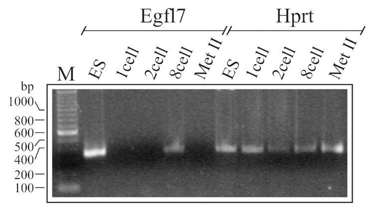
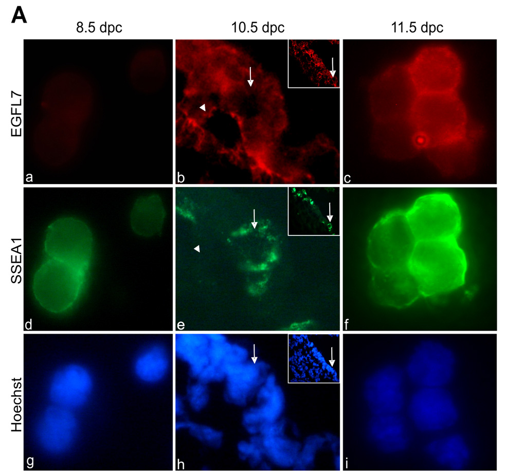
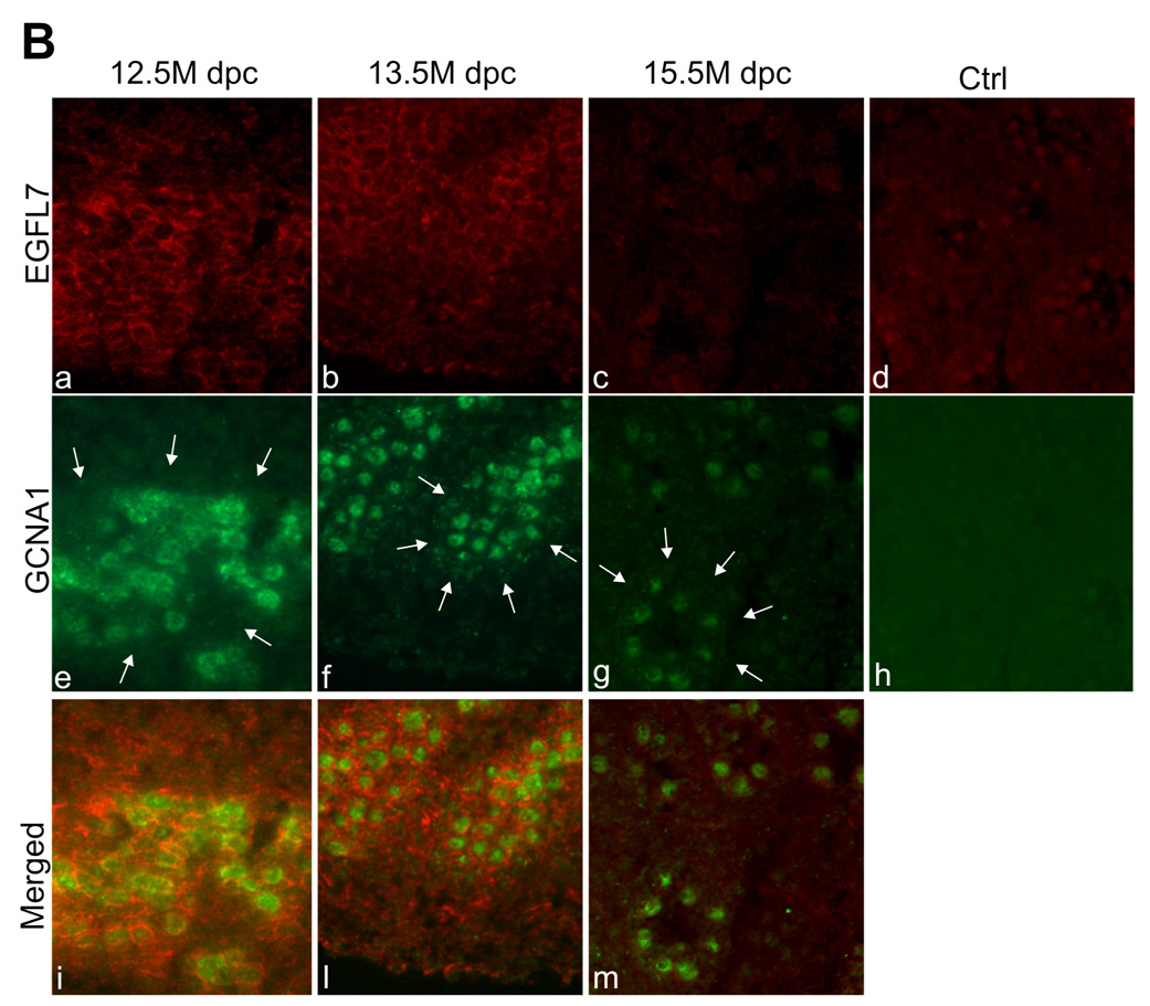
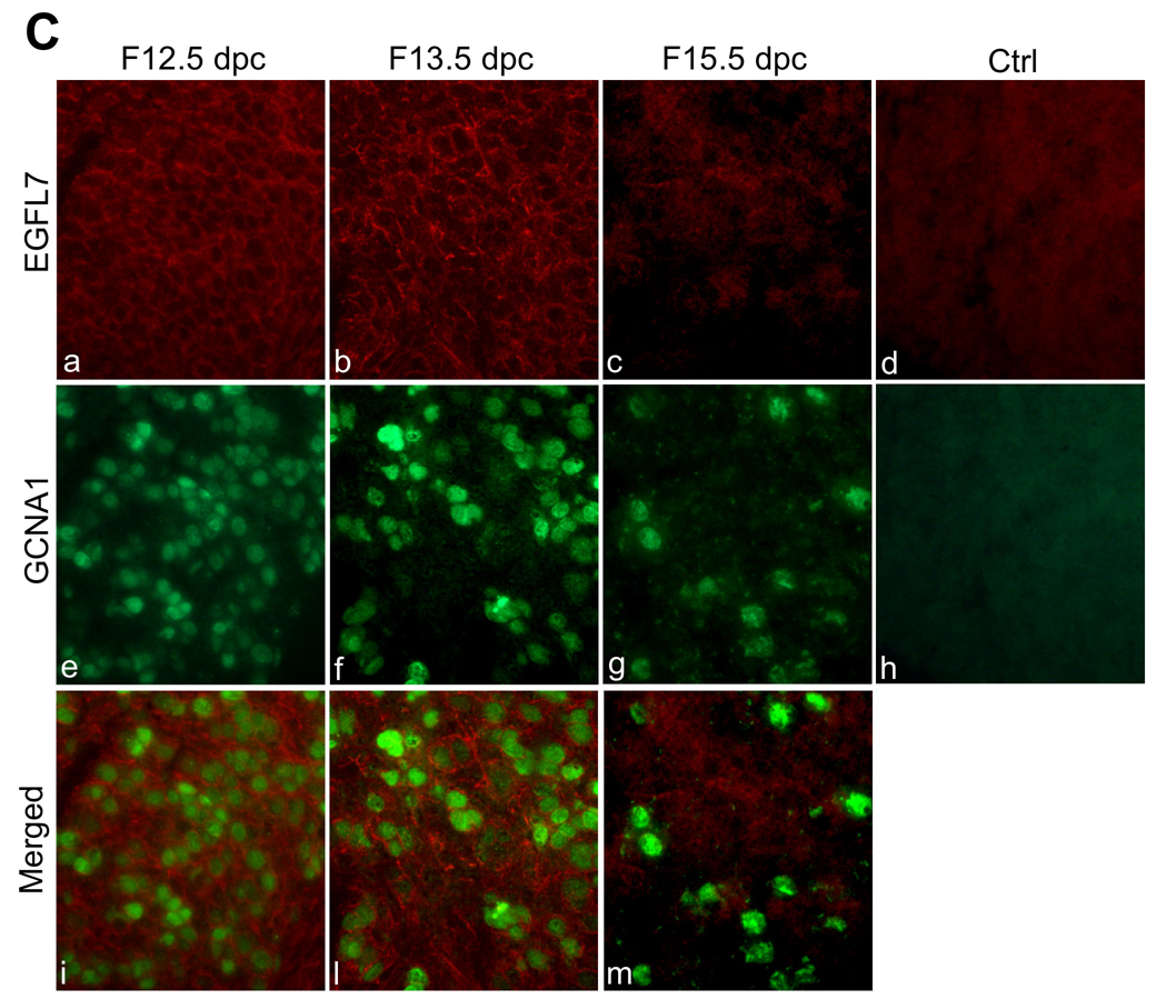
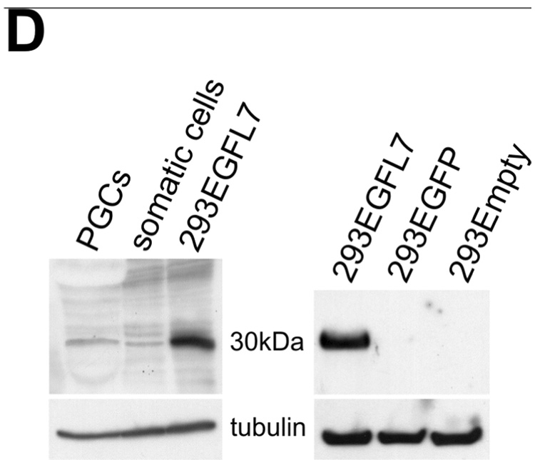
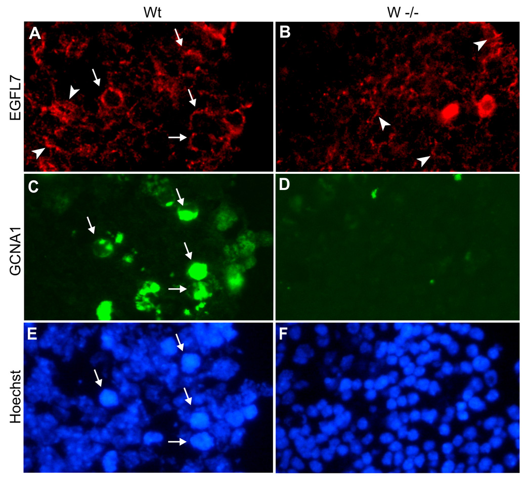
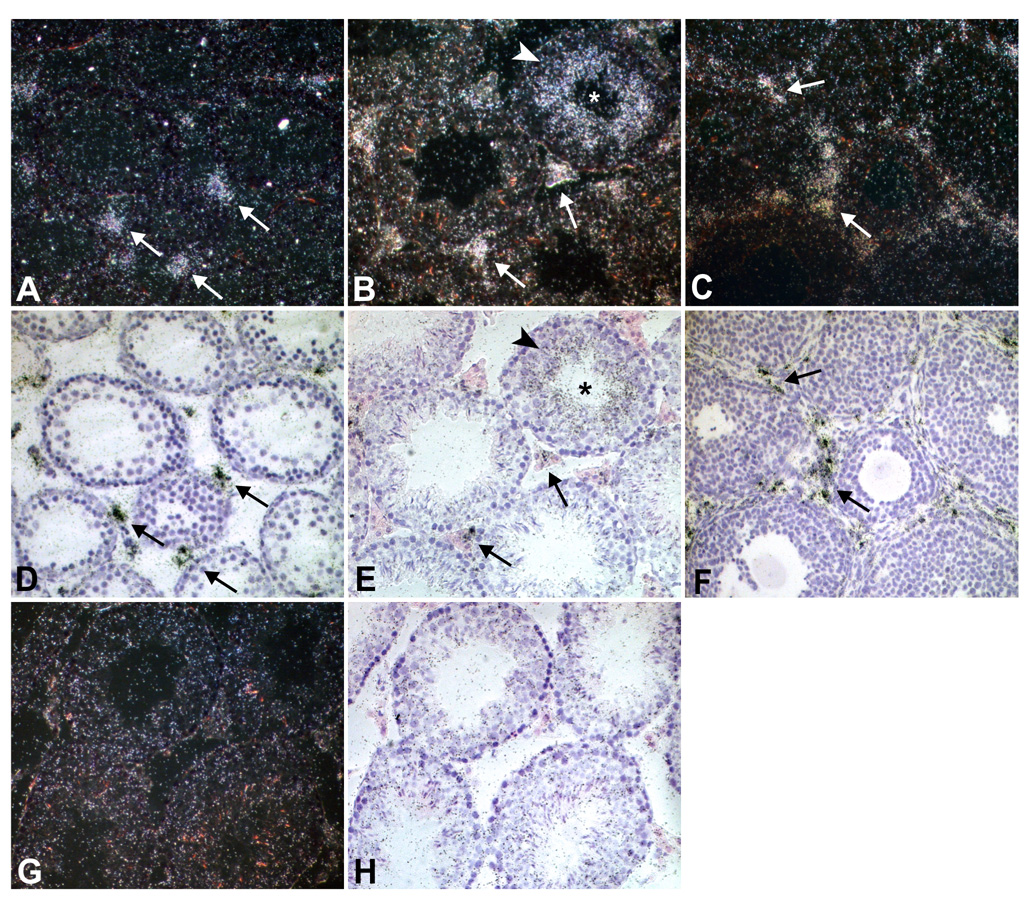
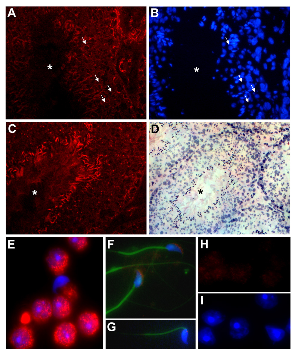
We have previously reported the isolation and characterization of a novel endothelial-restricted gene, Egfl7, that encodes a secreted protein of about 30-kDa. We and others demonstrated that Egfl7 is highly expressed by endothelial cells during embryonic development and becomes down-regulated in the adult vasculature. In the present paper, we show that during mouse embryonic development, Egfl7 is also expressed by primordial germ cells (PGC). Expression is down-regulated when PGCs differentiate into pro-spermatogonia and oogonia, and by 15.5 dpc Egfl7 can no longer be detected in the germ line of both sexes. Notably, Egfl7 is again transiently up-regulated in germ cells of the adult testis. In contrast, expression in the ovary remains limited to the vascular endothelium. Our results provide the first evidence of a non-endothelial expression of EGFL7 and suggest distinctive roles for Egfl7 in vascular development and germ cell differentiation.


Figures

Figure 1
Figure 1
RT-PCR analysis of Egfl7 expression in pre-implantation mouse embryos. Using intron spanning primers for Egfl7, a band of 348 bp was amplified from RNA isolated from ES cells and 8-cell stage embryos, but not from 1- and 2-cell stage embryos, and ovulated oocytes. Hprt primers were used as an amplification control.
Figure 2
Figure 2. Expression of EGFL7 in primordial germ cells (PGCs), gonocytes and oogonia
A: immunofluorescence analysis of EGFL7 and SSEA1 expression in isolated PGCs at 8.5 dpc (a, d, g) and 11.5 dpc (c, f, i) and in sagittal sections of 10.5 dpc embryos (b, e, h). Alkaline phosphatase staining was used to identify sections containing PGCs; adjacent sections were then incubated with antibodies. Arrows point to a migrating germ cell identified by the SSEA-1 antibody (e) that appears negative for EGFL7 (b); an EGFL7-positive and SSEA1-negative blood vessel is shown in b and e, respectively (arrowheads). The inserts show migrating PGCs and the arrows identify the cell shown at a higher magnification in b, e and h. a, b, c: EGFL7; d, e, f: SSEA1; g, h, i: Hoechst. Magnification 100x; B: Immunofluorescence analysis of EGFL7 and GCNA1 on cryostat sections of embryonic mouse testis from 12.5 dpc, 13.5 dpc (longitudinal sections) and 15.5 dpc (cross section). Arrows indicate location of testicular cords. a, b, c: EGFL7; e, f, g: GCNA1; i,l,m,: merge of EGFL7 and GCNA1; d and h: section of 15.5 dpc testis incubated with non-specific rabbit IgG and rat IgM, respectively. Magnification 40x. C: Immunofluorescence analysis of EGFL7 and GCNA1 on cryostat sections of embryonic mouse ovaries from 12.5dpc up to 15.5 dpc. a, b, c: EGFL7; e ,f ,g: GCNA1; i,l,m: merge of EGFL7 and GCNA1; d and h: section of 15.5 dpc ovaries incubated with non-specific rabbit IgG and rat IgM, respectively. Magnification 40x. D: Western blot analysis of EGFL7 expression in PGCs isolated from 11.5 dpc embryos. Somatic cells represent the flow through fraction of the immuno-magnetic purification and presumably contain endothelial cells. Cell extract from 293 cells transiently transfected with an Egfl7 expression vector, the empty vector and an unrelated vector (pEGFP-C1), were used as positive and negative controls, respectively. Anti-tubulin antibody was used as a loading control.
Figure 3
Figure 3
Immunofluorescence analysis of EGFL7 in gonads from 13.5 dpc White-spotting (W) females. Germ cells, identified by using the anti-GCNA1 antibody, were present in the wild type but not in the W−/− homozygous mutants (C and D). Wild type germ cells were also positive for EGFL7 (compare A and B: some of the germ cells were indicated by arrows). No evident differences in the vascular EGFL7 were observed (arrowheads depict some of the capillaries). A–B: EGFL7; C–D: GCNA1; E–F: Hoechst. Magnification 40X.
Figure 4
Figure 4
In situ hybridization analysis of Egfl7 in mouse testes and ovaries. A: dark-field image of a pre-puberal mouse testis section hybridized with an Egfl7 antisense probe showing expression of Egfl7 in the vasculature surrounding the tubules (arrows); B: adult mouse testis section hybridized with an Egfl7 antisense probe. The grains form a ring (arrowhead) surrounding the lumen of seminiferous tubules (*), a presumptive localization of post-meiotic germ cells; in situ signal is also present at low levels on the interstitial vasculature (arrows). C: Egfl7 in situ hybridization analysis of adult mouse ovaries showing Egfl7 expression limited to the vasculature surrounding the follicles (arrows). D, E, F: bright-field images of A, B and C, respectively. G and H sections of adult testis hybridized with an Egfl7 sense control probe. Magnification 20X.
Figure 5
Figure 5
Immuno-fluorescence analysis of EGFL7 expression in adult mouse testis and isolated adult germ cells. A-D: cross sections of adult mouse testis showing two seminiferous tubules (A,B and C,D, respectively); arrows in A and B indicate few of the EGFL7 positive spermatids. The tubule in C shows EGFL7 expression on spermatozoa; A, C: anti-EGFL7 antibody; B: Hoechst staining; D: hematoxilin and eosin counterstaining of section shown in C. Due to the H&E staining procedure, the tubule in D appears slightly shrunk compared to the corresponding image in C. Asterisks indicate the lumen of the tubules. E: spermatids isolated from adult testes by elutriation show strong immunoreactivity to the EGFL7 antibody. F: EGFL7 was also detected on the head of caudal sperm cells, possibly associated with the acrosomal vesicle; sperm tails were stained using FITC-phalloidin. G: control slide of sperm cells incubated with non-specific rabbit IgG and stained with FITC-phalloidin. H: control slides of spermatids incubated with non-specific rabbit IgG. I: Hoechst staining of H. Magnification in A–D: 40X. Magnification in E–I: 100X.

Similar articles

Cited by

References

    1. Campagnolo L, Leahy E, Chitnis S, Koschnick S, Fitch MJ, Fallon JT, Loskutoff D, Taubman MB, Stuhlmann H. EGFL7 is a chemoattractant for endothelial cells and is up-regulated in angiogenesis and arterial injury. Am. J. Path. 2005;167:275–284. - PMC - PubMed
    1. De Felici M, McLaren A. Isolation of mouse primordial germ cells. Exp. Cell Res. 1982;142:476–482. - PubMed
    1. De Felici M. Cell biology: a laboratory handbook. 2nd ed. Vol I. New York: Academic Press; 1998. Isolation and culture of germ cells from the mouse embryo; pp. 73–85.
    1. De Felici M. Regulation of primordial germ cell development in the mouse. Int. J. Dev. Biol. 44:575–580. - PubMed
    1. Enders GC, May JJ., 2nd Developmentally regulated expression of a mouse germ cell nuclear antigen examined from embryonic day 11 to adult in male and female mice. Dev. Biol. 1994;63:331–340. - PubMed

Publication types

LinkOut - more resources