. 2008 Sep;22(9):3234-46.
doi: 10.1096/fj.07-105676. Epub 2008 Jun 12.

Detection of migration of locally implanted AC133+ stem cells by cellular magnetic resonance imaging with histological findings

Affiliations

Detection of migration of locally implanted AC133+ stem cells by cellular magnetic resonance imaging with histological findings

Ali S Arbab et al. FASEB J. 2008 Sep.

Abstract

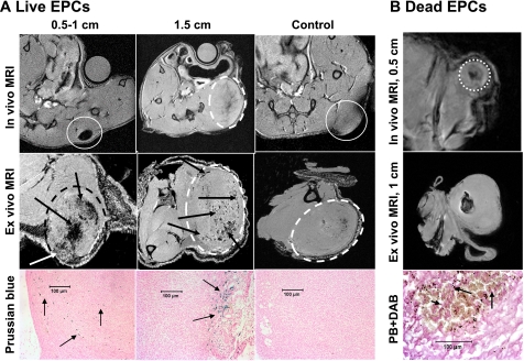
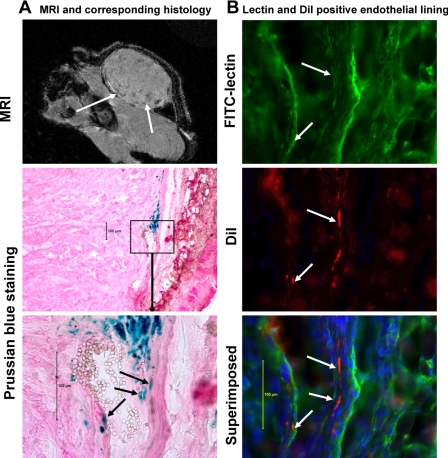
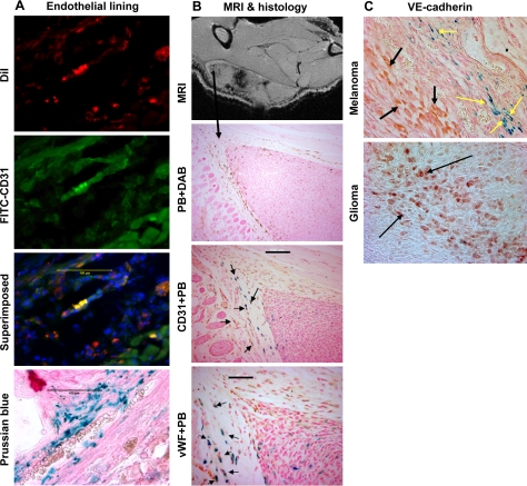
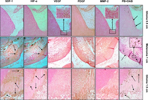
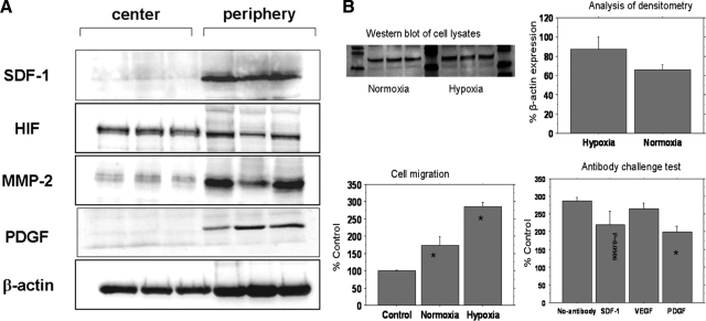
This study investigated the factors responsible for migration and homing of magnetically labeled AC133(+) cells at the sites of active angiogenesis in tumor. AC133(+) cells labeled with ferumoxide-protamine sulfate were mixed with either rat glioma or human melanoma cells and implanted in flank of nude mice. An MRI of the tumors including surrounding tissues was performed. Tumor sections were stained for Prussian blue (PB), platelet-derived growth factor (PDGF), hypoxia-inducible factor-1alpha (HIF-1alpha), stromal cell derived factor-1 (SDF-1), matrix metalloproteinase-2 (MMP-2), vascular endothelial growth factor (VEGF), and endothelial markers. Fresh snap-frozen strips from the central and peripheral parts of the tumor were collected for Western blotting. MRIs demonstrated hypointense regions at the periphery of the tumors where the PB(+)/AC133(+) cells were positive for endothelial cells markers. At the sites of PB(+)/AC133(+) cells, both HIF-1alpha and SDF-1 were strongly positive and PDGF and MMP-2 showed generalized expression in the tumor and surrounding tissues. There was no significant association of PB(+)/AC133(+) cell localization and VEGF expression in tumor cells. Western blot demonstrated strong expression of the SDF-1, MMP-2, and PDGF at the peripheral parts of the tumors. HIF-1alpha was expressed at both the periphery and central parts of the tumor. This work demonstrates that magnetically labeled cells can be used as probes for MRI and histological identification of administered cells.


Figures

Figure 1.
Figure 1.
Schematic representation of tumor sections used to extract total protein for Western blot analysis of expression of different angiogenic and chemoattractant factors in the tumor and the surrounding tissues. At first, 2-mm-thick sections from the center of the tumors (1.5 cm in size) were excised (left), and these sections were further cut into 5 separate pieces, as depicted at right. After a 2-mm portion of the tumor tissues was cut from all sides (marked as peripheral part), the leftover tumor tissues were considered the center part of the tumor. C, central; P, peripheral.
Figure 2.
Figure 2.
In vivo and ex vivo MRI images of implanted tumors mixed with either live FePro-labeled and unlabeled (control) AC133+ cells or labeled dead AC133+ cells. A) In vivo and ex vivo MRI images of implanted rat glioma mixed with live FePro-labeled and unlabeled (control) AC133+ cells. Note the loss of central low signal intensity (due to large number of labeled cells) in tumor measuring 0.5–1 cm compared with that measuring 1.5 cm (arrows on ex vivo images). These low signal intensities are different from the low signal intensity seen in control tumor (ex vivo). Corresponding DAB-enhanced Prussian blue (PB) staining shows iron-positive cells both in the central and peripheral part in the small tumor but in the peripheral portion only in the 1.5-cm tumor, with no iron-positive cells in the tumor injected with unlabeled cells. White circles indicate tumors on in vivo and ex vivo MRI. B) In vivo and ex vivo MRI images of implanted human melanoma mixed with magnetically labeled dead AC133+ cells at a tumor size of 0.5 and 1 cm. Note the localized low signal intensity in both tumors at sizes of 0.5 and 1 cm. Corresponding DAB-enhanced Prussian blue staining shows iron particles scattered in the central part of the tumor. Note the morphology of dead iron-positive cells (round, arrows), which is different from that of live cells (elongated; see Fig. 3). White circle indicates tumor on in vivo MRI.
Figure 3.
Figure 3.
Migration and incorporation of locally administered magnetically labeled AC133+ cells in human melanoma tumor in mouse. A) Gradient echo MRI shows low signal intensity areas mostly at the periphery of the tumors (white arrows). Representative histology sections show incorporation of Prussian blue-positive cells (staining performed after fluorescent microscopic images were obtained; see below) along the margin of a blood vessel (note the red blood cells within the lumen). B) To prove whether implanted live FePro-labeled AC133+ cells incorporated in the formation of tumor angiogenesis (vasculature), tumor vasculature (endothelial lining) was delineated by FITC-labeled tomato lectin, and fluorescent microscopic images were obtained both at FITC (lectin) and rhodamine (DiI-labeled cells) channels. The nucleus of the cells was delineated by DAPI staining. After fluorescent microscopy, the same section was stained for Prussian blue (for iron-positive cells) and bright-field microscopic images were obtained to match the area of fluorescent positive cells. Fluorescent microscopic images show incorporation of locally implanted DiI-labeled AC133+ cells (white arrows) into the tumor vasculatures. Arrows indicate lectin-positive endothelial lining and DiI-positive AC133+ cells. Note the corresponding Prussian blue-stained image indicating the presence of iron and DiI dye in the same cells.
Figure 4.
Figure 4.
Immunohistochemistry showing endothelial markers in administered cells. A) Immunohistochemistry of randomly selected melanoma frozen sections show expression of human CD31 in the implanted tumor. DiI-positive cells (AC133+ cells, red), human CD31+ cells stained with FITC conjugated secondary antibody (green), and combined superimposed image of the section indicating DiI-positive cells transform into endothelial cells (yellow). Prussian blue staining of the consecutive section shows multiple iron-positive cells at the corresponding sites. Note the red blood cells within the lumen on Prussian blue section. Scale bars = 100 μm. B) Immunohistochemistry of randomly selected rat glioma paraffin sections shows expression of both human CD31 and vWF in the implanted tumor. Ex vivo MRI shows low signal intensity areas both in the center and at the periphery of a 1-cm tumor. DAB-enhanced Prussian blue staining shows migration of labeled AC133+ cells at the periphery of the tumor. Double staining, CD31 (brown) followed by Prussian blue (blue) of the consecutive section shows double-positive cells (black arrows). Double staining, vWF (brown) followed by Prussian blue (blue) of the consecutive section shows double-positive cells (black arrows). Expression of both vWF and CD31 in iron-positive cells indicates the maturation of AC133+ cells into endothelial cells. DAB-enhanced Prussian blue image is ×10. Scale bars = 50 μm (vWF+ PB); 100 μm (CD31+ PB). C) Immunohistochemistry of randomly selected human melanoma and rat glioma paraffin sections shows expression of VE-cadherin in the tumor cells as well as in Prussian blue-positive cells. Double staining, VE-cadherin (brown) followed by Prussian blue of a section from implanted human melanoma mixed with FePro-labeled AC133+ cells. Yellow arrows show VE-cadherin- and Prussian blue-positive cells that are placed along the lining of vessels. Black arrows show VE-cadherin-positive melanoma cells. VE-cadherin staining of glioma shows numerous positive tumor cells (dark brown, black arrows).
Figure 5.
Figure 5.
Expression of different angiogenic and chemoattractant factors at the sites of migrated labeled AC133+ cells in tumors. Top row: expression of different angiogenic and chemoattractant factors at the sites of migrated labeled AC133+ cells in rat glioma at a tumor size of 0.5 cm (consecutive sections). Prussian blue staining shows migration of cells at the periphery of the tumor (arrows). MMP-2 and VEGF staining show expression of these factors at the corresponding sites of cell migration (inset, brown cells). PDGF staining shows generalized expression both in the tumor and adjacent surrounding tissues. HIF-1α and SDF-1 staining do not show expression of the factors at the corresponding sites of migrated cells. Middle row: expression of different angiogenic and chemoattractant factors at the sites of migrated labeled AC133+ cells in human melanoma at a tumor size of 1 cm (consecutive sections). Prussian blue staining shows migration of cells at the periphery of the tumor and at the sites of invasion into surrounding muscles and tissues (arrows). MMP-2 staining shows mild expression of MMP-2 throughout the tumor and surrounding tissues. PDGF staining also shows generalized expression both in the tumor and adjacent surrounding tissues. VEGF staining shows no localized expression of these factors at the corresponding sites of cell migration. HIF-1α and SDF-1 staining show very strong localized expression of the factors at the corresponding sites of migrated cells (arrows). Inset: multiple DAB enhanced Prussian blue positive cells. Bottom row: Expression of different angiogenic and chemoattractant factors at the sites of migrated labeled AC133+ cells in rat glioma at a tumor size of 1.5 cm (consecutive sections). Prussian blue staining shows migration of cells at the periphery of the tumor and at the sites of invasion into surrounding muscles and tissues (arrows). MMP-2 staining shows mild expression of MMP-2 throughout the tumor and surrounding tissues. PDGF staining shows generalized expression both in the tumor and adjacent surrounding tissues. VEGF staining shows no localized expression of these factors at the corresponding sites of cell migration. HIF-1α and SDF-1 staining show very strong localized expression of the factors at the corresponding sites of migrated cells (arrows). All images ×10.
Figure 6.
Figure 6.
Proof that human cells contain SPIO nanoparticles. To determine whether the migrated (to the peripheral part of tumors) iron-positive cells are indeed of human origin, sections were stained with human specific anti mitochondrial antibody. After staining, all sections were imaged with a microscope at ×50, and 20–30 randomly selected areas were captured where most abundant positive cells were observed. All slides were then submerged in xylene to remove the cover slips, the sections were restained for iron by using standard Prussian blue staining procedures, and cover slips were emplaced. The sections were scanned under a microscope to determine the exact locations of human mitochondrial positive cells and photomicrographed to show the colocalization of double-positive cells. Randomly selected areas show abundant cells carrying human mitochondria (brown cells and arrows; A, C). The same areas show multiple iron-positive cells (B, D). Note the exact match of iron- and human mitochondria-positive cells (arrows; AD). A few representative cells are indicated by the arrows. Scale bars = 50 μm.
Figure 7.
Figure 7.
Host macrophages and iron-positive cells. To determine whether host macrophages phagocytose some iron-positive cells or released SPIO nanoparticles from labeled human cells, consecutive sections were stained with anti-mouse F4/80 antibody (specific for mouse macrophages), photomicrographed, and stained for iron. Consecutive sections were also used as negative control where primary antibody was omitted, but all other procedures were identical. Multiple localized F4/80-positive cells (brown cells) are seen (A), which show no iron on Prussian blue staining (B). Images from negative control section show no brown cells (C, D). Scale bars = 50 μm.
Figure 8.
Figure 8.
Western blot analysis of hypoxic cell lysate and cell migration studies. A) Western blot analysis of expression of different angiogenic and chemoattractant factors in whole-tissue lysates of implanted rat glioma tumors (samples from 3 different tumors). Forty micrograms of total tumor tissue protein was resolved by electrophoresis using 8–12% SDS-polyacrylamide gels. On transfer to the nitrocellulose membrane, proteins were detected by immunobloting, using specific antibodies against proteins of interest. Immunoblotting using specific antibodies against SDF-1, PDGF, and MMP-2, respectively, reveals high expression levels in the periphery of the tumor. Antibodies against HIF-1α reveal HIF-1α expression in the center and the periphery of the tumor, with higher levels of expression within the center of the tumor. Housekeeping protein β-actin was used as a control for loading. B) Effects of hypoxic condition on stem cell migration and HIF-1α expression. Top left: Western blots of whole-cell lysates show the expression of HIF-1α in tumor cells cultured under normoxic and hypoxic conditions (triplicate samples). Top right: analysis of densitometry of HIF-1α immunoblots, expressed as percentage of β-actin expression, shows higher HIF-1α expression in the hypoxic condition. Bottom left: cell migration graph compares migration of AC133+ cells toward the supernatants collected from normoxic and hypoxic tumor cells compared with control condition (*P<0.05). Cell migration to the hypoxic supernatant is higher than that to the normoxic supernatant (P<0.01). Bottom right: antibody challenge test compares migration of AC133+ cells toward the supernatants collected from normoxic and hypoxic tumor cells with and without 1 μg/ml of neutralizing anti-SDF-1, anti-VEGF, and anti-PDGF antibodies. A significant decrease in migration of cells is observed in wells treated with anti-PDGF (*P<0.01), and anti-SDF-1 antibody also inhibits cell migration.

Similar articles

Cited by

References

    1. Folkman J. Angiogenesis: an organizing principle for drug discovery? Nat Rev Drug Discov. 2007;6:273–286. - PubMed
    1. Shi Q, Rafii S, Wu M H, Wijelath E S, Yu C, Ishida A, Fujita Y, Kothari S, Mohle R, Sauvage L R, Moore M A, Storb R F, Hammond W P. Evidence for circulating bone marrow-derived endothelial cells. Blood. 1998;92:362–367. - PubMed
    1. Samejima N, Yamazaki K. A study on the vascular proliferation in tissues around the tumor in breast cancer. Jpn J Surg. 1988;18:235–242. - PubMed
    1. Ellis L M, Liu W, Ahmad S A, Fan F, Jung Y D, Shaheen R M, Reinmuth N. Overview of angiogenesis: Biologic implications for antiangiogenic therapy. Semin Oncol. 2001;28:94–104. - PubMed
    1. Liekens S, De Clercq E, Neyts J. Angiogenesis: regulators and clinical applications. Biochem Pharmacol. 2001;61:253–270. - PubMed

Publication types

MeSH terms