. 2008 Jul 18;103(2):194-202.
doi: 10.1161/CIRCRESAHA.108.178590. Epub 2008 Jun 12.

Engineering robust and functional vascular networks in vivo with human adult and cord blood-derived progenitor cells

Affiliations

Engineering robust and functional vascular networks in vivo with human adult and cord blood-derived progenitor cells

Juan M Melero-Martin et al. Circ Res. .

Abstract

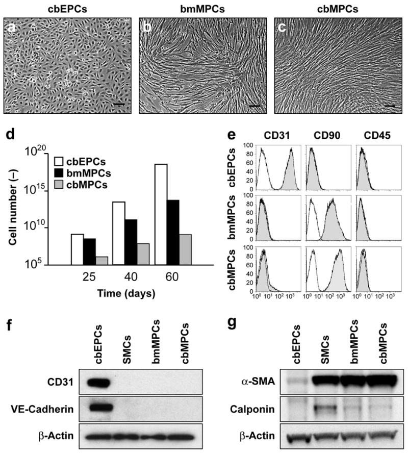
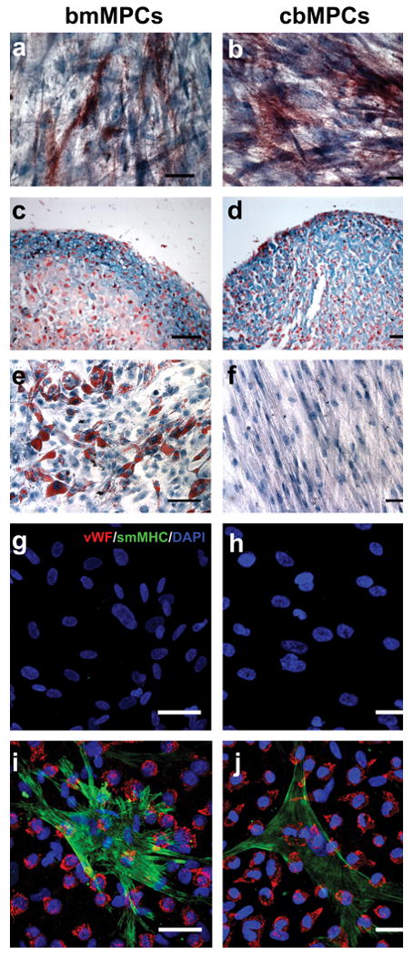
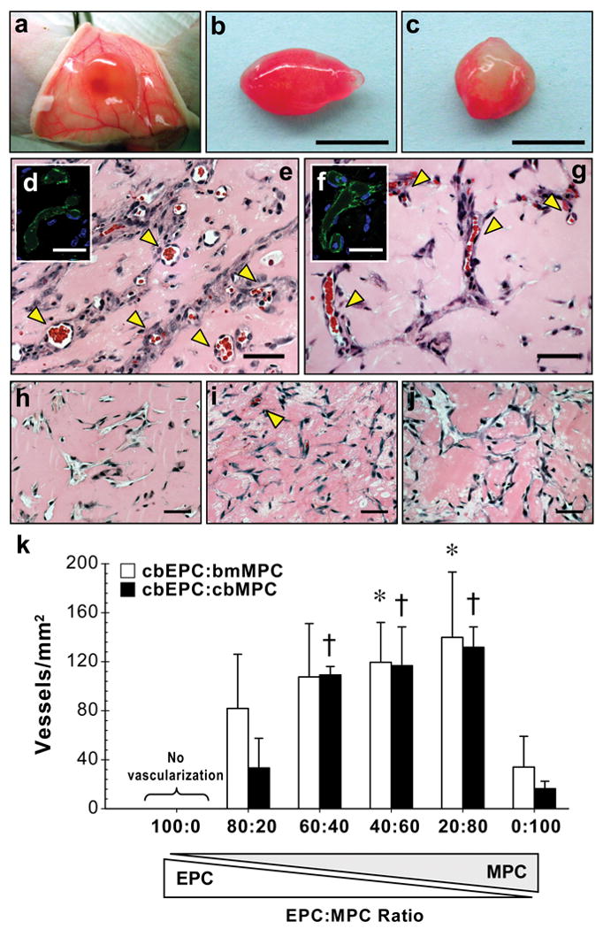
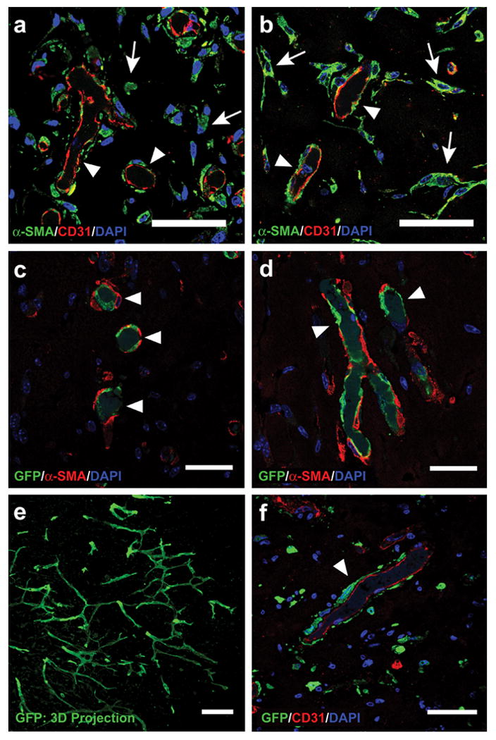
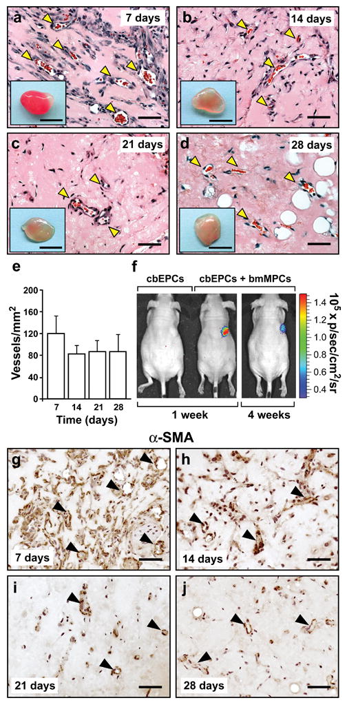
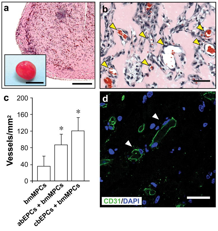
The success of therapeutic vascularization and tissue engineering will rely on our ability to create vascular networks using human cells that can be obtained readily, can be expanded safely ex vivo, and can produce robust vasculogenic activity in vivo. Here we describe the formation of functional microvascular beds in immunodeficient mice by coimplantation of human endothelial and mesenchymal progenitor cells isolated from blood and bone marrow. Evaluation of implants after 1 week revealed an extensive network of human blood vessels containing erythrocytes, indicating the rapid formation of functional anastomoses within the host vasculature. The implanted endothelial progenitor cells were restricted to the luminal aspect of the vessels; mesenchymal progenitor cells were adjacent to lumens, confirming their role as perivascular cells. Importantly, the engineered vascular networks remained patent at 4 weeks in vivo. This rapid formation of long-lasting microvascular networks by postnatal progenitor cells obtained from noninvasive sources constitutes an important step forward in the development of clinical strategies for tissue vascularization.


Conflict of interest statement

Disclosures: None.

Figures

Figure 1
Figure 1. Phenotypic characterization
of EPCs and MPCs. (a) cbEPCs presented typical cobblestone morphology, while both (b) bMPCs and (c) cbMPCs presented spindle morphology characteristic of mesenchymal cells in culture (scale bars, 100 μm). (d) cbEPCs and MPCs were serially passaged and their in vitro expansion potential estimated by the accumulative cell numbers obtained from 25 mL of either cord blood or bone marrow samples. (e) Flow cytometric analysis of cbEPCs, bmMPCs and cbMPCs for the endothelial marker CD31, mesenchymal marker CD90, and hematopoietic marker CD45. Solid gray histograms represent cells stained with fluorescent antibodies. Isotype-matched controls are overlaid in a black line on each histogram. Western blot analyses of cbEPCs, bmMPCs and cbMPCs for (f) endothelial markers CD31, and VE-cadherin, and (g) mesenchymal markers α-SMA, and Calponin. Expression of β-actin shows equal protein loading. SMCs isolated from human saphenous vein served as control.
Figure 2
Figure 2. Multilineage differentiation of MPCs
(a) bmMPCs and (b) cbMPCs differentiation into osteocytes was revealed by alkaline phosphatase staining. (c) bmMPCs and (d) cbMPCs differentiation into chondrocytes was revealed by the presence of glycosaminoglycans, detected by Alcian Blue staining. The presence of adipocytes was assessed by Oil Red O staining, and it was evident in (e) bmMPCs, but absent in (f) cbMPCs. Smooth muscle cell differentiation was evaluated by culturing MPCs in the absence or presence of cbEPCs (1:1 EPCs to MPCs ratio) for 7 days in EPC-medium. Induction of SMC phenotype was assessed by the expression of smMHC. Immunofluorescence staining with anti-vWF-Texas-Red and anti-smMHC-FITC, as well as nuclear staining with DAPI, revealed that smMHC was absent in both (g) bmMPCs and (h) cbMPCs, but it was induced in MPCs when co-cultured with cbEPCs (i, j). Scale bars correspond to 200 μm (e and f) and 50 μm (a-d and g-j).
Figure 3
Figure 3. Formation of vascular networks in vivo with EPCs and MPCs
A total of 1.9×10⁶ cells was resuspended in 200 μl of Matrigel using different ratios of cbEPCs and MPCs, and implanted on the back of six-week-old nu/nu mice by subcutaneous injection. Implants were harvested after 7 days and stained with H&E. (a, b) Macroscopic view of explanted Matrigel plugs seeded with 40% cbEPCs:60% bmMPCs and (c) 40% cbEPCs:60% cbMPCs (scale bars, 5 mm). (e, g) H&E staining revealed the presence of lumenal structures containing erythrocytes (yellow arrow heads) in implants where both cells types (cbEPCs and MPCs; 40:60) were used, but not in implants where (h) cbEPCs, (i) bmMPCs, and (j) cbMPCs were used alone (scale bars, 50 μm). Microvessels stained positive for human CD31 (d, f) (scale bars, 30 μm). Images are representative of implants harvested from at least four different mice. (k) Quantification of microvessel density was performed by counting erythrocyte-filled vessels in implants with ratios of 100:0, 80:20, 60:40, 40:60, 20:80 and 0:100 (cbEPCs:MPCs; n≥4 each condition). Each bar represents the mean ± S.D. (vessels/mm2) obtained from vascularized implants. * P < .05 compared to implants with bmMPCs alone (n=4). † P < .05 compared to implants with cbMPCs alone (n=4).
Figure 4
Figure 4. Specific location of EPCs and MPCs in the vascular bed
Matrigel implants containing cbEPCs and MPCs (40:60) were evaluated after one week. Implants with (a) bmMPCs and (b) cbMPCs produced lumenal structures that stained positive for human CD31, confirming that those lumens were formed by the implanted cells. In addition, α-SMA-expressing cells were detected both in the proximity (white arrows) and around the lumenal structures (white arrow heads), (scale bars, 50 μm). Implants that utilized GFP-labeled cbEPC and either (c) bmMPCs or (d) cbMPCs produced GFP-positive lumenal structures (white arrow heads) covered by α-SMA-expressing perivascular cells, confirming that cbEPCs were restricted to the luminal aspect of the vessels (scale bars, 30 μm). (e) Projections of whole-mount staining showed that the GFP-expressing cells formed extensive networks throughout the implants (scale bar 100 μm). (f) Implants that utilized GFP-labeled bmMPC and unlabeled cbEPCs resulted in human CD31-positive lumenal structures with GFP-expressing cells adjacent to lumens (white arrow heads), confirming the role of MPCs as perivascular cells (scale bars, 50 μm). Images are representative of implants harvested from four different mice.
Figure 5
Figure 5. Durability of the vascular bed
Matrigel implants containing cbEPCs and bmMPCs (40:60) were injected subcutaneously on the back of six-week-old nu/nu mice. H&E staining showed lumenal structures containing erythrocytes (yellow arrow heads) in implants after (a) 7, (b) 14, (c) 21, and (d) 28 days (scale bars, 50 μm; macroscopic view of explanted Matrigel plugs shown in insets; insets scale bars, 5 mm). (e) Quantification of microvessel density was performed by counting erythrocyte-filled vessels. Each bar represents the mean microvessel density value determined from four separate implants and mice ± S.D. (vessels/mm2). (f) Additional implants were prepared with luciferase-labeled cbEPC in the presence or absence of unlabeled bmMPCs. Mice were imaged using an IVIS Imaging System, and bioluminescence detected 30-40 min after intraperitoneal injection of luciferin. In implants where cbEPCs and bmMPCs were co-implanted, bioluminescence was detected at 1 (image Min=-2.91×10⁴., Max=2.67×10⁵) and 4 (image Min=-3.29×10⁴, Max=3.27×10⁵) weeks, but not in those where cbEPCs were used alone. Immunohistochemical staining of α-SMA in implants after (g) 7, (h) 14, (i) 21, and (j) 28 days, revealed that α-SMA-expressing cells were progressively restricted to perivascular locations (black arrow heads), (scale bars, 50 μm). Images are representative of implants harvested from four different mice.
Figure 6
Figure 6. Vascular network formation using adult progenitor cells
Matrigel implants containing 40% abEPCs and 60% bmMPCs (obtained from human adult peripheral blood and adult bone marrow samples respectively) were injected subcutaneously on the back of six-week-old nu/nu mice and evaluated after one week. (a, b) H&E staining showed an uniform and extensive presence of lumenal structures containing erythrocytes (yellow arrow heads) throughout the implants (a scale bar, 500 μm; macroscopic view of explanted Matrigel plug shown in inset; inset scale bar, 5 mm; b scale bar, 50 μm). (c) Quantification of microvessel density was performed in implants seeded with bmMPCs in the absence or presence of either abEPCs or cbEPCs by counting erythrocyte-filled vessels. Each bar represents the mean microvessel density determined from four separate implants and mice ± S.D. (vessels/mm2). * P < .05 compared to implants with bmMPCs alone (n=4). (d) Microvessels from implants containing 40% abEPCs and 60% bmMPCs stained positive for human CD31 (white arrow heads), confirming that those lumens were formed by the implanted cells (scale bars, 30 μm). Images are representative of implants harvested from four different mice.

Comment in

  • Therapeutic vasculogenesis: it takes two.
    Loffredo F, Lee RT. Loffredo F, et al. Circ Res. 2008 Jul 18;103(2):128-30. doi: 10.1161/CIRCRESAHA.108.180604. Circ Res. 2008. PMID: 18635829 Free PMC article. No abstract available.

References

    1. Jain RK, Au P, Tam J, Duda DG, Fukumura D. Engineering vascularized tissue. Nat Biotechnol. 2005;23:821–823. - PubMed
    1. Isner JM, Asahara T. Angiogenesis and vasculogenesis as therapeutic strategies for postnatal neovascularization. J Clin Invest. 1999;103:1231–1236. - PMC - PubMed
    1. Isner JM, Pieczek A, Schainfeld R, Blair R, Haley L, Asahara T, Rosenfield K, Razvi S, Walsh K, Symes JF. Clinical evidence of angiogenesis after arterial gene transfer of phVEGF165 in patient with ischaemic limb. Lancet. 1996;348:370–374. - PubMed
    1. Lee H, Cusick RA, Browne F, Ho Kim T, Ma PX, Utsunomiya H, Langer R, Vacanti JP. Local delivery of basic fibroblast growth factor increases both angiogenesis and engraftment of hepatocytes in tissue-engineered polymer devices. Transplantation. 2002;73:1589–1593. - PubMed
    1. Li X, Tjwa M, Moons L, Fons P, Noel A, Ny A, Zhou JM, Lennartsson J, Li H, Luttun A, Ponten A, Devy L, Bouche A, Oh H, Manderveld A, Blacher S, Communi D, Savi P, Bono F, Dewerchin M, Foidart JM, Autiero M, Herbert JM, Collen D, Heldin CH, Eriksson U, Carmeliet P. Revascularization of ischemic tissues by PDGF-CC via effects on endothelial cells and their progenitors. J Clin Invest. 2005;115:118–127. - PMC - PubMed

Publication types

MeSH terms