. 2008 Nov;39(5):610-8.
doi: 10.1165/rcmb.2007-0322OC. Epub 2008 Jun 12.

Thy-1 promoter hypermethylation: a novel epigenetic pathogenic mechanism in pulmonary fibrosis

Affiliations

Thy-1 promoter hypermethylation: a novel epigenetic pathogenic mechanism in pulmonary fibrosis

Yan Y Sanders et al. Am J Respir Cell Mol Biol. 2008 Nov.

Abstract

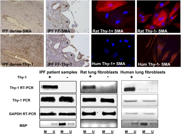
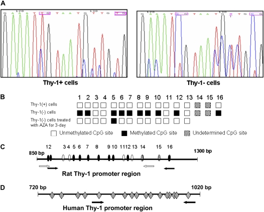
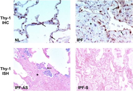
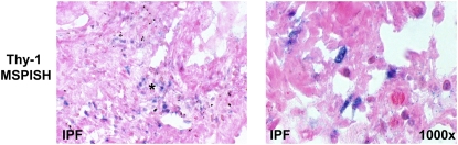
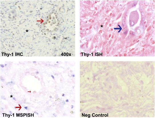
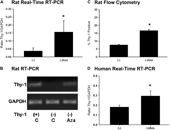
Mechanisms regulating myofibroblastic differentiation of fibroblasts within fibroblastic foci in idiopathic pulmonary fibrosis (IPF) remain unclear. Epigenetic processes, including DNA methylation, produce heritable but potentially reversible changes in DNA or its associated proteins and are prominent in development and oncogenesis. We have shown that Thy-1 suppresses myofibroblastic differentiation of lung fibroblasts and that fibroblasts in fibroblastic foci are Thy-1(-). Epigenetic down-regulation of Thy-1 has been demonstrated in cellular transformation and clinical cancer. We hypothesized that epigenetic regulation of Thy-1 affects the lung fibroblast fibrogenic phenotype. RT-PCR, methylation-specific PCR (MSP), and bisulfite genomic sequencing were used to determine the methylation status of the Thy-1 promoter in Thy-1(+) and Thy-1(-) lung fibroblasts, and MSP-in situ hybridization (MSPISH) was performed on fibrotic tissue. Thy-1 gene expression is absent in Thy-1(-) human and rat fibroblasts despite intact Thy-1 genomic DNA. Cytosine-guanine islands in the Thy-1 gene promoter are hypermethylated in Thy-1(-), but not Thy-1(+), fibroblasts. RT-PCR and MSP demonstrate that, in IPF samples in which Thy-1 expression is absent, the Thy-1 promoter region is methylated, whereas in lung samples retaining Thy-1 expression, the promoter region is unmethylated. MSPISH confirms methylation of the Thy-1 promoter in fibroblastic foci in IPF. Treatment with DNA methyltransferase inhibitors restores Thy-1 expression in Thy-1(-) fibroblasts. Epigenetic regulation of Thy-1 is a novel and potentially reversible mechanism in fibrosis that may offer the possibility of new therapeutic options.


Figures

<b>Figure 1.</b>
Figure 1.
Upper left: The four micrographs demonstrating immunohistochemistry staining (original magnification: ×200) for α-smooth muscle actin (SMA) and Thy-1 on serial sections from patients with idiopathic pulmonary fibrosis (IPF), demonstrating areas of dense fibrosis (IPF dense) or fibroblastic foci (IPF FF). FF are indicated by asterisks. Upper right: Four fluorescence micrographs demonstrating rat or human Thy-1(+) or Thy-1(−) lung fibroblasts stained with antibody to SMA conjugated to Cy3 (original magnification: ×400). Nuclei are counterstained with Hoechst reagent. PCR and RT-PCR of Thy-1 from Thy-1(+) and Thy-1(−) rat and human lung fibroblasts and representative samples from patients with IPF (positive or negative for Thy-1 expression) are shown. Lower panel: DNA gels stained with ethidium bromide, demonstrating results of RT-PCR analysis of Thy-1 expression, PCR amplification of Thy-1 genomic DNA, RT-PCR for glyceraldehyde-3-phosphate dehydrogenase (GAPDH) as a control, and methylation-specific PCR (MSP) using primers described in Table 2 and depicted in Figures 2C and 2D. MSP products are shown in the lowermost panels. M and U refer to primers specific for methylated and unmethylated promoter fragments, respectively.
<b>Figure 2.</b>
Figure 2.
Methylation status of Thy-1 promoter in Thy-1(−) and Thy-1(+) rat lung fibroblasts. (A) Bisulfite genomic sequencing (BGS) of Thy-1 promoter region using bisulfite modified DNA. In this representative portion of the promoter sequence, two CpG sites are methylated in Thy-1(−) rat fibroblasts, as indicated by T to C shift (pink boxes) (see Materials and Methods). (B) Schematic illustration of the rat Thy-1 promoter region subjected to methylation analysis, based on sequence data from Thy-1(+) cells and Thy-1(−) cells +/− treatment with AZA for 3 days. White boxes: unmethylated CpG; black boxes: methylated CpG; spotted boxes: undetermined sequence. MSP primers located at number 1 and 2 CpG islands of Forward primer and number 16 CpG of the Reverse primer. (C) The CpG island of the rat Thy-1 promoter region as determined by BGS and MSP; white arrows indicate the positions of the rat BGS primers, and black arrows indicate positions of the MSP primers. Ovals indicate CpG sites (white are unmethylated; black are methylated) as determined by BGS. Spotted ovals indicate additional CpG sites of unknown methylation status, which are within the amplified MSP product but not within region sequenced for BGS; the last one of these (number 16) is positioned within the reverse MSP primer. (D) The CpG island of human Thy-1 promoter region; mRNA starts at 1017 bp. Arrows indicate the positions of the MSP primers. Gray diamonds are CpG sites within the MSP primer sequences. Spotted diamonds indicate CpG islands (methylation status unknown.
<b>Figure 3.</b>
Figure 3.
Thy-1 expression by immunohistochemistry (IHC) (upper panels) and in situ hybridization (ISH) (middle panels) in normal (NL) and IPF lung samples. Arrows indicate Thy-1 expression in elongated alveolar septal cells in NL and in abnormal epithelium overlying fibroblastic foci (asterisk) in IPF. AS and S (middle panels) refer to antisense and sense (negative control) probes for ISH, respectively. Lower panels: Thy-1 promoter hypermethylation in IPF demonstrated by MSP-in situ hybridization (MSP-ISH). Blue nuclear signal indicates hypermethylation signal in elongated cells within fibroblastic foci (asterisk). (Original magnification: ×400, except where indicated.)
<b>Figure 4.</b>
Figure 4.
IHC, ISH, and MSP-ISH on serial sections from the same patient. Upper left: IHC shows Thy-1 protein in epithelial cells (red arrow) overlying fibroblastic focus (asterisk). Upper right: ISH shows Thy-1 mRNA expressed in epithelial cells (blue arrow). Lower left: MSP-ISH shows Thy-1 promoter methylation in fibroblastic cells within fibroblastic foci (large red arrow) but absent from epithelial cells (small arrow). (Original magnification: ×1,000, except where indicated.)
<b>Figure 5.</b>
Figure 5.
Increased Thy-1 expression in Thy-1(−) cells after demethylation treatment. (A) Histogram of real-time RT-PCR results for Thy-1 expression in rat lung fibroblasts by standard curve method; 5 μM AZA treated Thy-1(−) rat lung fibroblasts (3 d) compared with control (DMSO). (B) Representative RT-PCR results. (C) Thy-1 expression on cell surface by flow cytometry in Thy-1(−) rat lung fibroblasts and Thy-1(−) cells treated with AZA for 3 days. Cells were stained with FITC-conjugated anti-CD90 (Thy-1) antibody. (D) Histogram of real-time RT-PCR results for Thy-1 expression in human lung fibroblasts; 5 μM AZA treated Thy-1(−) human lung fibroblasts (3 days) compared with control (−). Experiments were performed in triplicate, and data are expressed as the ratio of Thy-1 to GAPDH by standard curve method. *P < 0.05 versus Thy-1(−) by ANOVA.
<b>Figure 6.</b>
Figure 6.
Expression of DNA methyltransferases (DNMTs) and methyl-CpG-binding domain proteins (MBD) by real-time RT-PCR in Thy-1(+) and Thy-1(−) rat lung fibroblasts. (A) DNMT1. (B) DNMT3a. (C) DNMT3b. (D) MBD1. (E) Mecp2. Experiments were performed in triplicate, and data are expressed as ratio of target to GAPDH by standard curve method. Primers and RT-PCR conditions were as described in Materials and Methods.

References

    1. Strieter RM, Gomperts BN, Keane MP. The role of CXC chemokines in pulmonary fibrosis. J Clin Invest 2007;117:549–556. - PMC - PubMed
    1. Pardo A, Selman M. Idiopathic pulmonary fibrosis: new insights in its pathogenesis. Int J Biochem Cell Biol 2002;34:1534–1538. - PubMed
    1. Kuhn C, Boldt J, King TE, Crouch E, Vartio T, McDonald JA. An immunohistochemical study of architectural remodeling and connective tissue synthesis in pulmonary fibrosis. Am Rev Respir Dis 1989;140:1693–1703. - PubMed
    1. Phan SH. Fibroblast phenotypes in pulmonary fibrosis. Am J Respir Cell Mol Biol 2003;29(3, Suppl)S87–S92. - PubMed
    1. Rege TA, Hagood JS. Thy-1 as a regulator of cell-cell and cell-matrix interactions in axon regeneration, apoptosis, adhesion, migration, cancer, and fibrosis. FASEB J 2006;20:1045–1054. - PubMed

Publication types

MeSH terms