Review
. 2008 Jul;47 Suppl 4(Suppl 4):iv2-8.
doi: 10.1093/rheumatology/ken177.

Structural basis of growth-related gain and age-related loss of bone strength

Affiliations
Review

Structural basis of growth-related gain and age-related loss of bone strength

E Seeman. Rheumatology (Oxford). 2008 Jul.

Abstract

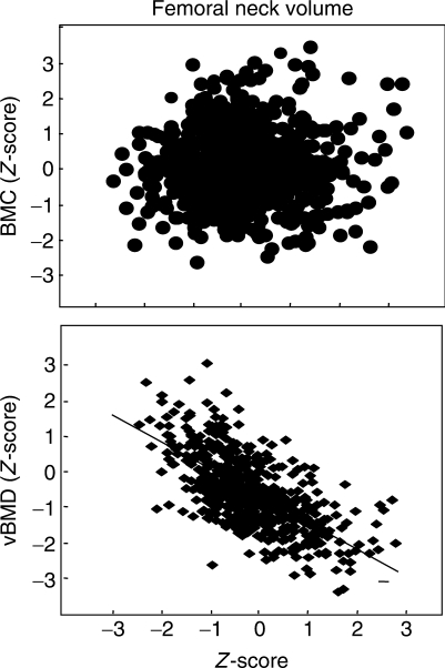
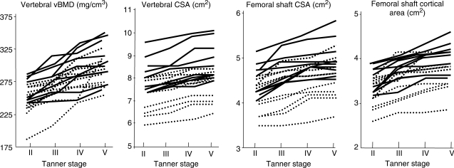
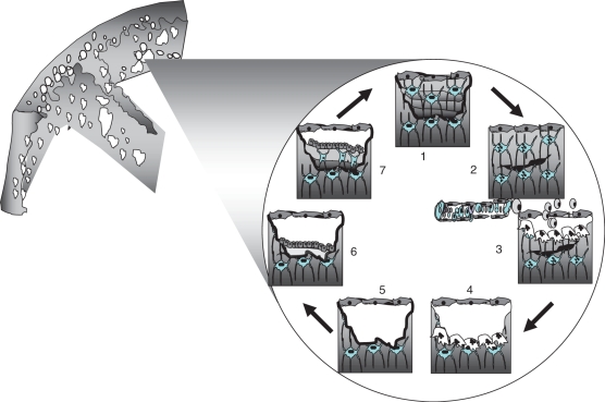
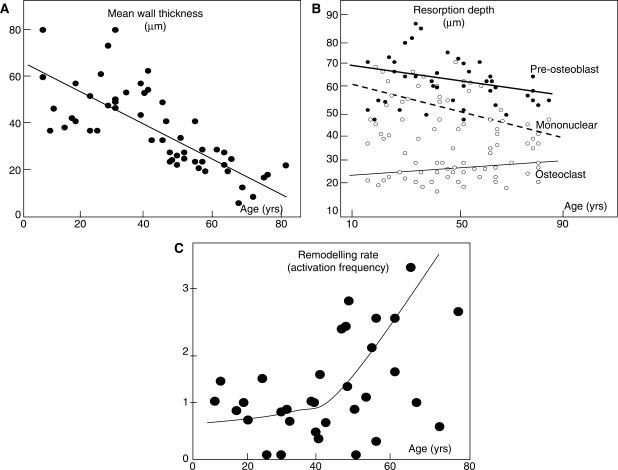
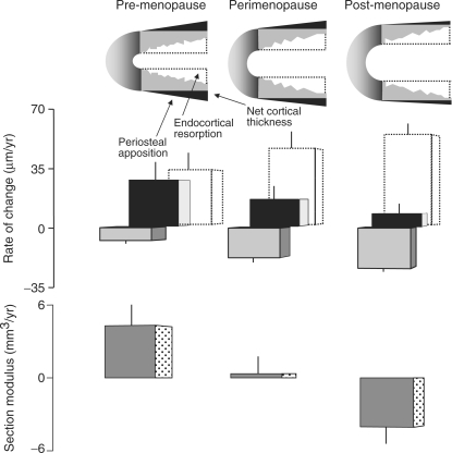
If bone strength was the only requirement of skeleton, it could be achieved with bulk, but bone must also be light. During growth, bone modelling and remodelling optimize strength, by depositing bone where it is needed, and minimize mass, by removing it from where it is not. The population variance in bone traits is established before puberty and the position of an individual's bone size and mass tracks in the percentile of origin. Larger cross-sections have a comparably larger marrow cavity, which results in a lower volumetric BMD (vBMD), thereby avoiding bulk. Excavation of a marrow cavity thus minimizes mass and shifts the cortex radially, increasing rigidity. Smaller cross-sections are assembled by excavating a smaller marrow cavity leaving a relatively thicker cortex producing a higher vBMD, avoiding the fragility of slenderness. Variation in cellular activity around the periosteal and endocortical envelopes fashions the diverse shapes of adjacent cross-sections. Advancing age is associated with a decline in periosteal bone formation, a decline in the volume of bone formed by each basic multicellular unit (BMU), continued resorption by each BMU, and high remodelling after menopause. Bone loss in young adulthood has modest structural and biomechanical consequences because the negative BMU balance is driven by reduced bone formation, remodelling is slow and periosteal apposition continues shifting the thinned cortex radially. But after the menopause, increased remodelling, worsening negative BMU balance and a decline in periosteal apposition accelerate cortical thinning and porosity, trabecular thinning and loss of connectivity. Interstitial bone, unexposed to surface remodelling becomes more densely mineralized, has few osteocytes and greater collagen cross-linking, and accumulates microdamage. These changes produce the material and structural abnormalities responsible for bone fragility.

Keywords: Ageing; Bone; Fragility; Growth; Modelling; Remodelling; Strength.


Figures

F<sc>ig</sc>. 1.
Fig. 1.
There is no association between the BMC Z-score and the volume of a femoral neck (including marrow volume), so larger cross-sections are assembled with relatively less mass and have a lower apparent vBMD. E. Seeman with permission.
F<sc>ig</sc>. 2.
Fig. 2.
Femoral neck size and shape varies along its length. Similar amounts of bone are used to assemble each cross-section, despite varying total CSA, shape and proportions of cortical and trabecular bone. Adjacent to the femoral shaft, the femoral neck is elliptical and the bone is mainly cortical with varying cortical thickness (Ct.Th) at each point around the perimeter. At the mid-femoral neck and adjacent to the femoral head where the femoral shaft (FS) is more circular, there is more trabecular bone and reciprocally less cortical bone, which is similar in thickness around the perimeters. Adapted from Zebaze et al. [8] with permission of the American Society for Bone and Mineral Research.
F<sc>ig</sc>. 3.
Fig. 3.
Variances in vertebral vBMD and CSA, femoral shaft total CSA and cortical area are established before puberty in girls. Individual values track retaining their percentile of origin during 3 yrs. Adapted from Loro et al. [9] with permission from the Endocrine Society.
F<sc>ig</sc>. 4.
Fig. 4.
(1) Osteocytes are connected by processes to each other and to lining cells on the endosteal surface. (2) Damage to osteocytic processes by a microcrack produces osteocyte apoptosis. The distribution of apoptotic osteocytes provides information needed to target osteoclasts to the damage. (3) Osteoclast precursors may be delivered from the marrow via the circulation. (4) Osteoclasts resorb damage and bone. (5) The reversal phase and formation of a cement line. (6) Osteoblasts deposit osteoid. (7) Some osteoblasts are entombed in osteoid and differentiate into osteocytes reconstructing the osteocytic canalicular network. E. Seeman, with permission.
F<sc>ig</sc>. 5.
Fig. 5.
Bone loss is the result of: (A) a reduction in the volume of bone formed in each basic metabolic unit—a reduction in mean wall thickness with age. Adapted from Lips et al. [33] with kind permission from Springer Science and Business Media. (B) A fall or little change in the volume of bone resorbed in each basic metabolic unit reflected in little age-related change in erosion depth as defined by the distance from the bone surface to the bottom of the erosion pit as lined by pre-osteoblasts, mononuclear cells or osteoclast surfaces (Adapted from Ericksen [35]) with permission from the Endocrine Society. (C) Increased remodelling rate (activation frequency). Courtesy, J. Compston.
F<sc>ig</sc>. 6.
Fig. 6.
The amount of bone resorbed by endocortical resorption (open bar) increases with age. The amount deposited by periosteal apposition (black bar) decreases. The net effect is a decline in cortical thickness (grey bar). In pre-menopausal women, the thinner cortex is displaced radially increasing section modulus (Z). In perimenopausal women, Z does not decrease despite cortical thinning because periosteal apposition still produces radial displacement. In post-menopausal women, Z decreases because endocortical resorption continues, periosteal apposition declines and little radial displacement occurs. Adapted from Szulc et al. [32] with permission of the American Society for Bone and Mineral Research.

References

    1. Currey JD. Structure and mechanics. Princeton, NJ: Princeton University Press; 2002. Bones; pp. 1–380.
    1. Lanyon LE, Baggott DG. Mechanical function as an influence on the structure and form of bone. J Bone Joint Surg Br. 1976;58:436–43. - PubMed
    1. Turner CH. Bone strength: current concepts. Ann NY Acad Sci. 2006;1068:429–46. - PubMed
    1. Burr DB, Turner CH, Naick P, et al. Does microdamage accumulation affect the mechanical properties of bone? J Biomech. 1998;31:337–45. - PubMed
    1. Currey JD. Mechanical consequences of variation in the mineral content of bone. J Biomech. 1969;2:1–11. - PubMed