. 2008 Aug 22;283(34):23048-61.
doi: 10.1074/jbc.M800032200. Epub 2008 Jun 13.

The major chemical-detoxifying system of UDP-glucuronosyltransferases requires regulated phosphorylation supported by protein kinase C

Affiliations

The major chemical-detoxifying system of UDP-glucuronosyltransferases requires regulated phosphorylation supported by protein kinase C

Nikhil K Basu et al. J Biol Chem. .

Abstract

Finding rapid, reversible down-regulation of human UDP-glucuronosyltransferases (UGTs) in LS180 cells following curcumin treatment led to the discovery that UGTs require phosphorylation. UGTs, distributed primarily in liver, kidney, and gastrointestinal tract, inactivate aromatic-like metabolites and a vast number of dietary and environmental chemicals, which reduces the risk of toxicities, mutagenesis, and carcinogenesis. Our aim here is to determine relevant kinases and mechanism(s) regulating phosphorylation of constitutive UGTs in LS180 cells and 10 different human UGT cDNA-transfected COS-1 systems. Time- and concentration-dependent inhibition of immunodetectable [(33)P]orthophosphate in UGTs and protein kinase Cepsilon (PKCepsilon), following treatment of LS180 cells with curcumin or the PKC inhibitor calphostin-C, suggested UGT phosphorylation is supported by active PKC(s). Immunofluorescent and co-immunoprecipitation studies with UGT-transfected cells showed co-localization of UGT1A7His and PKCepsilon and of UGT1A10His and PKCalpha or PKCdelta. Inhibition of UGT activity by PKCepsilon-specific antagonist peptide or by PKCepsilon-targeted destruction with PKCepsilon-specific small interference RNA and activation of curcumin-down-regulated UGTs with typical PKC agonists verified a central PKC role in glucuronidation. Moreover, in vitro phosphorylation of nascent UGT1A7His by PKCepsilon confirms it is a bona fide PKC substrate. Finally, catalase or herbimycin-A inhibition of constitutive or hydrogen peroxide-activated-UGTs demonstrated that reactive oxygen species-related oxidants act as second messengers in maintaining constitutive PKC-dependent signaling evidently sustaining UGT phosphorylation and activity. Because cells use signal transduction collectively to detect and respond appropriately to environmental changes, this report, combined with our earlier demonstration that specific phospho-groups in UGT1A7 determined substrate selections, suggests regulated phosphorylation allows adaptations regarding differential phosphate utilization by UGTs to function efficiently.


Figures
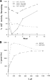
FIGURE 1.
FIGURE 1.
A, time course of human UGT1A7 or UGT1A10 activity expressed in COS-1 cells following treatment with curcumin and calphostin-C. Cells, untreated or treated with 200 μm curcumin or 500 nm calphostin-C as shown, were harvested; cell extracts were assayed in vitro as described under “Experimental Procedures.” Experiments, carried out as described under “Experimental Procedures,” were repeated thrice in triplicates; standard errors ranged from ±2to ±5%. The MTT assay did not show toxicity after 8 h. B, in vitro glucuronidation of curcumin by UGT1A7 and UGT1A10 expressed in COS-1 cells using increasing concentrations. Experiments, carried out as described under “Experimental Procedures,” were repeated thrice in triplicates; standard errors ranged from ±2to ±3%.
FIGURE 2.
FIGURE 2.
Curcumin or calphostin-C treatment of UGT-transfected COS-1 cells. 72 h after transfection with a pSVL-based UGT, COS-1 cells were treated with curcumin or calphostin-C; cell homogenates and substrates were as shown and used according to “Experimental Procedures.” Homogenates were subjected to Western blot analysis with anti-UGT-CE (11) according to “Experimental Procedures.” *, p ≤ 0.01; **, p ≤ 0.001.
FIGURE 3.
FIGURE 3.
Inhibition of UGT activity in LS180 cells by curcumin or calphostin-C treatment. A, Western blot analysis of curcumin treated-cells used UGT and PKCε antibodies in parallel with glucuronidation activity. Extracts (25 μg of protein) from curcumin (50 μm)-treated or control cells were resolved in an SDS 4–15% PAGE system. Antibody toward UGT-CE (52–55 kDa), the protein backbone of PKCε (92–96 kDa), or phosphoserine-729-PKCε was added to separate Western blots and processed as described under “Experimental Procedures.” Panel 4 shows in vitro glucuronidation of capsaicin with control values of 1882 ± 96 pmol/mg of protein/h. Experiments were repeated more than five times. Standard errors for data range between ±1 and ±5%. **, p ≤ 0.001. B, [33P]orthophosphate labeling of both PKCε and UGT proteins with parallel inhibition of labeling and UGT activity following curcumin treatment of LS180 cells. All cells were exposed to [33P]orthophosphate (5 mCi/ml) for 8 h and 50 μm curcumin for 0, 1, 3, 5, or 8 has described under “Experimental Procedures.” From solubilized cellular extract, immunocomplexes with anti-UGT-CE (panel 4) and with anti-PKCε (panel 2) were washed as described under “Experimental Procedures” before resolution on SDS-7.5% PAGE. Also, PKCε (panel 1) and UGT (panel 3) were analyzed by Western blots. Processed gels were exposed to x-ray film for 48 h. Experiments were repeated three times. UGT activity was toward capsaicin with control values of 1857 ± 74 pmol/mg of protein/h (panel 5); eugenol generated a similar profile (data not shown). C, concentration-dependent effects of calphostin-C on [33P]orthophosphate incorporation into UGTs in LS180 cells. Cells were allowed to incorporate the label for 8 h with calphostin-C treatment over the final hour as described under “Experimental Procedures” before immunocomplexing and Western blotting as described under C (panels 1–4) and determining UGT activity toward capsaicin (panel 5). *, p ≤ 0.01; **, p ≤ 0.001.
FIGURE 4.
FIGURE 4.
Co-localization of PKCα with calnexin (A) or calreticulin (B) in control COS-1 cells. Cells were processed for co-immunofluorescence as described under “Experimental Procedures.” For calnexin and PKCα co-localization, calnexin was visualized with donkey anti-rabbit FITC conjugate, and PKCα was visualized with donkey anti-mouse TRITC conjugate. Assembly of fluorescence images (panels 1 and 2) indicates fusion-generated yellow immunofluorescence (panel 4) outside the nuclei (panel 5). For calreticulin and PKCα co-localization, calreticulin was detected with donkey anti-rabbit FITC conjugate, and PKCα was detected with donkey anti-mouse TRITC conjugate. Assembly of fluorescence images (panels 1 and 2) indicates fusion-generated yellow immunofluorescence (panel 4) outside the nuclei (panel 5). Co-localization of PKCα or PKCδ with UGT1A10His expressed in COS1 cells. Co-localization was determined by immunofluorescence that probed UGT1A10 with donkey anti-rabbit FITC conjugate and PKCα with donkey anti-mouse TRITC conjugate as described under “Experimental Procedures.” C, assembly of fluorescence images (panels 1 and 2) indicates fusion, which generated yellow immunofluorescence (panel 4) outside the nuclei (panel 5). D, co-localization of PKC δ and UGT1A10His expressed in COS-1. Co-localization was analyzed by immunofluorescence with PKCδ detected with donkey anti-rabbit FITC conjugate, and UGT1A10His was detected with donkey anti-mouse TRITC as described under “Experimental Procedures.” Assembly of fluorescence images (panels 1 and 2) merged to generate yellow fluorescence (panel 4) outside the nuclei as shown (panel 5). Background immunofluorescence images in non-transfected cells are shown in supplemental Fig. S4. Scale bars represent 20 μm(A) or 10 μm(B–D).
FIGURE 5.
FIGURE 5.
A, specificity of anti-UGT-CE. Non-transfected and UGT1A7His- and -1A10His-transfected COS-1 cells were processed for immunoprecipitation with anti-His as described under “Experimental Procedures.” Western blots prepared after resolution of samples in a 4–15% SDS-PAGE system as described under “Experimental Procedures” were probed with anti-UGT-CE. B, co-immunoprecipitation of PKCε and UGT1A7His expressed in COS-1 cells. Upon terminating the experiment in which UGT1A7His-transfected COS-1 cells had been untreated or treated with 75μm curcumin (as shown above) or calphostin-C for 1 h, 60,000 × g supernatants generated as described under “Experimental Procedures” were immunocomplexed with anti-His, trapped with protein A-Sepharose for resolution in a SDS-10% PAGE system, and transblotted onto a nitrocellulose membrane. Proteins migrated as follows: β-COP, 110 kDa; PKCε, 96 kDa; and UGT1A7His, 58 kDa. The membrane was probed according to “Experimental Procedures” with different primary antibodies as follows: β-COP (ε-RACK), phosphoserine-PKCε, PKCε, phosphoserine, UGT-CE, and His. C, co-immunoprecipitation of UGT1A10His, PKCα, and PKCδ. Co-immunoprecipitation of PKCα and PKCδ expressed in UGT1A10-transfeceted COS-1 cells, which had been treated 1 h each with curcumin or calphostin-C as shown, was carried out with 60,000 × g supernatants as described in the legend for B. Supernatants were immunocomplexed with anti-His, trapped with protein A-Sepharose for resolution in a 4 to 15% SDS-PAGE system, and transblotted onto a nitrocellulose membrane.
FIGURE 6.
FIGURE 6.
A, effect of calyculin-A on curcumin-inhibited UGT activity in LS180 cells. Calyculin-A (6 nm) was added with or without curcumin or 1 h after curcumin (50 μm); its effect on control or inhibited cells was assessed as shown. (Calyculin-A is a protein phosphatase 1 inhibitor.) Standard errors for UGT activity range between ±1 and ±4% in three experiments performed in triplicates. B, effects of PKC agonists on curcumin (50 μm)-inhibited UGT were examined with either 30 μm DAG, 2.0 μm PMA, or 100 μg/ml PS as shown. Control activity was 1817 ± 88 pmol/mg of protein/h. Standard errors for UGT activity range between ±2 and ±5% in three separate experiments. C, effect of PKCε-specific translocation inhibitor on UGT activity. Non-conjugated 8-amino acid peptide (Oct) (175 μm) or its scrambled (Scr) control was introduced into saponin-permeabilized (18) LS180 cells as described under “Experimental Procedures.” Also, the rationale for PKC isozyme-specific inhibition is described under “Experimental Procedures” and/or “Results.” Control activity was 1952 ± 102 pmol/mg of protein/h. UGT assays in panels A–D used capsaicin and eugenol (not shown). *, p ≤ 0.01; **, p ≤ 0.001. D, effect of silencing PKCε on UGT1A7 activity. siRNA specific for PKCε (100 nm) or control siRNA (100 nm) was transfected into COS-1 cells following expression of UGT1A7 for 24 h; incubation continued for 48 h before harvesting cells for glucuronidation of eugenol or my cophenolic acid as shown (see Ref. 51) in a 2-h incubation. For Western blot analysis, samples were solubilized and fractionated by centrifugation as previously described (7), and 60,000 × g supernatants were resolved in a 4 to 15% SDS-PAGE system and transblotted. For Western analysis, anti-PKCε, anti-UGT-CE, and anti-β-actin were used as previously described (7). *, p ≤ 0.01; **, p ≤ 0.001. E, in vitro transcription, translation, and phosphorylation of UGT1A7His by PKCε. pcDNA3.1-UGT1A7His construct was linearized and transcribed for synthesis of nascent protein to undergo in vitro PKCε-dependent phosphorylation using [γ-33P]ATP with as described under “Experimental Procedures”; accordingly, aliquots of the kinased samples were counted. Affinity-purified control and experimental samples of [33P]UGTHis were analyzed in duplicate gels. One was dried for autoradiography and one was used for Western blot with anti-UGT-CE as described under “Experimental Procedures.” Samples are defined at the bottom of E. **, p ≤ 0.001.
FIGURE 7.
FIGURE 7.
Effects of ROS on UGT activity in LS180 cells. A, control LS180 cells were treated with 0.4 and 4.0 μg/ml catalase for 40 min or with 1.0, 2.0, and 5.0 μm herbimycin-A for 40 min. *, p ≤ 0.01; **, p ≤ 0.001. B, cells were treated with 0.5 or 1.0 mm H2O2 for 5 and 10 min. Cells under B were preincubated with 4.0 μg/ml catalase (Cat'ase) or 5.0 μm herbimycin-A (Herb) for 30 min before the addition of 1.0 mm H2O2 for an additional 5 or 10 min. Cells were also treated with 50 μm curcumin for 30 min or 250 nm calphostin-C for 30 min before addition of 1.0 mm H2O2 for 10 min. UGT activity was measured with eugenol with control value 8431 ± 141 pmol/mg of protein/h. A similar profile was observed with capsaicin as substrate. *, p ≤ 0.01; **, p ≤ 0.001.
FIGURE 8.
FIGURE 8.
Proposed model for effects of agonists and antagonists on PKC activity that controls UGT activity. The PKC schematic illustrates two modes of regulation: 1) well-established upstream phosphoinositide-dependent kinase-1 and tyrosine kinase(s) phosphorylate conserved serine/threonine and tyrosines (20, 21, 36, 39), respectively, in the catalytic domain generating putative competent and active PKC, and 2) the allosteric long term mediator (PMA) or lipid-derived short term agonists, DAG and PS (Fig. 6B), activate competent PKC via the regulatory domain (36) triggering attachment to specific cellular structures. Although the schematic suggests direct phosphorylation of ER-bound UGT by PKC based on parallel curcumin/calphostin-C inhibition of phosphoserine/radiolabeling with [33P]orthophosphate and UGT activity (Fig. 3, A–C), the fact that UGT1A7 acquired new substrate activity due to loss of serine 432 phosphorylation by PKCε translocation inhibitor peptide strongly suggests that PKCε directly phosphorylates UGT1A7 (7). Curcumin or calphostin-C inhibits attachment of PKC to molecular structures (7, 33, 36) (Fig. 5, B and C). Further, cellular oxidants and ROS can activate PKC via disruption of oxygen-sensitive zinc-complexed cysteine thiols in the regulatory domain (10, 47) and/or via stimulation of tyrosine phosphorylation of PKC (Fig. 7B). Consistent with PKC activation via H2O2-stimulated tyrosine phosphorylation, which is inhibited by herbimycin-A (20, 21), we observed inhibition of both constitutive and peroxide-stimulated UGT by both agents (Fig. 7, A and B). Inasmuch as cellular exposure to high concentrations of oxidized antioxidants, such as curcumin and nordihydroguaiaretic acid (4), can disrupt critical cysteines in the catalytic domain (10, 43, 47), the inhibitory effects of 50 μm curcumin, but not 25 μm (not shown), on both PKC phosphorylation and UGT activity are consistent with curcumin antioxidant action. Low levels (1.0–10 μm) of antioxidant polyphenols can act chemopreventively to scavenge oxidants (52). Potentially, UGTs with high Km values (≥10 μm) (4) and PKC isozymes with differential inhibitory sensitivities (Fig. 2) to polyphenols enable the PKC-UGT signaling pathway to adapt to different conditions and remain functional to beneficial chemopreventive action.

Similar articles

Cited by

References

    1. Dutton, G. J. (1980) in Glucuronidation of Drugs and Other Compounds (Dutton, G. J., ed) pp. 69–78, CRC Press, Boca Raton, FL
    1. Ritter, J. K., Crawford, J. M., and Owens, I. S. (1991) J. Biol. Chem. 266 1043–1047 - PubMed
    1. Wells, P. G., Mackenzie, P. I., Chowdhury, J. R., Guillemette, C., Gregory, P. A., Ishii, Y., Hansen, A. J., Kessler, F. K., Kim, P. M., Chowdhury, N. R., and Ritter, J. K. (2004) Drug Metab. Dispos. 32 281–290 - PubMed
    1. Basu, N. K., Ciotti, M., Hwang, M. S., Kole, L., Mitra, P. S., Cho, J. W., and Owens, I. S. (2004) J. Biol. Chem. 279 1429–1441 - PubMed
    1. Basu, N. K., Kole, L., and Owens, I. S. (2003) Biochem. Biophys. Res. Commun. 303 98–104 - PubMed

Publication types

LinkOut - more resources