. 2008 Sep 5;283(36):24534-45.
doi: 10.1074/jbc.M800006200. Epub 2008 Jun 13.

Analogs of tetrahydroisoquinoline natural products that inhibit cell migration and target galectin-3 outside of its carbohydrate-binding site

Affiliations

Analogs of tetrahydroisoquinoline natural products that inhibit cell migration and target galectin-3 outside of its carbohydrate-binding site

Alem W Kahsai et al. J Biol Chem. .

Abstract

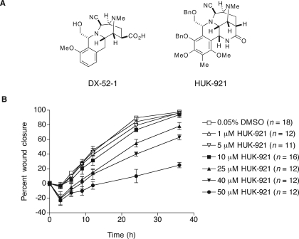
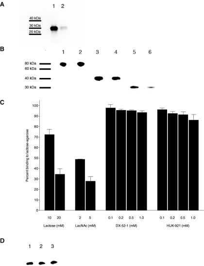
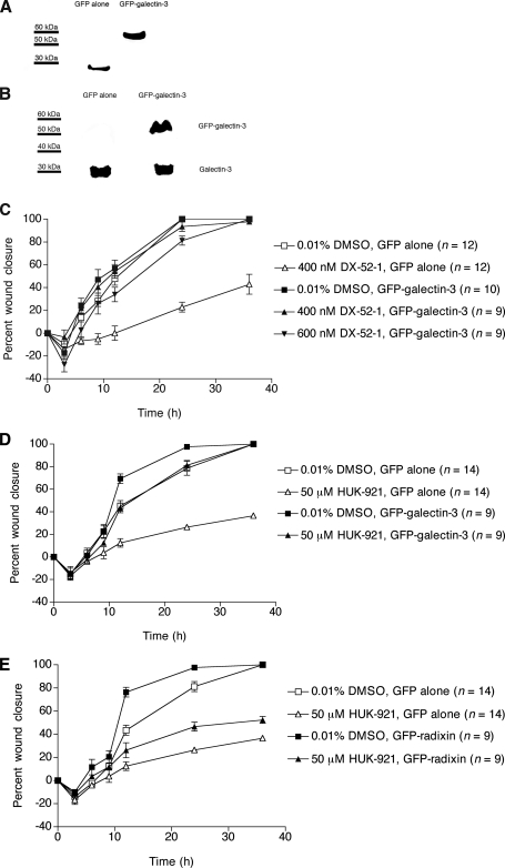
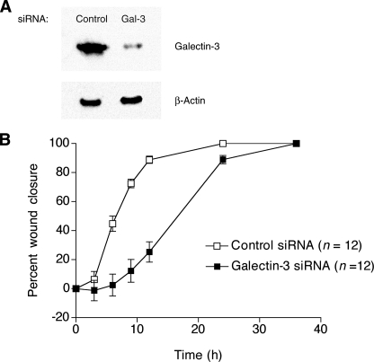
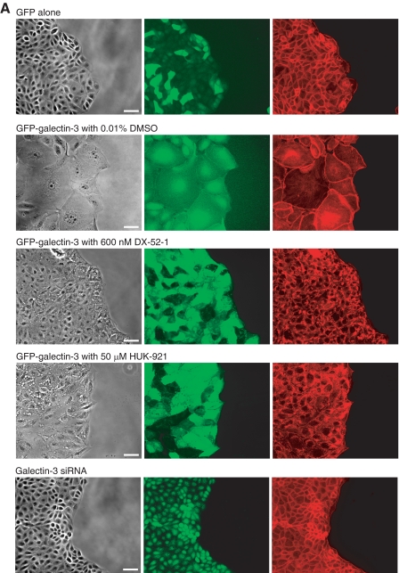
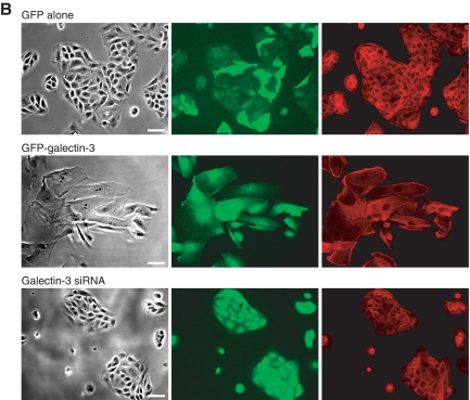
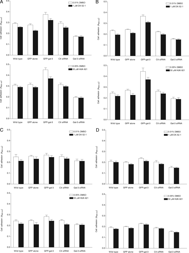
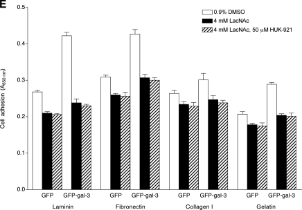
Cell migration is central to a number of normal and disease processes. Small organic molecules that inhibit cell migration have potential as both research probes and therapeutic agents. We have identified two tetrahydroisoquinoline natural product analogs with antimigratory activities on Madin-Darby canine kidney epithelial cells: a semisynthetic derivative of quinocarmycin (also known as quinocarcin), DX-52-1, and a more complex synthetic molecule, HUK-921, related to the naphthyridinomycin family. It has been assumed that the cellular effects of reactive tetrahydroisoquinolines result from the alkylation of DNA. We have reported previously that the primary target of DX-52-1 relevant to cell migration appears to be the membrane-cytoskeleton linker protein radixin. Here we extend the analysis of the protein targets of DX-52-1, reporting that the multifunctional carbohydrate-binding protein galectin-3 is a secondary target of DX-52-1 that may also be relevant to the antimigratory effects of both DX-52-1 and HUK-921. All known inhibitors of galectin-3 target its beta-galactoside-binding site in the carbohydrate recognition domain. However, we found that DX-52-1 and HUK-921 bind galectin-3 outside of its beta-galactoside-binding site. Intriguingly HUK-921, although a less potent inhibitor of cell migration than DX-52-1, had far greater selectivity for galectin-3 over radixin, exhibiting little binding to radixin, both in vitro and in cells. Overexpression of galectin-3 in cells led to a dramatic increase in cell adhesion on different extracellular matrix substrata as well as changes in cell-cell adhesion and cell motility. Galectin-3-overexpressing cells had greatly reduced sensitivity to DX-52-1 and HUK-921, and these compounds caused a change in localization of the overexpressed galectin-3 and reversion of the cells to a more normal morphology. The converse manipulation, RNA interference-based silencing of galectin-3 expression, resulted in reduced cell-matrix adhesion and cell migration. In aggregate, the data suggest that DX-52-1 and HUK-921 inhibit a carbohydrate binding-independent function of galectin-3 that is involved in cell migration.


Figures

FIGURE 1.
FIGURE 1.
Structures and antimigratory activities of analogs of reactive tetrahydroisoquinoline natural products. A, chemical structures of DX-52-1 and HUK-921. Me, methyl; Bn, benzyl. B, HUK-921 inhibits cell sheet migration during wound closure in MDCK cell monolayers. Final dimethyl sulfoxide (DMSO) carrier solvent concentration was 0.05% for each treatment (compound was added 30 min prior to wounding). Error bars represent S.E. for the indicated number of wounds. The IC50 for inhibition of wound closure at 24 h by HUK-921 is 32.5 μm as calculated from the data presented in this figure, whereas the corresponding value for DX-52-1 is 140 nm (18).
FIGURE 2.
FIGURE 2.
DX-52-1 and HUK-921 bind galectin-3 non-competitively with known carbohydrate ligands of galectin-3. A, DX-52-1 covalently and specifically binds galectin-3. Recombinant galectin-3 was incubated with 10 μm biotinylated DX-52-1 (lane 1) or 10 μm biotinylated DX-52-1 plus 500 μm free, non-biotinylated DX-52-1 competitor added simultaneously (lane 2). Following SDS-PAGE and transfer to polyvinylidene difluoride, the blot was probed with streptavidin-horseradish peroxidase. The degree of competition of biotinylated DX-52-1 by free DX-52-1 was calculated as 84.3 ± 3.5% (mean ± S.E.) from three independent experiments. B, HUK-921 competes with biotinylated DX-52-1 for binding to galectin-3 but not radixin. Lane 1, full-length radixin with 10 μm biotinylated DX-52-1; lane 2, full-length radixin with 10 μm biotinylated DX-52-1 plus 500 μm HUK-921; lane 3, C-terminal domain fragment of radixin with 10 μm biotinylated DX-52-1; lane 4, C-terminal domain fragment of radixin with 10 μm biotinylated DX-52-1 plus 500 μm HUK-921; lane 5, galectin-3 with 10 μm biotinylated DX-52-1; lane 6, galectin-3 with 10 μm biotinylated DX-52-1 plus 500 μm HUK-921. This experiment was done similarly to that shown in A except that potential competition of the binding of biotinylated DX-52-1 to galectin-3 and radixin by HUK-921 added simultaneously was evaluated. Based on quantitation of the degree of binding from the intensity of the bands from three independent experiments, we calculated the percent competition between biotinylated DX-52-1 and HUK-921 as 16.5 ± 6.8% for full-length radixin, 29.5 ± 6.9% for the C-terminal domain fragment of radixin, and 84.3 ± 4.2% for galectin-3 (means ± S.E. in all cases). C, percent binding of galectin-3 to lactose-agarose beads in the presence of the indicated concentrations of lactose, LacNAc, DX-52-1, or HUK-921. The extent of galectin-3 binding to lactose-conjugated agarose in the presence of DMSO is defined as 100% binding. (Every individual experiment was performed along with a parallel DMSO control, and each value was normalized to its parallel control.) Error bars represent S.E. for four to seven independent experiments, quantitated from Western blots, as described under “Experimental Procedures.” D, lactose and LacNAc do not compete with biotinylated DX-52-1 for binding to galectin-3. Lane 1, galectin-3 with 20 μm biotinylated DX-52-1; lane 2, galectin-3 with 20 μm biotinylated DX-52-1 plus 20 mm lactose; lane 3, galectin-3 with 20 μm biotinylated DX-52-1 plus 5 mm LacNAc. Based on quantitation of the intensity of bands from three independent experiments, we found only very weak competition between 20 μm biotinylated DX-52-1 and 20 mm lactose or 5 mm LacNAc and calculated the percent competition values as 25.0 ± 5.2 and 20.4 ± 2.4% (means ± S.E.), respectively.
FIGURE 3.
FIGURE 3.
Overexpression of galectin-3 results in strongly reduced sensitivity to the antimigratory activities of DX-52-1 and HUK-921, whereas overexpression of radixin only weakly reduces sensitivity to HUK-921. A, Western blot analysis with an anti-GFP antibody to probe whole-cell lysates from MDCK cells stably expressing GFP alone or GFP-galectin-3. B, Western blot analysis with an anti-galectin-3 antibody showing levels of GFP-galectin-3 and endogenous galectin-3 in MDCK cells stably expressing GFP alone or GFP-galectin-3. C, overexpression of galectin-3 markedly decreases the sensitivity of MDCK cells to the inhibitory effect of DX-52-1 on cell sheet migration in the wound closure assay. D, overexpression of galectin-3 markedly decreases the sensitivity of MDCK cells to the antimigratory activity of HUK-921. E, overexpression of radixin only mildly decreases the sensitivity of MDCK cells to the antimigratory activity of HUK-921 in contrast to previous results with DX-52-1 (18). Error bars represent S.E. for the indicated number of wounds in C–E.
FIGURE 4.
FIGURE 4.
RNAi-based silencing of galectin-3 expression results in a reduced rate of cell migration. A, Western blot analysis of whole-cell lysates from MDCK cells stably expressing either an inert, control siRNA or an effective galectin-3-specific (Gal-3) siRNA prepared as described under “Experimental Procedures.” B, knockdown of galectin-3 results in a decreased rate of cell sheet migration during wound closure in MDCK cell monolayers. Error bars represent S.E. for the indicated number of wounds. Note that, unlike knockdown of galectin-3 or treatment with DX-52-1 or HUK-921, treatment with LacNAc had little or no effect on cell migration in this system.
FIGURE 5.
FIGURE 5.
Galectin-3-overexpressing cells are highly spread and display reduced cell-cell contact, and treatment with DX-52-1 or HUK-921 causes a change in localization of GFP-galectin-3 and reversion of cells to a more normal morphology, whereas galectin-3 knockdown cells display reduced membrane protrusion. A, images of cells expressing GFP alone, GFP-galectin-3 in the presence or absence of DX-52-1 or HUK-921, or a galectin-3-specific siRNA, all 4 h after wounding of MDCK cell monolayer treatment (compounds were added 30 min prior to wounding). From left to right, each row consists of a phase-contrast image, a GFP fluorescence image, and a second fluorescence image showing TRITC-phalloidin staining of filamentous actin. Scale bars, 50 μm. B, images of cells expressing GFP alone, GFP-galectin-3, or a galectin-3-specific siRNA plated at low density on fibronectin-coated glass coverslips and then imaged 48 h later. From left to right, each row consists of a phase-contrast image, a GFP fluorescence image, and a second fluorescence image showing TRITC-phalloidin staining of filamentous actin. Scale bars, 50 μm. (Note that the shRNA expression vector harbors a GFP cassette, as described under “Experimental Procedures,” so the galectin-3 knockdown cells also display GFP fluorescence.)
FIGURE 5.
FIGURE 5.
Galectin-3-overexpressing cells are highly spread and display reduced cell-cell contact, and treatment with DX-52-1 or HUK-921 causes a change in localization of GFP-galectin-3 and reversion of cells to a more normal morphology, whereas galectin-3 knockdown cells display reduced membrane protrusion. A, images of cells expressing GFP alone, GFP-galectin-3 in the presence or absence of DX-52-1 or HUK-921, or a galectin-3-specific siRNA, all 4 h after wounding of MDCK cell monolayer treatment (compounds were added 30 min prior to wounding). From left to right, each row consists of a phase-contrast image, a GFP fluorescence image, and a second fluorescence image showing TRITC-phalloidin staining of filamentous actin. Scale bars, 50 μm. B, images of cells expressing GFP alone, GFP-galectin-3, or a galectin-3-specific siRNA plated at low density on fibronectin-coated glass coverslips and then imaged 48 h later. From left to right, each row consists of a phase-contrast image, a GFP fluorescence image, and a second fluorescence image showing TRITC-phalloidin staining of filamentous actin. Scale bars, 50 μm. (Note that the shRNA expression vector harbors a GFP cassette, as described under “Experimental Procedures,” so the galectin-3 knockdown cells also display GFP fluorescence.)
FIGURE 6.
FIGURE 6.
Effects of galectin-3 overexpression, galectin-3 knockdown, or treatment with DX-52-1, HUK-921, or LacNAc on adhesion of cells to different ECM proteins. Shown are the levels of attachment of MDCK cells that were not manipulated (“wild type”) and cells expressing GFP alone, GFP-galectin-3 (GFP-gal-3), a control (Ctl) siRNA, or a galectin-3 specific (Gal-3) siRNA to fibronectin (A), laminin (B), collagen I (C), and gelatin (hydrolyzed collagen; D) in the presence or absence of DX-52-1 or HUK-921, all 2 h after plating. E, levels of attachment of MDCK cells expressing GFP alone or GFP-galectin-3 (GFP-gal-3) in the presence or absence of LacNAc, with or without co-treatment with HUK-921, 2 h after plating. Error bars represent S.E. for three independent experiments in each case.
FIGURE 6.
FIGURE 6.
Effects of galectin-3 overexpression, galectin-3 knockdown, or treatment with DX-52-1, HUK-921, or LacNAc on adhesion of cells to different ECM proteins. Shown are the levels of attachment of MDCK cells that were not manipulated (“wild type”) and cells expressing GFP alone, GFP-galectin-3 (GFP-gal-3), a control (Ctl) siRNA, or a galectin-3 specific (Gal-3) siRNA to fibronectin (A), laminin (B), collagen I (C), and gelatin (hydrolyzed collagen; D) in the presence or absence of DX-52-1 or HUK-921, all 2 h after plating. E, levels of attachment of MDCK cells expressing GFP alone or GFP-galectin-3 (GFP-gal-3) in the presence or absence of LacNAc, with or without co-treatment with HUK-921, 2 h after plating. Error bars represent S.E. for three independent experiments in each case.

References

    1. Disanza, A., Steffen, A., Hertzog, M., Frittoli, E., Rottner, K., and Scita, G. (2005) CMLS Cell. Mol. Life Sci. 62 955–970 - PMC - PubMed
    1. Li, S., Guan, J. L., and Chien, S. (2005) Annu. Rev. Biomed. Eng. 7 105–150 - PubMed
    1. Vicente-Manzanares, M., Webb, D. J., and Horwitz, A. R. (2005) J. Cell Sci. 118 4917–4919 - PubMed
    1. Stossel, T. P., Fenteany, G., and Hartwig, J. H. (2006) J. Cell Sci. 119 3261–3264 - PubMed
    1. Ananthakrishnan, R., and Ehrlicher, A. (2007) Int. J. Biol. Sci. 3 303–317 - PMC - PubMed

Publication types

MeSH terms

LinkOut - more resources