Case Reports
. 2008 Aug;131(Pt 8):1990-2001.
doi: 10.1093/brain/awn114. Epub 2008 Jun 12.

Mutation of FIG4 causes a rapidly progressive, asymmetric neuronal degeneration

Affiliations
Case Reports

Mutation of FIG4 causes a rapidly progressive, asymmetric neuronal degeneration

Xuebao Zhang et al. Brain. 2008 Aug.

Abstract

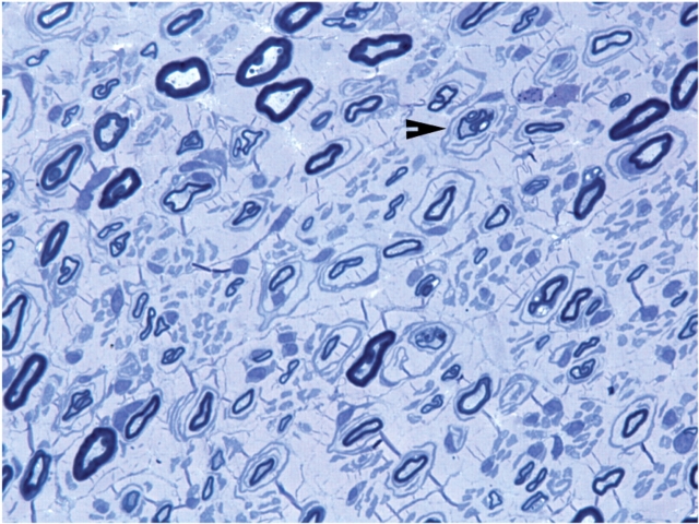
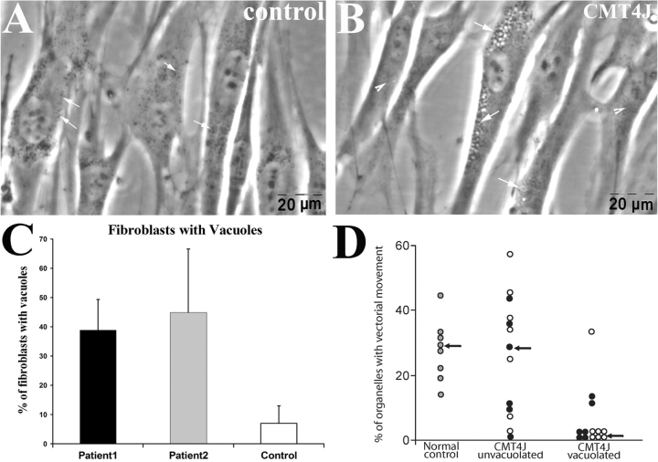
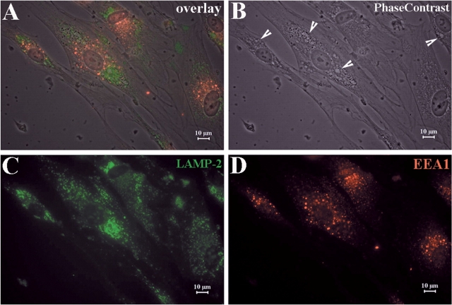
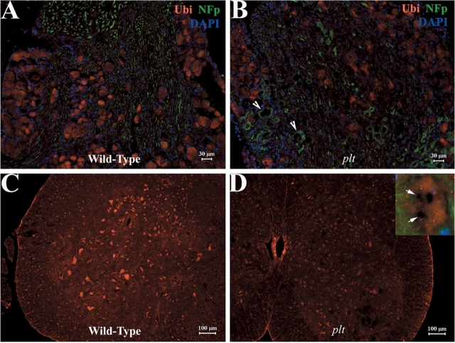
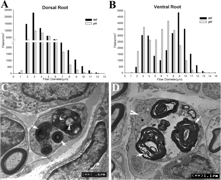
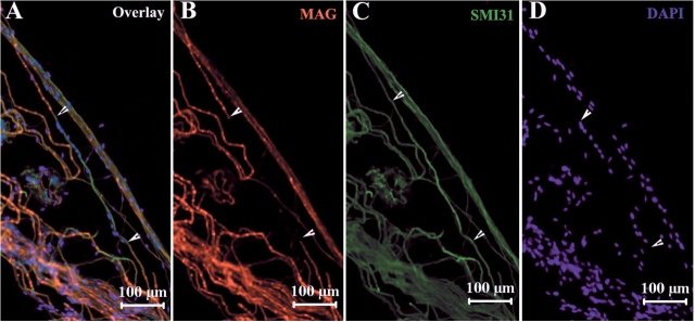
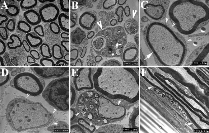
Recessive Charcot-Marie-Tooth disease type-4J (CMT4J) and its animal model, the pale tremor mouse (plt), are caused by mutations of the FIG4 gene encoding a PI(3,5)P(2) 5-phosphatase. We describe the 9-year clinical course of CMT4J, including asymmetric, rapidly progressive paralysis, in two siblings. Sensory symptoms were absent despite reduced numbers of sensory axons. Thus, the phenotypic presentation of CMT4J clinically resembles motor neuron disease. Time-lapse imaging of fibroblasts from CMT4J patients demonstrates impaired trafficking of intracellular organelles because of obstruction by vacuoles. Further characterization of plt mice identified axonal degeneration in motor and sensory neurons, limited segmental demyelination, lack of TUNEL staining and lack of accumulation of ubiquitinated protein in vacuoles of motor and sensory neurons. This study represents the first documentation of the natural history of CMT4J. Physical obstruction of organelle trafficking by vacuoles is a potential novel cellular mechanism of neurodegeneration.


Figures

Fig. 1
Fig. 1
Loss of myelinated nerve fibres in sural biopsy of a patient with CMT4J. A semithin section with toluidine blue staining was performed on the sural nerve biopsy of the patient 2. The number of myelinated nerve fibres is severely reduced, and there is a large amount of collagen in the extracellular matrix. Arrowhead: onion bulbs and thinly myelinated nerve fibres.
Fig. 2
Fig. 2
Vacuolation in FIG4-deficient human fibroblasts. Fibroblasts were isolated from skin biopsies of patients with CMT4J and normal control. Cells were imaged under phase-contrast lens. (A) Cultured fibroblasts from the control contained occasional vacuoles. Intracellular organelles appear as ‘black-dots’ (Waterman-Storer et al., 1999) and are scattered in the cytoplasm (arrows). (B) Excessive vacuoles in fibroblasts from patients (arrows). (C) Nineteen to twenty two fields from cultures of each human subject were randomly selected under 40× magnification, and the cell number and number of cells with vacuoles were counted. Means ± SD values: 7% ± 6 vacuolated control cells; 39% ± 10 vacuolated cells from patient 1; 45% ± 22 vacuolated cells from patient 2. (D) Time lapse imaging was carried out on regions of fibroblasts containing many vacuoles (vacuolated) or lacking vacuoles (non-vacuolated). The percentage of intracellular organelles exhibiting vectorial movement was measured as described in Methods section (Schrader, 2001) (p = 0.01 for filled circles; p = 0.02 for open circles; compared with non-vacuolated area).
Fig. 3
Fig. 3
Vacuoles contain markers for late- endosomes and lysosomes. (A) Fibroblasts from human skin biopsies were cultured on glass slides. IHC was then performed on these slides. (B) Numerous vacuoles were visualized under phase contrast imaging. (C) Cells were stained with antibodies against late-endosome-lysosome marker LAMP-2. (D) Cells were stained with antibodies against early endosome marker EEA1. Vacuoles were co-localized with LAMP-2, but not EEA1. This finding is consistent with the observation in mouse fibroblasts (Chow et al., 2007) and suggests that the vacuoles are derived from dysfunctional late-endosomes or lysosomes, and not from early endosomes.
Fig. 4
Fig. 4
NF and ubiquitin do not accumulate in neuronal vacuoles. Sections of mouse spinal cord and DRG were examined with IHC technique. (A–D) Immunoreactivity against ubiquitin was evenly distributed in the cytoplasm of motor and sensory neurons. Ubiquitinated inclusions were not present in vacuoles. (A–B) Antibodies against NFp produced a strong staining in the axons but not in neuronal cytoplasm. Some axons appeared irregular and enlarged (B, arrows). Large motor neurons in the anterior horn were reduced in number (5D). Inset: A spinal motor neuron at high magnification lacking NFp and ubiquitin in vacuoles (arrows).
Fig. 5
Fig. 5
Axonal degeneration in plt mice. Morphometric analysis was performed on the semithin sections of dorsal and ventral roots of 6-week-old plt (n = 4) and wild-type (n = 4) mice (total number of counted myelinated nerve fibres = 3679 for dorsal roots and 2480 for ventral roots in 4 plt mice and 4300 for dorsal roots and 2611 for ventral roots in wild-type mice). Sensory nerve fibre loss in the dorsal roots involved both small and large diameter myelinated fibres (A). In contrast, axonal loss in the ventral roots mainly affected the large diameter fibres. Small diameter myelinated nerve fibre numbers were increased (B). We have also examined femoral, saphenous, tibial and sural nerves under light microscopy (semithin sections) and EM. Active axonal degeneration was conspicuous. Examples are shown in Figs C and D. In (C), a degenerated nerve fibre is in the centre of the picture. Myelin debris is localized in the Schwann cell cytoplasm (arrows). An axon-like residue is visible (arrowhead), presumably from a degenerated axon. A similar nerve fibre is shown in (D). Again, myelin debris is prominent (arrows in D). The axon is still preserved, but appears irregular with condensed cytoskeletons and intra-axonal organelles (arrowhead).
Fig. 6
Fig. 6
Segmental demyelination in teased sciatic nerve fibres from plt mice. Mouse sciatic nerves were teased into individual glass slides and stained with IHC techniques. (A) Myelin specific protein MAG was strongly expressed in Schmidt-Lantemann incisures. MAG staining was absent in some segments of myelinated nerve fibres (e.g. between arrowheads in A, B). Labelling for NFp was intact in the same segment (C), demonstrate segmental demyelination. Many nuclei were lined up along this segment of the axon (D) derived from invading macrophages or remyelinating Schwann cells. Demyelination was not observed in wild-type nerve fibres.
Fig. 7
Fig. 7
Demyelination confirmed by ultrastructure. Peripheral nerves were prepared into ultrathin sections and examined under EM. (A) Sciatic nerves from wild-type mice exhibited myelinated nerve fibres of various diameter as well as non-myelinated fibres. (B) Sciatic nerves from plt mice showed significantly reduced density of myelinated nerve fibres. Macrophages were present in dorsal roots but less frequently in other nerves (arrowheads). Macrophage cytoplasm contained debris, presumably from engulfed myelin. Nerve fibres with thin myelin (arrow in C) or absence of myelin (D) were observed. Many had basal lamina redundancy (arrowheads in C and D), a pathological change suggesting remyelination. (E) Myelin debris was occasionally observed in Schwann cell cytoplasm (arrowheads), suggesting active demyelination/remyelination. (F) Myelination debris in a segment of myelinated nerve fibre (between arrows) can also be seen in longitudinal sections of nerves. Vacuoles were restricted to neuronal soma and not present in axons or Schwann cells. The ultrastructural changes are consistent with the IHC findings in Fig. 6 supporting segmental demyelination in plt mice.

References

    1. Bai YH, Ianokova E, Pu Q, Ghandour K, Levinson R, Martin JJ, et al. R69C Mutation in P0 Gene alters myelination and ion channel subtypes. Arch Neurol. 2006;63:1787–94. - PubMed
    1. Ballar P, Shen Y, Yang H, Fang S. The role of a novel p97/valosin-containing protein-interacting motif of gp78 in endoplasmic reticulum-associated degradation. J Biol Chem. 2006;281:35359–68. - PubMed
    1. Ballatore C, Lee VM, Trojanowski JQ. Tau-mediated neurodegeneration in Alzheimer's disease and related disorders. Nat Rev Neurosci. 2007;8:663–72. - PubMed
    1. Baloh RH, Schmidt RE, Pestronk A, Milbrandt J. Altered axonal mitochondrial transport in the pathogenesis of Charcot-Marie-Tooth disease from mitofusin 2 mutations. J Neurosci. 2007;27:422–30. - PMC - PubMed
    1. Barber RP, Phelps PE, Houser CR, Crawford GD, Salvaterra PM, Vaughn JE. The morphology and distribution of neurons containing choline acetyltransferase in the adult rat spinal cord: an immunocytochemical study. J Comp Neurol. 1984;229:329–46. - PubMed

Publication types