

Insights into plant cell wall degradation from the genome sequence of the soil bacterium Cellvibrio japonicus
- PMID: 18556790
- PMCID: PMC2493263
- DOI: 10.1128/JB.01701-07
Insights into plant cell wall degradation from the genome sequence of the soil bacterium Cellvibrio japonicus
Abstract
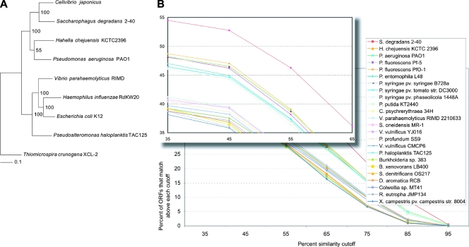
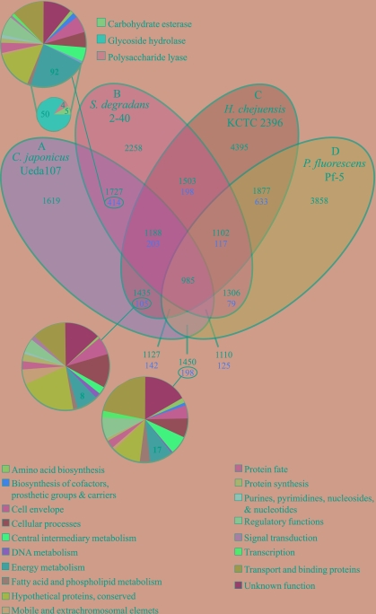
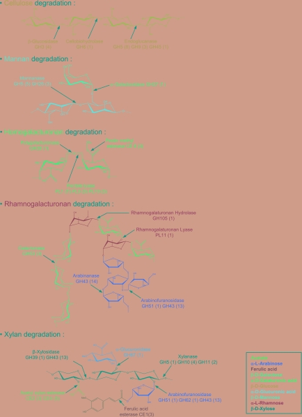
The plant cell wall, which consists of a highly complex array of interconnecting polysaccharides, is the most abundant source of organic carbon in the biosphere. Microorganisms that degrade the plant cell wall synthesize an extensive portfolio of hydrolytic enzymes that display highly complex molecular architectures. To unravel the intricate repertoire of plant cell wall-degrading enzymes synthesized by the saprophytic soil bacterium Cellvibrio japonicus, we sequenced and analyzed its genome, which predicts that the bacterium contains the complete repertoire of enzymes required to degrade plant cell wall and storage polysaccharides. Approximately one-third of these putative proteins (57) are predicted to contain carbohydrate binding modules derived from 13 of the 49 known families. Sequence analysis reveals approximately 130 predicted glycoside hydrolases that target the major structural and storage plant polysaccharides. In common with that of the colonic prokaryote Bacteroides thetaiotaomicron, the genome of C. japonicus is predicted to encode a large number of GH43 enzymes, suggesting that the extensive arabinose decorations appended to pectins and xylans may represent a major nutrient source, not just for intestinal bacteria but also for microorganisms that occupy terrestrial ecosystems. The results presented here predict that C. japonicus possesses an extensive range of glycoside hydrolases, lyases, and esterases. Most importantly, the genome of C. japonicus is remarkably similar to that of the gram-negative marine bacterium, Saccharophagus degradans 2-40(T). Approximately 50% of the predicted C. japonicus plant-degradative apparatus appears to be shared with S. degradans, consistent with the utilization of plant-derived complex carbohydrates as a major substrate by both organisms.
Figures
References
-
- Barrangou, R., C. Fremaux, H. Deveau, M. Richards, P. Boyaval, S. Moineau, D. A. Romero, and P. Horvath. 2007. CRISPR provides acquired resistance against viruses in prokaryotes. Science 3151709-1712. - PubMed
-
- Bhat, M. K. 2000. Cellulases and related enzymes in biotechnology. Biotechnol. Adv. 18355-383. - PubMed
-
- Blake, A. W., L. McCartney, J. E. Flint, D. N. Bolam, A. B. Boraston, H. J. Gilbert, and J. P. Knox. 2006. Understanding the biological rationale for the diversity of cellulose-directed carbohydrate-binding modules in prokaryotic enzymes. J. Biol. Chem. 28129321-29329. - PubMed
Publication types
MeSH terms
Substances
Associated data
- Actions
Grants and funding
LinkOut - more resources
Full Text Sources
Other Literature Sources
Molecular Biology Databases
