Comparative Study
. 2008 Dec;125(4):549-57.
doi: 10.1111/j.1365-2567.2008.02869.x. Epub 2008 Jun 13.

FADD and the NF-kappaB family member Bcl-3 regulate complementary pathways to control T-cell survival and proliferation

Affiliations
Comparative Study

FADD and the NF-kappaB family member Bcl-3 regulate complementary pathways to control T-cell survival and proliferation

Svetla Rangelova et al. Immunology. 2008 Dec.

Abstract

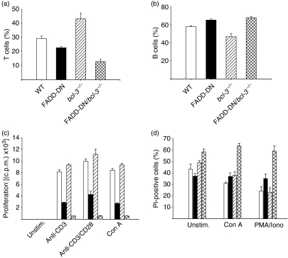
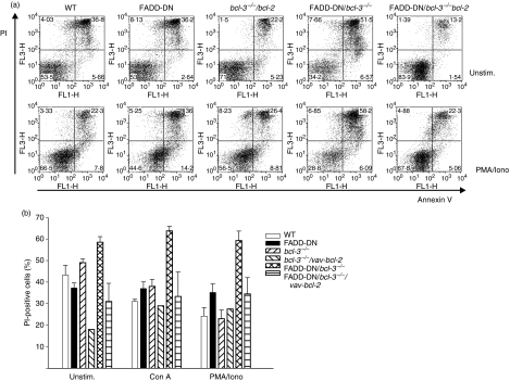
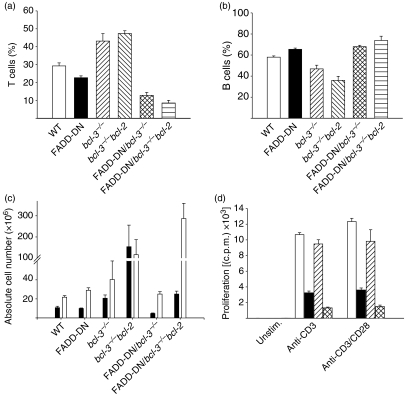
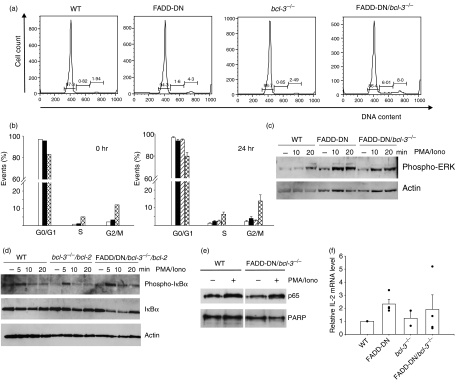
Fas-associated protein with death domain/mediator of receptor induced toxicity (FADD/MORT1) was first described as a transducer of death receptor signalling but was later recognized also to be important for proliferation of T cells. B-cell lymphoma 3 (Bcl-3) is a relatively little understood member of the nuclear factor (NF)-kappaB family of transcription factors. We recently found that Bcl-3 is up-regulated in T cells from mice where FADD function is blocked by a dominant negative transgene (FADD-DN). To understand the importance of this, we generated FADD-DN/bcl-3(-/-) mice. Here, we report that T cells from these mice show massive cell death and severely reduced proliferation in response to T-cell receptor (TCR) stimulation in vitro. Transgenic co-expression of Bcl-2 (FADD-DN/bcl-3(-/-)/vav-bcl-2 mice) rescued the survival but not the proliferation of T cells. FADD-DN/bcl-3(-/-) mice had normal thymocyte numbers but reduced numbers of peripheral T cells despite an increase in cycling T cells in vivo. However, activation of the classical NF-kappaB and extracellular regulated kinase (ERK) pathways and expression of interleukin (IL)-2 mRNA upon stimulation were normal in T cells from FADD-DN/bcl-3(-/-) mice. These data suggest that FADD and Bcl-3 regulate separate pathways that both contribute to survival and proliferation in mouse T cells.


Figures

Figure 1
Figure 1
Loss of peripheral T cells, proliferation defect and enhanced cell death in T cells in Fas-associated protein with death domain/B-cell lymphoma 3 (FADD-DN/bcl-3−/−) mice. (a, b) Percentages of T and B cells in spleens were assessed by staining for CD3 and B220, followed by flow cytometry. Data represent mean ± standard error of the mean (SEM) for four mice of each genotype. (c) T cells were purified by magnetic sorting from spleens of wild-type (WT) mice (open bars), FADD-DN transgenic mice (filled bars), bcl-3−/− mice (hatched bars) and FADD-DN/bcl-3−/− mice (cross-hatched bars). Purified T cells (3 × 10⁵) were stimulated with the mitogens indicated. Proliferation was measured as incorporation of 3H-thymidine at 48 hr. One representative experiment is shown (mean ± standard deviation). A similar reduction of proliferation of T cells from FADD-DN/bcl-3−/− mice for the various stimuli was seen in at least five experiments. (d) T cells were purified by magnetic sorting from spleens of WT mice (open bars), FADD-DN transgenic mice (filled bars), bcl-3−/− mice (hatched bars) and FADD-DN/bcl-3−/− mice (cross-hatched bars). Purified T cells (3 × 10⁵) were cultured without stimulation or stimulated with the mitogens indicated for 24 hr, and cell death was measured as uptake of propidium iodide (PI). Data represent mean ± SEM for at least three mice of each genotype analysed in independent experiments. Con A, concanavalin A; c.p.m., counts per minute; Iono, ionomycin; PMA, phorbol 12-myristate 13-acetate; Unstim., unstimulated.
Figure 2
Figure 2
B-cell lymphoma 2 (Bcl-2) over-expression blocks the abnormally increased death of T cells from Fas-associated protein with death domain (FADD-DN)/bcl-3−/− mice. T cells were purified as described in the text from spleens of mice with the genotypes indicated. Purified T cells (3 × 10⁵) were cultured without stimulation (Unstim.) or stimulated with the agents indicated overnight. Cell death was measured by staining with annexin V and propidium iodide (PI). (a) Examples of cultures of unstimulated (top) and phorbol 12-myristate 13-acetate/ionomycin (PMA/Iono)-stimulated cells (bottom). (b) Summary of the data obtained for several mice. Data represent means for two bcl-3−/−/vav-bcl-2 mice [individual values: unstimulated, 19 and 17%; concanavalin A (Con A), 33 and 25%; PMA/Iono, 29 and 26%] and mean ± standard error of the mean for three or four FADD-DN-bcl-3−/−/vav-bcl-2 mice. For comparison, data for the other genotypes are reproduced from Fig. 1(d).
Figure 3
Figure 3
Cell numbers and proliferation defect of T cells from Fas-associated protein with death domain/B-cell lymphoma 3 (FADD-DN/bcl-3−/−)/vav-bcl-2 mice. (a–c) Comparison of relative numbers of T cells (a) and B cells (b) and of absolute lymphocyte numbers in spleens from mice of the various genotypes [(c) filled bars: T cells; open bars: B cells]. Data represent mean ± standard error of the mean for four mice (a, b) or five or six mice (c) with the exception of bcl-3−/−/vav-bcl-2 mice (three mice). Data from single- and double-mutant mice are reproduced from Fig. 1 for better comparison. (d) T cells were purified by magnetic sorting from spleens of wild-type (WT) mice (open bars), FADD-DN mice (filled bars), bcl-3−/−/vav-bcl-2 mice (hatched bars) and FADD-DN/bcl-3−/−/vav-bcl-2 mice (cross-hatched bars). Purified T cells (3 × 10⁵) were stimulated with the agents indicated. Proliferation was measured as incorporation of 3H-thymidine at 48 hr. One representative experiment is shown (mean ± standard deviation). A similar reduction of proliferation of T cells from FADD-DN/bcl-3−/−/vav-bcl-2 mice was seen in five experiments. c.p.m., counts per minute.
Figure 4
Figure 4
Increased numbers of cycling cells and normal signalling events in T cells from Fas-associated protein with death domain/B-cell lymphoma 3 (FADD-DN/bcl-3−/−) mice. (a) Cell cycle analysis of purified T cells from spleens of mice of the indicated genotypes. Cells were fixed in ethanol, stained with propidium iodide (PI) and analysed by flow cytometry. Cells with an apparent sub-G1 DNA content were gated out, and percentages of cells in the cell cycle phases and of sub-G1 cells are given. (b) Summary of cell cycle analyses. Cells from wild-type (WT) mice (open bars), FADD-DN mice (filled bars), bcl-3−/− mice (hatched bars) and FADD-DN/bcl-3−/− mice (cross-hatched bars) were analysed as in (a), either immediately after isolation (0 hr, left) or after 24 hr in culture without stimulation (24 hr, right). Data represent the mean for two mice (0 hr) or mean ± standard error of the mean (SEM) for four mice of each genotype (24 hr). (c) Purified T cells from mice of the genotypes indicated were stimulated with phorbol 12-myristate 13-acetate/ionomycin (PMA) plus ionomycin (Iono) for the times shown. Cells were lysed and analysed by western blotting for IκB phosphorylation and degradation. Similar results were obtained in three experiments with FADD-DN/bcl-3−/− mice and two with FADD-DN/bcl-3−/−/vav-bcl-2 mice. (d) Purified T cells from mice of the genotypes indicated were stimulated with PMA plus ionomycin for the times shown. Cells were lysed and analysed by western blotting for levels of phospho-extracellular regulated kinase (ERK). Similar results were obtained in two separate experiments. (e) Purified T cells of the genotypes indicated were either stimulated with PMA plus ionomycin for 15 min or were left untreated. Nuclear extracts were prepared and analysed by western blotting for nuclear translocation of p65. Probing with an antibody to poly-ADP-ribose-polymerase (PARP) served as a control for the loading and purity of nuclear fractionation. Similar results were obtained in two independent experiments. (f) Expression of interleukin (IL)-2 mRNA was analysed by reverse transcriptase–polymerase chain reaction (RT-PCR). T cells from mice of the genotypes indicated were stimulated for 4 hr with PMA plus ionomycin, and RNA was extracted and analysed by quantitative PCR for expression of IL-2 mRNA. In each experiment, values for WT mice were set to one, and relative values are shown. Columns represent means ± SEM for two (bcl-3−/−) or four (all other genotypes) mice from independent experiments. The individual values are shown as dots.

References

    1. Ashkenazi A, Dixit VM. Death receptors: signaling and modulation. Science. 1998;281:1305–8. - PubMed
    1. Chang DW, Xing Z, Pan Y, Algeciras-Schimnich A, Barnhart BC, Yaish-Ohad S, Peter ME, Yang X. c-FLIP(L) is a dual function regulator for caspase-8 activation and CD95-mediated apoptosis. EMBO J. 2002;21:3704–14. - PMC - PubMed
    1. Micheau O, Thome M, Schneider P, Holler N, Tschopp J, Nicholson DW, Briand C, Grutter MG. The long form of FLIP is an activator of caspase-8 at the Fas death-inducing signaling complex. J Biol Chem. 2002;277:45162–71. - PubMed
    1. Yeh WC, Pompa JL, McCurrach ME, et al. FADD: essential for embryo development and signaling from some, but not all, inducers of apoptosis. Science. 1998;279:1954–8. - PubMed
    1. Zhang J, Cado D, Chen A, Kabra NH, Winoto A. Fas-mediated apoptosis and activation-induced T-cell proliferation are defective in mice lacking FADD/Mort1. Nature. 1998;392:296–300. - PubMed

Publication types

MeSH terms

LinkOut - more resources