. 2008 Jul;69(2):453-65.
doi: 10.1111/j.1365-2958.2008.06296.x.

Two variable active site residues modulate response regulator phosphoryl group stability

Affiliations

Two variable active site residues modulate response regulator phosphoryl group stability

Stephanie A Thomas et al. Mol Microbiol. 2008 Jul.

Abstract

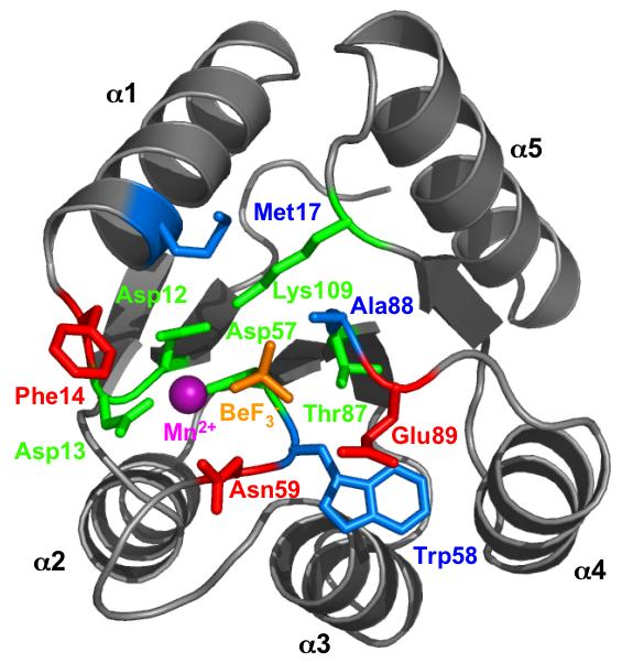
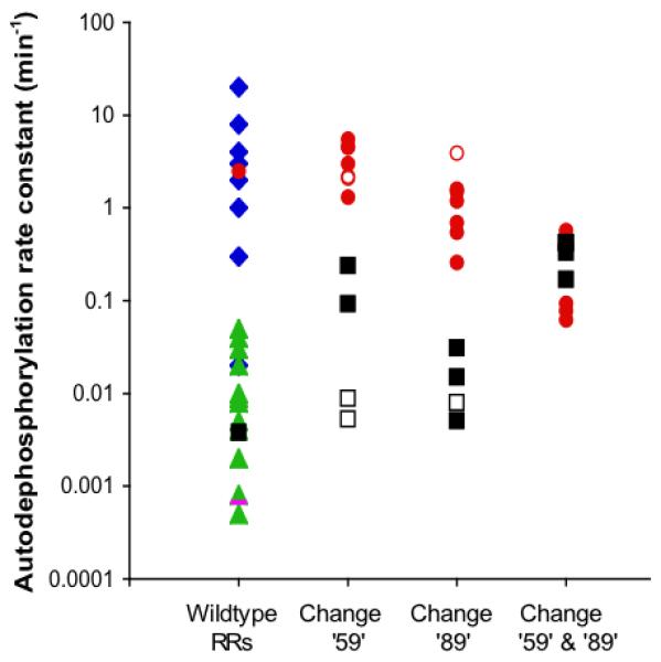
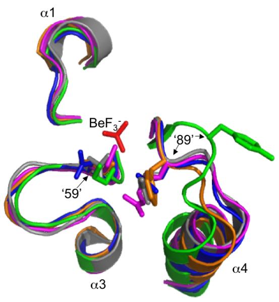
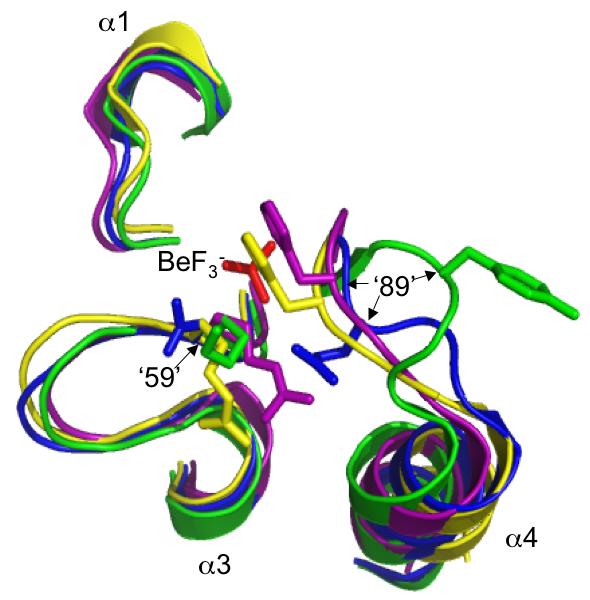
Many signal transduction networks control their output by switching regulatory elements on or off. To synchronize biological response with environmental stimulus, switching kinetics must be faster than changes in input. Two-component regulatory systems (used for signal transduction by bacteria, archaea and eukaryotes) switch via phosphorylation or dephosphorylation of the receiver domain in response regulator proteins. Although receiver domains share conserved active site residues and similar three-dimensional structures, rates of self-catalysed dephosphorylation span a >or= 40,000-fold range in response regulators that control diverse biological processes. For example, autodephosphorylation of the chemotaxis response regulator CheY is 640-fold faster than Spo0F, which controls sporulation. Here we demonstrate that substitutions at two variable active site positions decreased CheY autodephosphorylation up to 40-fold and increased the Spo0F rate up to 110-fold. Particular amino acids had qualitatively similar effects in different response regulators. However, mutant proteins matched to other response regulators at the two key variable positions did not always exhibit similar autodephosphorylation kinetics. Therefore, unknown factors also influence absolute rates. Understanding the effects that particular active site amino acid compositions have on autodephosphorylation rate may allow manipulation of phosphoryl group stability for useful purposes, as well as prediction of signal transduction kinetics from amino acid sequence.


Figures

Fig. 1
Fig. 1
Receiver domain active site structure. E. coli CheY bound to the phosphoryl group analog BeF3- (orange) and Mn2+ (magenta) is shown (pdb 1FQW) (Lee et al., 2001). Conserved active site residues, green; variable active site residues tested experimentally, red; variable active site residues not tested in this work, blue. Figure created using PyMOL (DeLano, 2002).
Fig. 2
Fig. 2
Response regulator autodephosphorylation rate constants. Note logarithmic scale. Column 1 shows wildtype values from Table 1: chemotaxis proteins (CheB or CheY) from various species, blue diamonds; E. coli CheY, red circle; transcriptional regulators, green triangles; B. subtilis Spo0F, black square. The rate constant for spontaneous dephosphorylation of the model acyl phosphate compound acetyl phosphate (Goudreau et al., 1998) is shown as a magenta bar for comparison. Each solid circle or square in columns 2-4 respectively represents a mutant CheY (red circle) or Spo0F (black square) protein from this study with an amino acid substitution at position ‘59’ (column 2), ‘89’ (column 3) or both (column 4). Open circles or squares represent previously published mutant proteins cited in the text.
Fig. 3
Fig. 3
Orientations of residues at positions ‘59’ and ‘89’ in various activated receiver domain structures. Sections of superimposed backbones are displayed for (A) CheY·BeF3- (blue), Spo0F·BeF3- (green), PhoB·BeF3- (magenta), ArcA·BeF3- (grey), and Spo0A·PO32- (orange) and (B) CheY·BeF3- (blue), Spo0F·BeF3- (green), DctD·BeF3- (yellow), FixJ·PO32- (purple). For clarity, only the β1-α1, β3-α3, and β4-α4 loops surrounding the BeF3-/PO32- group (red) are shown. Sidechains are shown for position ‘59’, located at the C-terminal end of β3, and position ‘89’, on the β4-α4 loop. Note that the distance between positions ‘59’ and ‘89’ in Spo0F is greater than the ∼4 Å distance observed in other receiver domains (distances not shown). The specific software, method of superimposition, literature references, and pdb files used to create this figure are described in Experimental procedures.
Fig. 3
Fig. 3
Orientations of residues at positions ‘59’ and ‘89’ in various activated receiver domain structures. Sections of superimposed backbones are displayed for (A) CheY·BeF3- (blue), Spo0F·BeF3- (green), PhoB·BeF3- (magenta), ArcA·BeF3- (grey), and Spo0A·PO32- (orange) and (B) CheY·BeF3- (blue), Spo0F·BeF3- (green), DctD·BeF3- (yellow), FixJ·PO32- (purple). For clarity, only the β1-α1, β3-α3, and β4-α4 loops surrounding the BeF3-/PO32- group (red) are shown. Sidechains are shown for position ‘59’, located at the C-terminal end of β3, and position ‘89’, on the β4-α4 loop. Note that the distance between positions ‘59’ and ‘89’ in Spo0F is greater than the ∼4 Å distance observed in other receiver domains (distances not shown). The specific software, method of superimposition, literature references, and pdb files used to create this figure are described in Experimental procedures.

References

    1. Ames SK, Frankema N, Kenney LJ. C-terminal DNA binding stimulates N-terminal phosphorylation of the outer membrane protein regulator OmpR from Escherichia coli. Proc Natl Acad Sci U S A. 1999;96:11792–11797. - PMC - PubMed
    1. Anderson JC, Clarke EJ, Arkin AP, Voigt CA. Environmentally controlled invasion of cancer cells by engineered bacteria. J Mol Biol. 2006;355:619–627. - PubMed
    1. Appleby JL, Bourret RB. Proposed signal transduction role for conserved CheY residue Thr87, a member of the response regulator active-site quintet. J Bacteriol. 1998;180:3563–3569. - PMC - PubMed
    1. Ashby MK. Survey of the number of two-component response regulator genes in the complete and annotated genome sequences of prokaryotes. FEMS Microbiol Lett. 2004;231:277–281. - PubMed
    1. Bachhawat P, Swapna GV, Montelione GT, Stock AM. Mechanism of activation for transcription factor PhoB suggested by different modes of dimerization in the inactive and active states. Structure. 2005;13:1353–1363. - PMC - PubMed

Publication types