Serotonin type 1D receptors (5HTR) are differentially distributed in nerve fibres innervating craniofacial tissues
- PMID: 18557979
- PMCID: PMC2682350
- DOI: 10.1111/j.1468-2982.2008.01635.x
Serotonin type 1D receptors (5HTR) are differentially distributed in nerve fibres innervating craniofacial tissues
Abstract
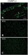
We tested the hypothesis that the 5HT(1D)R, the primary antinociceptive target of triptans, is differentially distributed in tissues responsible for migraine pain. The density of 5HT(1D)R was quantified in tissues obtained from adult female rats with Western blot analysis. Receptor location was assessed with immunohistochemistry. The density of 5HT(1D)R was significantly greater in tissues known to produce migraine-like pain (i.e. circle of Willis and dura) than in structures in which triptans have no antinociceptive efficacy (i.e. temporalis muscle). 5HT(1D)R-like immunoreactivity was restricted to neuronal fibres, where it colocalized with calcitonin gene-related peptide and tyrosine hydroxylase immunoreactive fibres. These results are consistent with our hypothesis that the limited therapeutic profile of triptans could reflect its differential peripheral distribution and that the antinociceptive efficacy reflects inhibition of neuropeptide release from sensory afferents. An additional site of action at sympathetic efferents is also suggested.
Figures


Similar articles
-
Peptidergic nociceptors of both trigeminal and dorsal root ganglia express serotonin 1D receptors: implications for the selective antimigraine action of triptans.J Neurosci. 2003 Nov 26;23(34):10988-97. doi: 10.1523/JNEUROSCI.23-34-10988.2003. J Neurosci. 2003. PMID: 14645495 Free PMC article.
-
Distribution of 5-HT(1B), 5-HT(1D) and 5-HT(1F) receptor expression in rat trigeminal and dorsal root ganglia neurons: relevance to the selective anti-migraine effect of triptans.Brain Res. 2010 Nov 18;1361:76-85. doi: 10.1016/j.brainres.2010.09.004. Epub 2010 Sep 9. Brain Res. 2010. PMID: 20833155
-
Colocalization of CGRP with 5-HT1B/1D receptors and substance P in trigeminal ganglion neurons in rats.Eur J Neurosci. 2001 Jun;13(11):2099-104. doi: 10.1046/j.0953-816x.2001.01586.x. Eur J Neurosci. 2001. PMID: 11422450
-
[Triptans in migraine--here and now (15 years after they were implemented in therapy)].Neurol Neurochir Pol. 2005;39(4 Suppl 1):S68-77. Neurol Neurochir Pol. 2005. PMID: 16419574 Review. Polish.
-
Is selective 5-HT1F receptor agonism an entity apart from that of the triptans in antimigraine therapy?Pharmacol Ther. 2018 Jun;186:88-97. doi: 10.1016/j.pharmthera.2018.01.005. Epub 2018 Jan 17. Pharmacol Ther. 2018. PMID: 29352859 Review.
Cited by
-
Ionic mechanisms underlying inflammatory mediator-induced sensitization of dural afferents.J Neurosci. 2010 Jun 9;30(23):7878-88. doi: 10.1523/JNEUROSCI.6053-09.2010. J Neurosci. 2010. PMID: 20534836 Free PMC article.
-
The complex actions of sumatriptan on rat dural afferents.Cephalalgia. 2012 Jul;32(10):738-49. doi: 10.1177/0333102412451356. Epub 2012 Jun 18. Cephalalgia. 2012. PMID: 22711897 Free PMC article.
-
Serotonin, 5HT1 agonists, and migraine: new data, but old questions still not answered.Curr Opin Support Palliat Care. 2014 Jun;8(2):137-42. doi: 10.1097/SPC.0000000000000044. Curr Opin Support Palliat Care. 2014. PMID: 24670810 Free PMC article. Review.
-
Orofacial pain.Pain. 2011 Mar;152(3 Suppl):S25-S32. doi: 10.1016/j.pain.2010.12.024. Epub 2011 Feb 2. Pain. 2011. PMID: 21292394 Free PMC article. Review. No abstract available.
-
From Mechanism to Cure: Renewing the Goal to Eliminate the Disease of Pain.Pain Med. 2018 Aug 1;19(8):1525-1549. doi: 10.1093/pm/pnx108. Pain Med. 2018. PMID: 29077871 Free PMC article. Review.
References
-
- Waeber C, Moskowitz MA. Therapeutic implications of central and peripheral neurologic mechanisms in migraine. Neurology. 2003;61:S9–20. - PubMed
-
- Ottani A, Ferraris E, Giuliani D, Mioni C, Bertolini A, Sternieri E, Ferrari A. Effect of sumatriptan in different models of pain in rats. Eur J Pharmacol. 2004;497:181–6. - PubMed
-
- Cumberbatch MJ, Hill RG, Hargreaves RJ. Differential effects of the 5HT1B/1D receptor agonist naratriptan on trigeminal versus spinal nociceptive responses. Cephalalgia. 1998;18:659–63. - PubMed
-
- Dao TT, Lund JP, Remillard G, Lavigne GJ. Is myofascial pain of the temporal muscles relieved by oral sumatriptan? A cross-over pilot study. Pain. 1995;62:241–4. - PubMed
Publication types
MeSH terms
Substances
Grants and funding
LinkOut - more resources
Full Text Sources
Other Literature Sources
Medical

