Review
. 2008 Sep;1781(9):448-58.
doi: 10.1016/j.bbalip.2008.05.005. Epub 2008 Jun 17.

Sphingosine-1-phosphate lyase in development and disease: sphingolipid metabolism takes flight

Affiliations
Review

Sphingosine-1-phosphate lyase in development and disease: sphingolipid metabolism takes flight

Henrik Fyrst et al. Biochim Biophys Acta. 2008 Sep.

Abstract

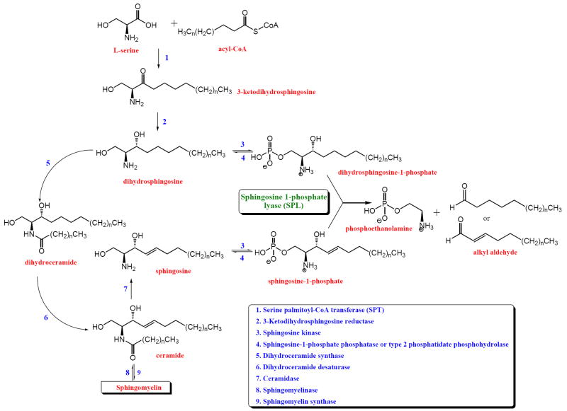
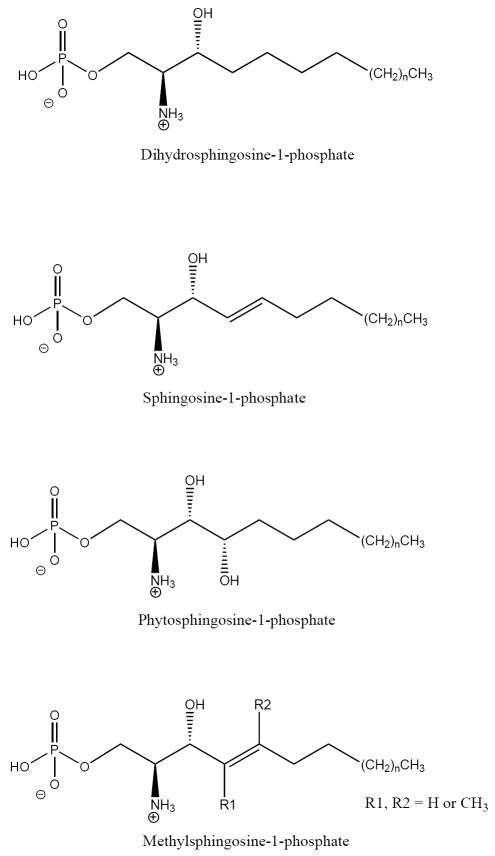
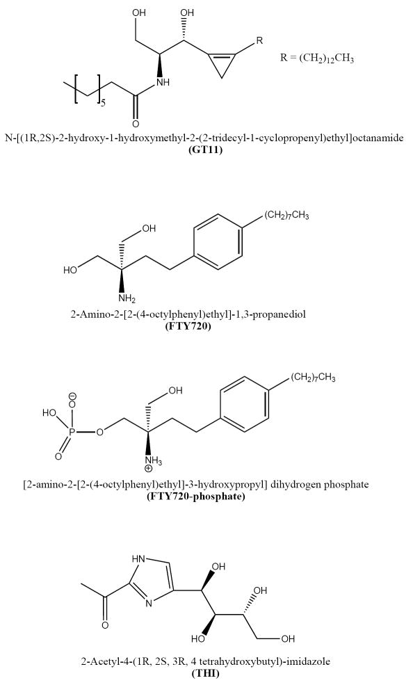
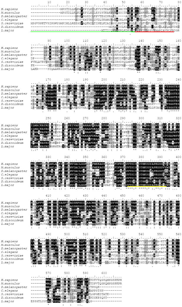
Sphingosine-1-phosphate lyase (SPL) is a highly conserved enzyme that catalyses the final step of sphingolipid degradation, namely the irreversible cleavage of the carbon chain at positions 2-3 of a long-chain base phosphate (LCBP), thereby yielding a long-chain aldehyde and phosphoethanolamine. LCBPs are potent signaling molecules involved in cell proliferation, survival, migration, cell-cell interactions and cell stress responses. Therefore, tight regulation of LCBP signaling is required for proper cell function, and perturbations of this system can lead to alterations in biological processes including development, reproduction and physiology. SPL is a key enzyme in regulating the intracellular and circulating levels of LCBPs and is, therefore, gaining attention as a putative target for pharmacological intervention. This review provides an overview of our current understanding of SPL structure and function, mechanisms involved in SPL regulation and the role of SPL in development and disease.


Figures

Figure 1
Figure 1
Sphingolipid metabolism. LCBs formed from either sphingolipid de novo synthesis (step 1, 2) or from sphingolipid degradation (step 9, 7) can be phosphorylated by sphingosine kinases (step 3). The formed LCBPs can then be dephosphorylated by sphingosine-1-phosphate phosphatase or type 2 phosphatidate phosphohydrolase activity (step 4). Alternatively, LCBPs can be irreversible cleaved by SPL, thereby yielding an alkyl aldehyde and phosphoethanolamine.
Figure 2
Figure 2
LCBPs metabolized by SPL. Dihydrosphingosine-1-phosphate, sphingosine-1-phosphate and phytosphingosine-1-phosphate are the major LCBPs found in mammals and yeast and the naturally occurring D-erythro isomer of these compounds is metabolized by SPL. SPL also metabolizes certain forms of LCBPs containing methyl groups at C4 or C5. SPL shows broad specificity towards the chain length of the LCBP and medium and long chain compounds are used as substrates.
Figure 3
Figure 3
SPL inhibitors. Several sphingolipid analogs inhibit enzyme activity and lately compounds such as the ceramide analog GT11, the LCB analog FTY720 and the LCBP analog FTY720-phosphate has been shown to inhibit SPL. Moreover, pyridoxal-phosphate analogs including THI, has also been shown to inhibit SPL.
Figure 4
Figure 4
Sequence alignment of SPL. ClustalW alignment of Homo sapiens, Mus musculus, Drosophila melanogaster, Caenorhabditis elegans, Saccharomyces cerevisiae, Dictyostelium discoideum and Leishmania major SPL. The underlined regions indicate the ER luminal domain (green), the transmembrane spanning domain (red) and the predicted pyridoxal-phosphate binding domain (yellow). Black shading indicates identical residues. Gray shading indicates biochemically similar residues.
Figure 5
Figure 5
SPL expression in murine thymic epithelium and intestinal mucosa. β-galactosidase staining was performed as described on glutaraldehyde-fixed, frozen sections of SPL reporter mouse tissues [108]. SPL expression can be appreciated in the cytoplasm of the epithelial cells but not lymphocytes of the thymus (a) and in the differentiated enterocytes in the villus tips of the jejunum (b).
Figure 5
Figure 5
SPL expression in murine thymic epithelium and intestinal mucosa. β-galactosidase staining was performed as described on glutaraldehyde-fixed, frozen sections of SPL reporter mouse tissues [108]. SPL expression can be appreciated in the cytoplasm of the epithelial cells but not lymphocytes of the thymus (a) and in the differentiated enterocytes in the villus tips of the jejunum (b).
Figure 6
Figure 6
Dorsal longitudinal flight muscle (DLM) abnormalities in the Drosophila SPL null mutant Sply. Whereas the hemithoraces of the Canton-S wild-type fly invariably contains six DLM fibers of equal proportion, Sply homozygotes exhibit a general pattern of missing fibers, asymmetric development and compensatory hypertrophy of remaining fibers. Shown here is a Sply homozygote with only four DLM fibers in the right hemithorax. The upper DLM fiber in each row is marked with an arrow.
Figure 7
Figure 7
SPL downregulation in intestinal tumorigenesis. Immunohistochemistry with a murine SPL-specific antibody used as described previously shows robust SPL expression in normal differentiated epithelial cells of the villus of the small intestine (brown staining) in the ApcMin/+ mouse model of colon cancer, whereas undifferentiated adenomatous areas in the submucosa do not express SPL [60]. (Left) hematoxylin & eosin; (Right) immunohistochemistry.

References

    1. Hannun YA, Luberto C, Argraves KM. Enzymes of sphingolipid metabolism: from modular to integrative signaling. Biochemistry. 2001;40:4893–4903. - PubMed
    1. Gómez-Muñoz A, Kong J, Parhar K, Wang S, Gangoiti P, González M, Eivemark S, Salh B, Duronio V, Steinbrecher U. Ceramide-1-phosphate promotes cell survival through activation of the phosphatidylinositol 3-kinase/protein kinase B pathway. FEBS Lett. 2005;579:3744–3750. - PubMed
    1. Wijesinghe D, Lamour N, Gomez-Munoz A, Chalfant C. Ceramide kinase and ceramide-1-phosphate. Methods Enzymol. 2007;434:265–292. - PubMed
    1. Gardell S, Dubin A, Chun J. Emerging medicinal roles for lysophospholipid signaling. Trends Mol Med. 2006;12:65–75. - PubMed
    1. Karliner J. Mechanisms of cardioprotection by lysophospholipids. J Cell Biochem. 2004;92:1095–1103. - PubMed

Publication types

LinkOut - more resources