Review
. 2008 Jul 14;620(1-2):8-26.
doi: 10.1016/j.aca.2008.05.022. Epub 2008 May 18.

Sensitive optical biosensors for unlabeled targets: a review

Affiliations
Review

Sensitive optical biosensors for unlabeled targets: a review

Xudong Fan et al. Anal Chim Acta. .

Abstract

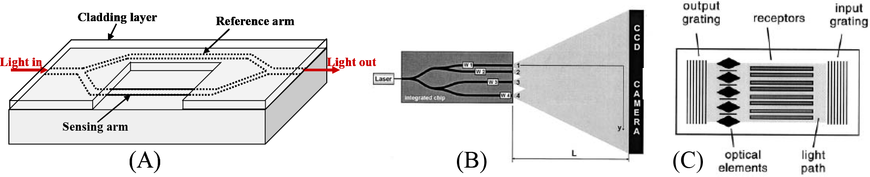
This article reviews the recent progress in optical biosensors that use the label-free detection protocol, in which biomolecules are unlabeled or unmodified, and are detected in their natural forms. In particular, it will focus on the optical biosensors that utilize the refractive index change as the sensing transduction signal. Various optical label-free biosensing platforms will be introduced, including, but not limited to, surface plasmon resonance, interferometers, waveguides, fiber gratings, ring resonators, and photonic crystals. Emphasis will be given to the description of optical structures and their respective sensing mechanisms. Examples of detecting various types of biomolecules will be presented. Wherever possible, the sensing performance of each optical structure will be evaluated and compared in terms of sensitivity and detection limit.


Figures

Figure 1.
Figure 1.
Conceptual illustration of an optical label-free biosensor.
Figure 2.
Figure 2.
Various SPR sensor configurations. (A) Prism coupling. (B) Waveguide coupling. (C) Optical fiber coupling. (D) Side polished fiber coupling. (E) Grating coupling. (F) Long-range and short-range surface plasmon (LRSP and SRSP). Reprinted with permission from [15, 20, 24, 44].
Figure 3.
Figure 3.
(A) Mach-Zehnder interferometer sensor. (B) Multichannel Young’s interferometer sensor. (C) Hartman interferometer sensor. Reprinted with permission from [90, 95].
Figure 4.
Figure 4.
Comparison of various waveguide structures.
Figure 5.
Figure 5.
Various ring resonator biosensors. (A) Silicon-on-insulator ring resonator. (B) Polymer ring resonator. (C) Microtoroid. (D) Glass ring resonator array. (E) Microsphere. (F) Capillary based opto-fluidic ring resonator (OFRR). Reprinted with permission from [134, 161, 165, 185, 186].
Figure 6.
Figure 6.
(A) D-shaped fiber with surface etched gratings. (B) FBG on an etched fiber. (C) Nanofiber loop. (D) Fiber-optic coupler biosensor. (E) Fiber Fabry-Perot cavity DNA sensor showing hollow segment. (F) Fiber Fabry-Perot cavity formed by two sets of FBGs. Reprinted with permission from [205, 207, 220, 227, 229].
Figure 7.
Figure 7.
Photonic crystal biosensor configurations. (A) Photonic crystal microcavity based biosensor. (B) Photonic crystal waveguide based biosensor. (C) Photonic crystal fiber based biosensor. (D) 1D photonic crystal resonators array for parallel detection. Reprinted with permission from [219, 238, 239, 246].

References

    1. Narayanaswamy R, Wolfbeis OS, Optical Sensors, Springer, New York, 2004.
    1. Moerner WE, Proc. Nat. Acad. Sci, 104 (2007) 12596. - PMC - PubMed
    1. Cox WG, L SV, Biotechniques 36 (2004) 114. - PubMed
    1. Jackson JD, Classical Electrodynamics, John Wiley & Sons, Hoboken, NJ, 1999.
    1. Prasad PN, Introduction to Biophotonics, John Wiley & Sons, Inc., Hoboken, New Jersey, 2003.

Publication types

LinkOut - more resources