

Increased expression and internalization of the endotoxin coreceptor CD14 in enterocytes occur as an early event in the development of experimental necrotizing enterocolitis
- PMID: 18558203
- PMCID: PMC2603609
- DOI: 10.1016/j.jpedsurg.2008.02.050
Increased expression and internalization of the endotoxin coreceptor CD14 in enterocytes occur as an early event in the development of experimental necrotizing enterocolitis
Abstract
Background: The early signaling events in the development of necrotizing enterocolitis (NEC) remain undefined. We have recently shown that the endotoxin (lipopolysaccharide [LPS]) receptor toll-like receptor 4 (TLR4) on enterocytes is critical in the pathogenesis of experimental NEC. Given that the membrane receptor CD14 is known to facilitate the activation of TLR4, we now hypothesize that endotoxemia induces an early upregulation of CD14 in enterocytes and that this participates in the early intestinal inflammatory response in the development of NEC.
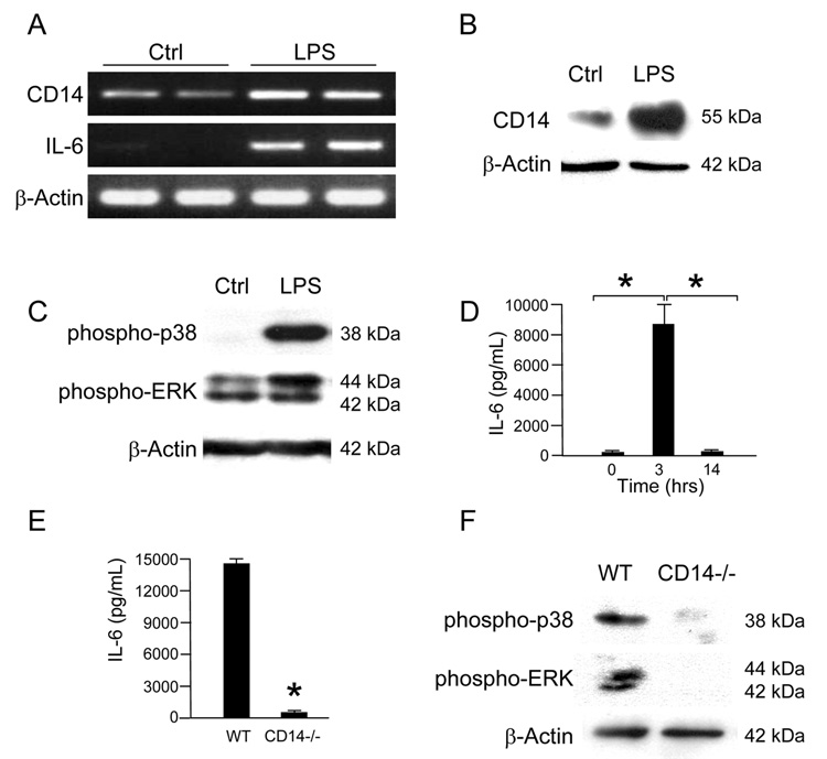
Methods: IEC-6 enterocytes were treated with LPS (50 microg/mL), and the subcellular localization of CD14 and TLR4 was assessed by confocal microscopy. C57/Bl6 or CD14-/- mice were treated with LPS (5 mg/kg), whereas experimental NEC was induced using a combination of gavage formula feeding and intermittent hypoxia. CD14 expression was determined by sodium dodecyl sulfate-polyacrylamide gel electrophoresis and reverse transcriptase-polymerase chain reaction, and interleukin 6 was quantified by enzyme-linked immunosorbent assay and reverse transcriptase-polymerase chain reaction.
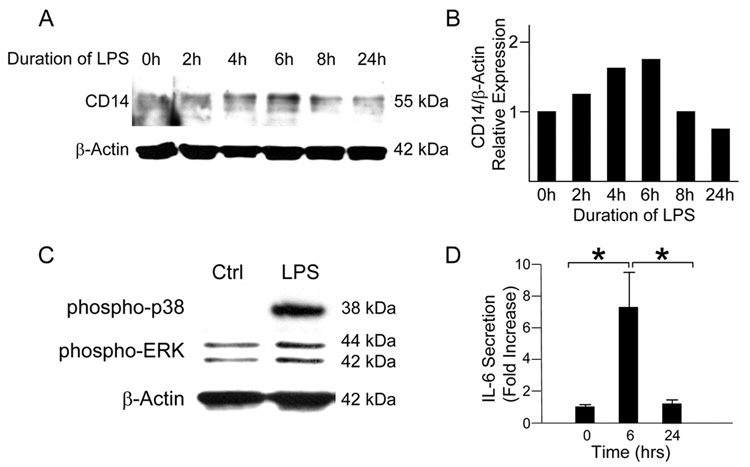
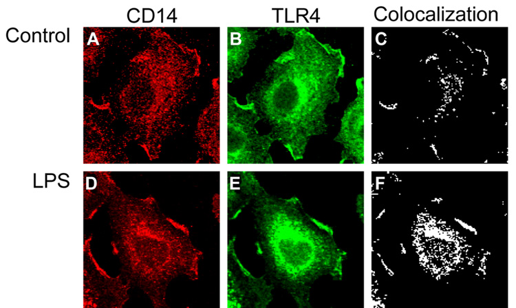
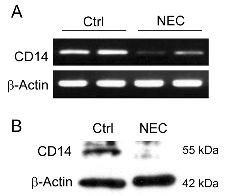
Results: Exposure of IEC-6 enterocytes to LPS led to an initial, transient increase in CD14 expression. The early increase in CD14 expression was associated with internalization of CD14 to a perinuclear compartment where increased colocalization with TLR4 was noted. The in vivo significance of these findings is suggested as treatment of mice with LPS led to an early increase in CD14 expression in the intestinal mucosa, whereas the persistent endotoxemia of experimental NEC was associated with decreased CD14 expression within enterocytes.
Conclusions: LPS signaling in the enterocyte is marked by an early, transient increase in expression of CD14 and redistribution of the receptor. This process may contribute to the early activation of the intestinal inflammatory response that is observed in the development of NEC.
Figures
Similar articles
-
Toll-like receptor-4 inhibits enterocyte proliferation via impaired beta-catenin signaling in necrotizing enterocolitis.Gastroenterology. 2010 Jan;138(1):185-96. doi: 10.1053/j.gastro.2009.09.045. Epub 2009 Sep 26. Gastroenterology. 2010. PMID: 19786028 Free PMC article.
-
Nucleotide-binding oligomerization domain-2 inhibits toll-like receptor-4 signaling in the intestinal epithelium.Gastroenterology. 2010 Sep;139(3):904-17, 917.e1-6. doi: 10.1053/j.gastro.2010.05.038. Epub 2010 May 24. Gastroenterology. 2010. PMID: 20580721 Free PMC article.
-
Increased expression and function of integrins in enterocytes by endotoxin impairs epithelial restitution.Gastroenterology. 2005 Apr;128(4):1012-22. doi: 10.1053/j.gastro.2005.01.052. Gastroenterology. 2005. PMID: 15825082
-
Innate immune signaling in the pathogenesis of necrotizing enterocolitis.Clin Dev Immunol. 2013;2013:475415. doi: 10.1155/2013/475415. Epub 2013 May 23. Clin Dev Immunol. 2013. PMID: 23762089 Free PMC article. Review.
-
Toll-Like Receptor-Mediated Intestinal Inflammatory Imbalance in the Pathogenesis of Necrotizing Enterocolitis.Cell Mol Gastroenterol Hepatol. 2018 Apr 6;6(2):229-238.e1. doi: 10.1016/j.jcmgh.2018.04.001. eCollection 2018. Cell Mol Gastroenterol Hepatol. 2018. PMID: 30105286 Free PMC article. Review.
Cited by
-
In vitro analysis of antigen induced T cell-monocyte conjugates by imaging flow cytometry.J Immunol Methods. 2018 Sep;460:93-100. doi: 10.1016/j.jim.2018.06.014. Epub 2018 Jul 4. J Immunol Methods. 2018. PMID: 29981305 Free PMC article.
-
Animal models of necrotizing enterocolitis: review of the literature and state of the art.Innov Surg Sci. 2018 Mar 10;3(2):87-92. doi: 10.1515/iss-2017-0050. eCollection 2018 Jun. Innov Surg Sci. 2018. PMID: 31579771 Free PMC article. Review.
-
Toll-like receptor regulation of intestinal development and inflammation in the pathogenesis of necrotizing enterocolitis.Pathophysiology. 2014 Feb;21(1):81-93. doi: 10.1016/j.pathophys.2013.11.007. Epub 2013 Dec 22. Pathophysiology. 2014. PMID: 24365655 Free PMC article.
-
Role of innate host defenses in susceptibility to early-onset neonatal sepsis.Clin Perinatol. 2010 Jun;37(2):307-37. doi: 10.1016/j.clp.2010.04.001. Clin Perinatol. 2010. PMID: 20569810 Free PMC article. Review.
-
TLRs, SNPs and VLBWs: oh my!J Perinatol. 2013 Oct;33(10):745-7. doi: 10.1038/jp.2013.94. J Perinatol. 2013. PMID: 24071961 Free PMC article. No abstract available.
References
-
- Lambert DK, Christensen RD, Henry E, et al. Necrotizing enterocolitis in term neonates: data from a multihospital health-care system. J Perinatol. 2007 - PubMed
-
- Lin PW, Stoll BJ. Necrotising enterocolitis. Lancet. 2006;368:1271–1283. - PubMed
-
- Warner BW, Warner BB. Role of epidermal growth factor in the pathogenesis of neonatal necrotizing enterocolitis. Semin Pediatr Surg. 2005;14:175–180. - PubMed
-
- Anand R, Leaphart CL, Mollen K, et al. The Role of the Intestinal Barrier in the Pathogenesis of Necrotizing Enterocolitis. Shock. 2007;27:124–133. - PubMed
Publication types
MeSH terms
Substances
Grants and funding
LinkOut - more resources
Full Text Sources
Research Materials
