. 2008 Sep 1;17(17):2738-51.
doi: 10.1093/hmg/ddn175. Epub 2008 Jun 16.

Accumulation of N-terminal mutant huntingtin in mouse and monkey models implicated as a pathogenic mechanism in Huntington's disease

Affiliations

Accumulation of N-terminal mutant huntingtin in mouse and monkey models implicated as a pathogenic mechanism in Huntington's disease

Chuan-En Wang et al. Hum Mol Genet. .

Abstract

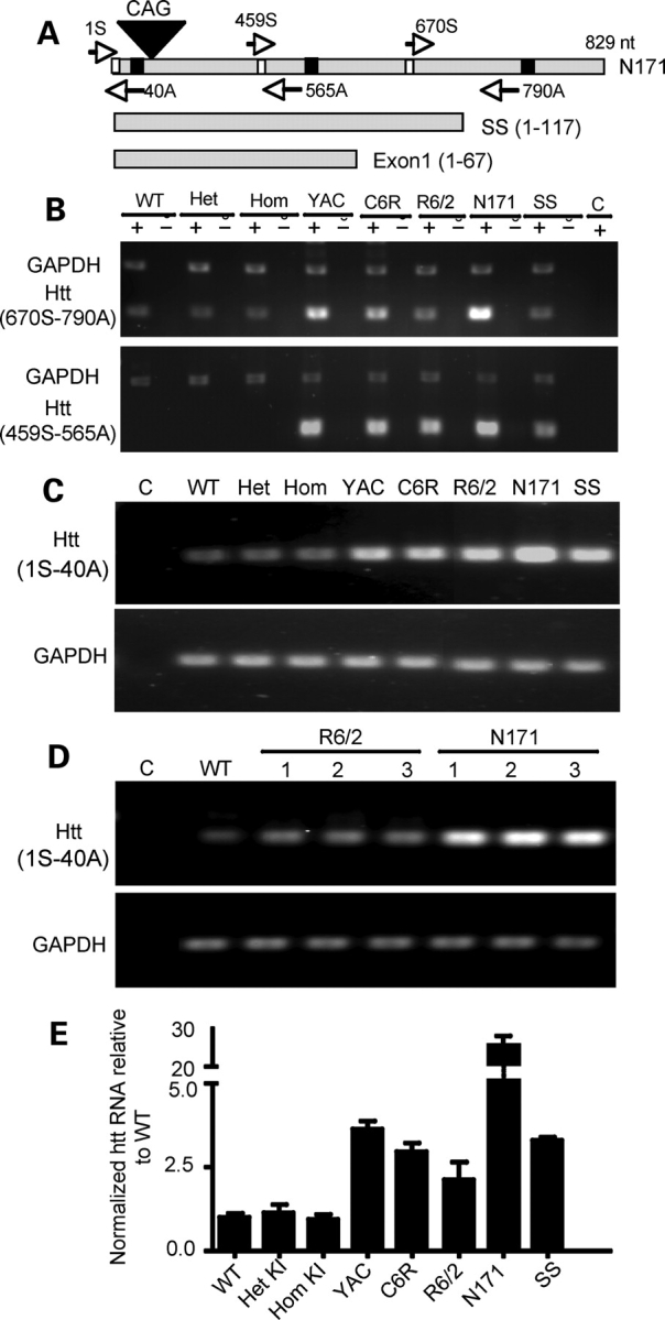
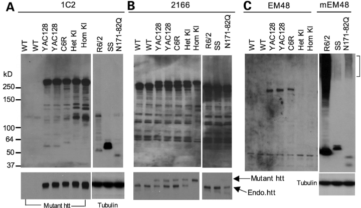
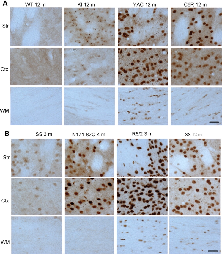
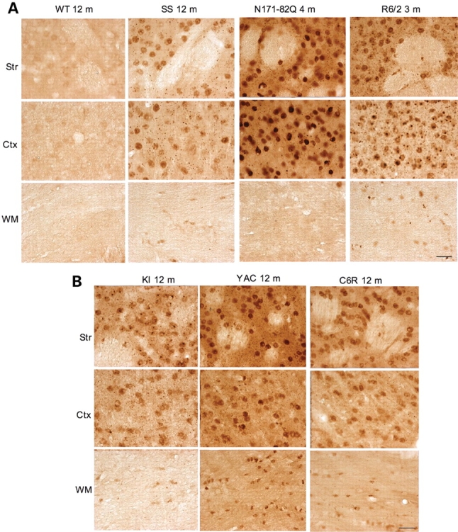
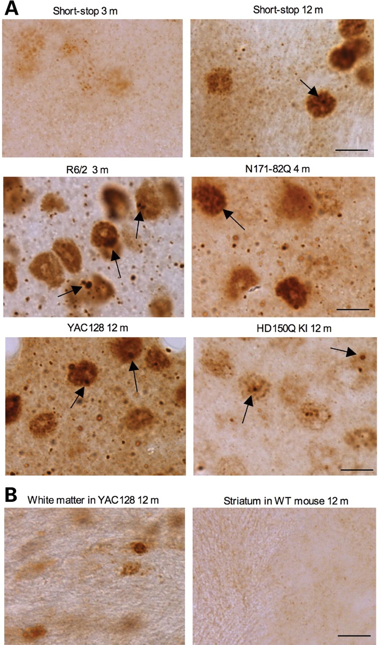
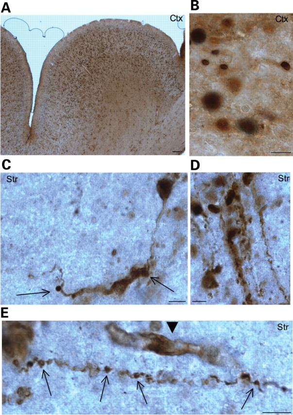
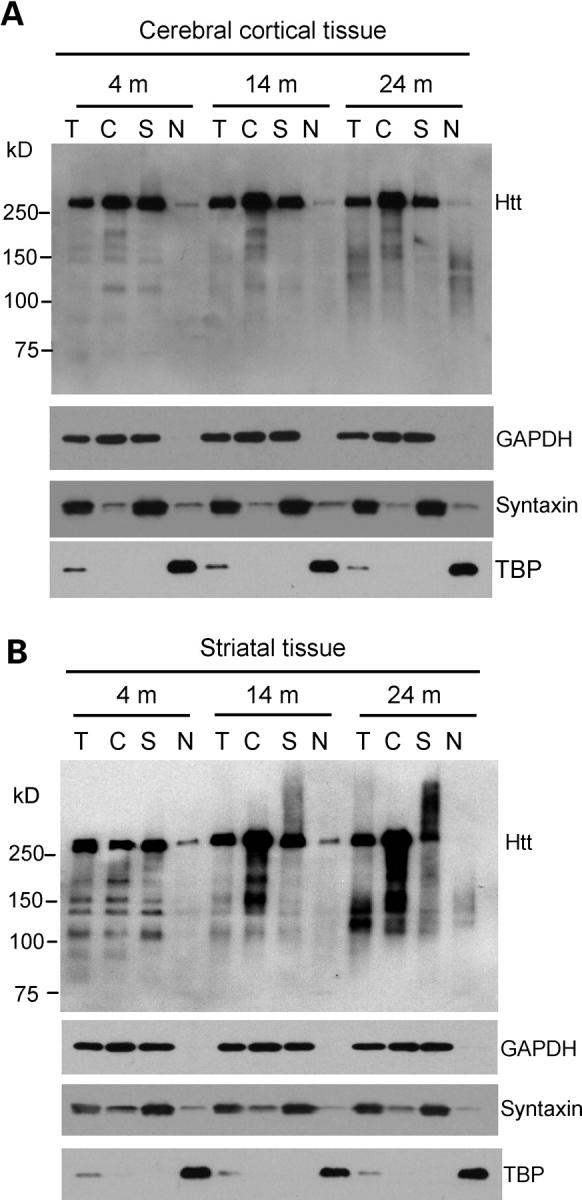
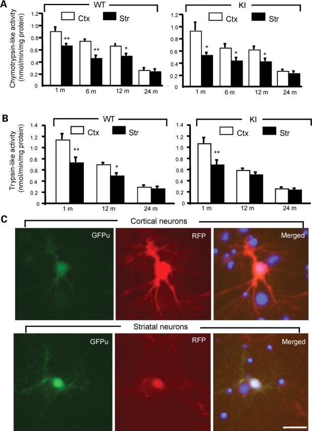
A number of mouse models expressing mutant huntingtin (htt) with an expanded polyglutamine (polyQ) domain are useful for studying the pathogenesis of Huntington's disease (HD) and identifying appropriate therapies. However, these models exhibit neurological phenotypes that differ in their severity and nature. Understanding how transgenic htt leads to variable neuropathology in animal models would shed light on the pathogenesis of HD and help us to choose HD models for investigation. By comparing the expression of mutant htt at the transcriptional and protein levels in transgenic mice expressing N-terminal or full-length mutant htt, we found that the accumulation and aggregation of mutant htt in the brain is determined by htt context. HD mouse models demonstrating more severe phenotypes show earlier accumulation of N-terminal mutant htt fragments, which leads to the formation of htt aggregates that are primarily present in neuronal nuclei and processes, as well as glial cells. Similarly, transgenic monkeys expressing exon-1 htt with a 147-glutamine repeat (147Q) died early and showed abundant neuropil aggregates in swelling neuronal processes. Fractionation of HD150Q knock-in mice brains revealed an age-dependent accumulation of N-terminal mutant htt fragments in the nucleus and synaptosomes, and this accumulation was most pronounced in the striatum due to decreased proteasomal activity. Our findings suggest that the neuropathological phenotypes of HD stem largely from the accumulation of N-terminal mutant htt fragments and that this accumulation is determined by htt context and cell-type-dependent clearance of mutant htt.


Figures

Figure 1.
Figure 1.
Transcript expression of mutant htt in various HD mouse models. (A) Primers used for RT–PCR. The forward primer 670S and the reverse primer 790A are capable of amplifying both mouse and human htt cDNAs, whereas the forward primer 459S and the reverse primer 565A are specific for human htt. Note that 790A cannot be used to amplify transgenic htt in SS and exon-1 htt. (B) RT–PCR results obtained with 670S and 790A primers for mouse and human htt (upper panel) or the primers (459S and 565A) specific to human htt (lower panel). RT–PCR was performed with (+) or without (−) reverse transcriptase. The same PCR reactions also included primers to amplify GAPDH. Control (C) is PCR without a reverse transcriptase. (C) Gel result of qRT–PCR using primers (1S and 40A). PCR of GAPDH was also performed to verify the quantity of cDNA used in each reaction (lower panel). (D) Repeating PCR of the brain tissues from different mice confirmed that N171-82Q transcripts are higher than transgenic htt transcripts in R6/2 mice (lower panel). (E) qRT–PCR data showing the relative levels (mean ± SE., n = 3) of mutant htt transcripts in the cerebral cortex tissues of various HD mice. KI, HD150Q knock-in; YAC, YAC128 transgenic mice; C6R, caspase-6-resistant YAC transgenic mice; SS, shortstop. Agarose gel [1.5% for (B) and 2.2% for (C–D)] electrophoresis was performed to reveal PCR products.
Figure 2.
Figure 2.
Western blot analysis of htt expression in the cerebral cortex of various HD mouse models. Whole cell lysates of cerebral cortex tissues from WT and different HD mouse models were analyzed via western blotting with 1C2 (A), 2166 (B), and EM48 (C) antibodies. KI, HD150Q knock-in; YAC, YAC128 transgenic mice; C6R, caspase-6-resistant YAC transgenic mice; SS, shortstop. R6/2 (1-month-old) and other HD mice (3-month-old) were examined. For 1C2 and 2166 immunoblots, short exposed blots (lower panels) are shown to distinguish between endogenous and transgenic htt (arrows). mEM48 was used for immunostaining of N-terminal htt fragments, and the blot was also probed with anti-tubulin.
Figure 3.
Figure 3.
Micrographs of EM48 immunohistochemistry. (A) EM48 staining of 12-month-old WT or HD mice expressing full-length mutant htt. KI, HD150Q knock-in; YAC, YAC128 transgenic mice; C6R, caspase-6-resistant YAC transgenic mice. (B) EM48 staining of transgenic mice expressing N-terminal mutant htt at the age of 3, 4, or 12 months as indicated. SS, shortstop transgenic mice; Ctx, cerebral cingulate cortex; Str, dorsomedial striatum; WM, white matter in the middle portion of the corpus callosum. Scale bars, 20 µm.
Figure 4.
Figure 4.
Micrographs of 1C2 immunohistochemistry. (A) 1C2 staining of WT or transgenic mice expressing N-terminal mutant htt (B) at the age of 3, 4, or 12 months as indicated. (B) 1C2 staining of 12-month-old HD mice expressing full-length mutant htt. KI, HD150Q knock-in; YAC, YAC128 transgenic mice; C6R, caspase-6-resistant YAC transgenic mice; SS, shortstop transgenic mice; Ctx, cerebral cingulate cortex; Str, dorsomedial striatum; WM, white matter in the middle portion of the corpus callosum. Scale bars, 20 µm.
Figure 5.
Figure 5.
High-magnification (×63) micrographs of EM48 immunohistochemistry. (A) Rabbit EM48 staining of the dorsomedial striatum of shortstop mouse (3‐ and 12‐month‐old), R6/2 mouse (3‐month‐old), N171-82Q mouse (4‐month‐old), HD150Q KI mouse (12‐month‐old), YAC128 mouse (12‐month‐old). (B) Rabbit EM48 staining of glial cells in the white matter of YAC128 mouse (12‐month‐old) and the striatum of WT mouse (12‐month‐old). Note that mutant htt forms nuclear inclusions (arrows) and neuropil aggregates that are out of neuronal cell bodies. Mutant htt also accumulates in the nuclei of glial cells. Scale bar, 10 µm.
Figure 6.
Figure 6.
Expression of N-terminal mutant htt in transgenic monkey brains. EM48 immunostaining of the cerebral cortex (A and B) and striatum (CE) of transgenic monkeys [rHD147Q-2 (A, B, C, E) and rHD147Q-3 (D)] that expressed exon-1 htt with 147Q. A low-magnification (×10 objective lens) graph is shown in (A), and high-magnification (×63 objective lens) graphs are shown in (B–E). Note that mutant htt is abundant in the nucleus and also forms aggregates in neuronal processes. Arrows indicate aggregates in the neuronal processes with a disrupted or swelling appearance. Arrowhead indicates a glial-like cell that was also labeled by EM48. Scale bars, 5 µm.
Figure 7.
Figure 7.
Accumulation of N-terminal htt fragments in the brains of HD150Q knock-in mice. (A and B) Western blotting of total cell lysates (T) and cytosolic (C), synaptosomal (S), and nuclear (N) fractions from the cerebral cortex (A) and striatum (B) of HD150Q KI mice at 4, 14 and 24 months of age. The blots were probed with 1C2 antibody for htt and antibodies for the cytoplasmic protein GAPDH, the synaptic protein syntaxin, or the nuclear protein TBP.
Figure 8.
Figure 8.
Decreased proteasomal activity in striatal neurons. (A and B) Biochemical assays of chymotrypsin-like (A) and trypsin-like (B) activity in homogenates from the brain cortex (Ctx) and striatum (Str) of WT and HD150Q KI mice at different ages. The data (mean ± SE) were obtained from 4–8 mice each group. (C) Expression of GFPu/RFP in cultured cortical and striatal neurons (DIV 12) from rat brains. The merged images (right panels) also show the nuclei of cells that did not express transgenic GFPu/RFP. The ratio of GFPu to RFP was described in the text. Scale bar, 10 µm. *P<0.05; **P<0.01 as compared to the cortex sample.

References

    1. Landles C., Bates G.P. Huntingtin and the molecular pathogenesis of Huntington's disease. Fourth in molecular medicine review series. EMBO Rep. 2004;5:958–963. - PMC - PubMed
    1. Gusella J.F., Macdonald M.E. Huntington's disease: seeing the pathogenic process through a genetic lens. Trends Biochem. Sci. 2006;31:533–540. - PubMed
    1. Li S.H., Li X.J. Huntingtin-protein interactions and the pathogenesis of Huntington's disease. Trends Genet. 2004;20:146–154. - PubMed
    1. Vonsattel J.P., Myers R.H., Stevens T.J., Ferrante R.J., Bird E.D., Richardson E.P., Jr Neuropathological classification of Huntington's disease. J. Neuropathol. Exp. Neurol. 1985;44:559–577. - PubMed
    1. Martin J.B., Gusella J.F. Huntington's disease. Pathogenesis and management. N. Engl. J. Med. 1986;315:1267–1276. - PubMed

Publication types

Substances