Review
. 2008:31:317-38.
doi: 10.1146/annurev.neuro.31.060407.125627.

Brain circuits for the internal monitoring of movements

Affiliations
Review

Brain circuits for the internal monitoring of movements

Marc A Sommer et al. Annu Rev Neurosci. 2008.

Abstract

Each movement we make activates our own sensory receptors, thus causing a problem for the brain: the spurious, movement-related sensations must be discriminated from the sensory inputs that really matter, those representing our environment. Here we consider circuits for solving this problem in the primate brain. Such circuits convey a copy of each motor command, known as a corollary discharge (CD), to brain regions that use sensory input. In the visual system, CD signals may help to produce a stable visual percept from the jumpy images resulting from our rapid eye movements. A candidate pathway for providing CD for vision ascends from the superior colliculus to the frontal cortex in the primate brain. This circuit conveys warning signals about impending eye movements that are used for planning subsequent movements and analyzing the visual world. Identifying this circuit has provided a model for studying CD in other primate sensory systems and may lead to a better understanding of motor and mental disorders.


Figures
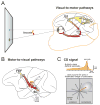
Figure 1
Figure 1
Visual and saccadic circuits in the monkey brain. A. Outline of the major components of the circuit for generating visually guided saccades. Visual signals (yellow) travel from area V1 (far right) through dorsal and ventral processing streams. A major intermediary region is LIP. Visual information from both streams converges in the FEF. Signals are sent downstream (red) to influence the SC and deeper motor areas that trigger a saccade. B. Candidate pathways for conveying saccade-related CD to cerebral cortex. The SC-MD-FEF pathway has been studied in detail. Other routes may exist as well (grey arrows). C. The CD signal in MD thalamic relay neurons that project to FEF. The MD neurons have a tonic increase in firing rate terminated by a burst of activity just before saccade initiation (top). Spatially, the neurons fire only for specific ranges of contralateral saccades (bottom).
Figure 2
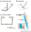
Figure 2
Double step task used to evaluate the influence of CD. A. The pattern of double step saccades (arrows) that would be expected with intact CD vs. total loss of CD. B. Inactivation of the putative CD pathway: the GABA agonist muscimol was injected at the site of MD relay neurons. C. Average accuracy deficit, measured as the horizontal shift in second saccade endpoints. There was a clear deficit for trials with contralateral first saccades (right), but no deficit following ipsilateral first saccades (left). D. Precision deficit in compensating for trial-by-trial variability in first saccades. Compensation is illustrated for second saccade vectors in an example session. The vectors are spread out in the vertical dimension for clarity, but their relative horizontal positions are maintained (as indicated by axis at bottom). Deficit across all sessions (n=22) is summarized with the bar graph (inset). Bars show means and SEs; on average there was a 58% deficit ([97%-39%]/97%).
Figure 3
Figure 3
Shifting visual receptive fields. A. Concept shown schematically. B. Example of an FEF neuron that shows the shift. A visual probe was flashed in the RF in some trials (blue) or in the FF in other trials (magenta). No probe appeared in control trials (black). Curves show mean and SE of average neuronal activity aligned to probe onset (left and center panels) or to saccade initiation (right panel).
Figure 4
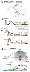
Figure 4
Shifting RFs: spatial. A. Neurons with shifting RFs were recorded in the FEF. B. The prediction was that shifting RFs would jump, as expected from a vector CD input. There should be a significant change in activity (asterisk) at the FF but no significant difference (n.s.d.) at the midpoint. C. (Top) Example FEF neuron shows a jump, not a spread of activity. (Bottom) The same result in the average of the FEF population (n=13 neurons). D. Experiment to see if shifting RFs travel toward the saccadic endpoint (“?” trajectory). The data support, instead, a trajectory parallel to the saccade (average data from n=12 neurons). See text for details.
Figure 5
Figure 5
Shifting RFs: timing. A. Shifting RF activity aligned to probe onset for an example neuron. Start of the shift is much later than the neuron's normal visual latency. Also shown are times of saccade onset in each trial (dots) corresponding to the eye position data (traces at bottom). B. Shifting RF activity aligned to saccade initiation. For this neuron, shift onset time and saccadic onset time were correlated with Pearson R = 0.97; for the population, R = 0.50 (both p < 0.01).
Figure 6
Figure 6
Shifting RFs: timing of CD influence. A. Shift onset times for four example FEF neurons. The orange trace is from the neuron in Fig. 5A, and the red trace is from the neuron in Figure 3B. The blue and green traces show data from two other neurons. Shift magnitudes are normalized to each other to facilitate comparison of timing. B. Distribution of shift onset times for the sample of FEF neurons (n=26). C. Distribution of CD onset times (the start of the saccade-related bursts) in the population of MD relay neurons that project to FEF.
Figure 7
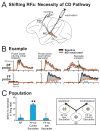
Figure 7
Shifting RFs: necessity of CD pathway. A. Inactivation of the CD pathway while an FEF neuron with a shifting RF is recorded. B. Example of a shifting RF that was impaired by MD inactivation. C. (Left) The deficit in the population of neurons. Reductions in activity were seen only in the FF, not the RF, and only for contralateral, not ipsilateral, saccades. (Right) Task configurations for testing shifting RFs that accompanied contralateral (contra) saccades vs. ipsilateral (ipsi) saccades. **, significant changes at p < 0.0001 level.
Figure 8
Figure 8
The forward model concept. CD (left) is combined with the current state of the system to generate a prediction of the sensory input (upper right). This prediction is compared with the actual, reafferent sensory input (lower right). Resultant discrepancies inform the brain about external influences and miscalibrations. Modified from Wolpert & Miall (1996) with permission from Elsevier.
Figure 9
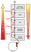
Figure 9
Hypothesized chain of saccadic CD in the primate brain. Descending visuomotor levels (right) extend from the cortical areas that detect a visual stimulus (top) to the brainstem motor neurons that contract the muscles (bottom). Ascending CD signals may branch from any of these levels (left). The pathway that branches from SC up to the FEF conveys a CD signal rich with spatial vector information, because that is the nature of the signal found in the SC and sent downstream from it. Other CD signals, branching from above and below the SC level, would reflect other aspects of movement. It may be more accurate to consider CD as a set of signals representing the state of each saccade-related motor level, in contrast to a unitary CD signal representing the saccade itself.

Similar articles

Cited by

References

    1. Andersen RA, Buneo CA. Intentional maps in posterior parietal cortex. Annu Rev Neurosci. 2002;25:189–220. - PubMed
    1. Andreasen NC, Nopoulos P, O'Leary DS, Miller DD, Wassink T, Flaum M. Defining the phenotype of schizophrenia: cognitive dysmetria and its neural mechanisms. Biol Psychiatry. 1999;46:908–20. - PubMed
    1. Batista AP, Buneo CA, Snyder LH, Andersen RA. Reach plans in eye-centered coordinates. Science. 1999;285:257–60. - PubMed
    1. Bell CC. Properties of a modifiable efference copy in an electric fish. J Neurophysiol. 1982;47:1043–56. - PubMed
    1. Bellebaum C, Daum I, Koch B, Schwarz M, Hoffmann KP. The role of the human thalamus in processing corollary discharge. Brain. 2005;128:1139–54. - PubMed

Publication types

LinkOut - more resources