Review
. 2008:31:563-90.
doi: 10.1146/annurev.neuro.31.060407.125631.

Signaling mechanisms linking neuronal activity to gene expression and plasticity of the nervous system

Affiliations
Review

Signaling mechanisms linking neuronal activity to gene expression and plasticity of the nervous system

Steven W Flavell et al. Annu Rev Neurosci. 2008.

Abstract

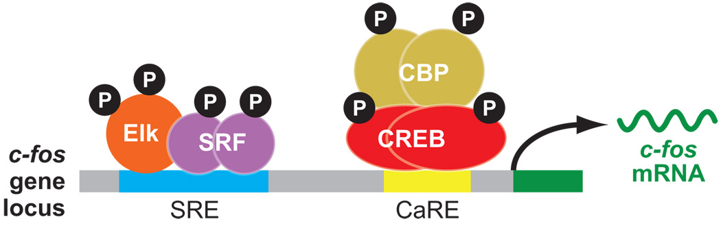
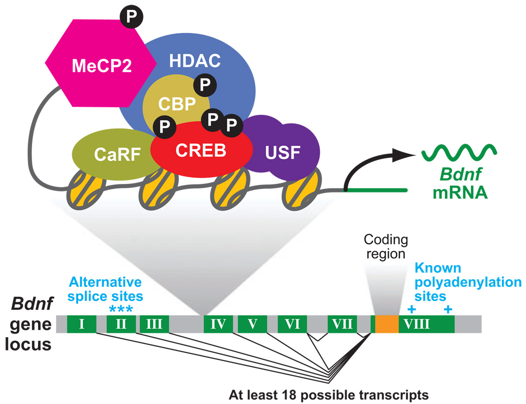
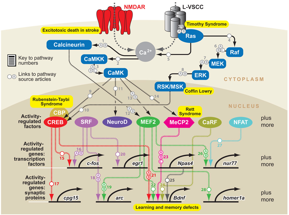
Sensory experience and the resulting synaptic activity within the brain are critical for the proper development of neural circuits. Experience-driven synaptic activity causes membrane depolarization and calcium influx into select neurons within a neural circuit, which in turn trigger a wide variety of cellular changes that alter the synaptic connectivity within the neural circuit. One way in which calcium influx leads to the remodeling of synapses made by neurons is through the activation of new gene transcription. Recent studies have identified many of the signaling pathways that link neuronal activity to transcription, revealing both the transcription factors that mediate this process and the neuronal activity-regulated genes. These studies indicate that neuronal activity regulates a complex program of gene expression involved in many aspects of neuronal development, including dendritic branching, synapse maturation, and synapse elimination. Genetic mutations in several key regulators of activity-dependent transcription give rise to neurological disorders in humans, suggesting that future studies of this gene expression program will likely provide insight into the mechanisms by which the disruption of proper synapse development can give rise to a variety of neurological disorders.


Figures

Figure 1
Figure 1
Regulatory mechanisms that control calcium-dependent c-fos transcription in neurons. At least two separate cis-acting regulatory elements are critical for calcium-dependent c-fos transcription: the CaRE and the SRE. These elements, as well as the protein complexes that are recruited to each of these elements, are shown. The transcribed region (dark green) and the c-fos mRNA produced by the c-fos gene (dark green) are also shown.
Figure 2
Figure 2
The genomic structure and transcriptional regulation of bdnf. The genomic structure of the mouse bdnf gene is shown here. Exons are depicted in dark green and their numbers are indicated by roman numerals; introns are shown in gray. At least six alternative promoters can be clearly detected at this gene (although more may exist—see text for a more detailed explanation). Each of these promoters controls the expression of a unique mRNA that consists of one unique exon (numbered I through VI) that is directly spliced to a common exon (exon VIII), which contains the entire bdnf coding region (orange). The diversity of bdnf transcripts is made even greater because the second exon can be spliced from three alternative splice sites. Moreover, transcripts initiated at exon VI can sometimes include an additional exon (exon VII). Finally, two alternative polyadenylation sites can be utilized within the 3′ UTR. Thus the bdnf gene can produce a large number of bdnf mRNAs that differ only in their 5′ and 3′ UTRs. The regulatory elements that are critical for calcium-dependent transcription from bdnf promoter IV are shown along with the protein complexes known to assemble at this specific promoter.
Figure 3
Figure 3
Signal transduction networks mediating neuronal activity-dependent gene expression. Calcium influx through either neurotransmitter receptors or voltage-gated calcium channels leads to the activation of many calcium-regulated signaling enzymes, which sets in motion several signal transduction cascades. These pathways converge on preexisting transcription factors in the nucleus and lead to their activation through direct posttranslational protein modifications. Several of the activity-regulated genes encode transcriptional regulators, which in turn promote the transcription of additional activity-regulated genes. Many other activity-regulated genes encode proteins that function in dendrites or at synapses and thereby coordinate activity-dependent dendritic and synaptic remodeling within the neuron. Genetic mutations in the genes that encode several of these signaling molecules give rise to neurological disorders in humans ( yellow boxes). Only a subset of the signaling pathways that mediate activity-dependent transcription are shown here.

Similar articles

Cited by

References

    1. Aid T, Kazantseva A, Piirsoo M, Palm K, Timmusk T. Mouse and rat BDNF gene structure and expression revisited. J. Neurosci. Res. 2007;85:525–535. - PMC - PubMed
    1. Aizawa H, Hu SC, Bobb K, Balakrishnan K, Ince G, et al. Dendrite development regulated by CREST, a calcium-regulated transcriptional activator. Science. 2004;303:197–202. - PubMed
    1. Altar CA, Laeng P, Jurata LW, Brockman JA, Lemire A, et al. Electroconvulsive seizures regulate gene expression of distinct neurotrophic signaling pathways. J. Neurosci. 2004;24:2667–2677. - PMC - PubMed
    1. Antonini A, Stryker MP. Development of individual geniculocortical arbors in cat striate cortex and effects of binocular impulse blockade. J. Neurosci. 1993;13:3549–3573. - PMC - PubMed
    1. Bailey CH, Chen M. Morphological basis of long-term habituation and sensitization in Aplysia. Science. 1983;220:91–93. - PubMed

Publication types

MeSH terms

Substances

LinkOut - more resources