. 2008 Jun;15(6):586-96.
doi: 10.1016/j.chembiol.2008.04.012.

Shape shifting leads to small-molecule allosteric drug discovery

Affiliations

Shape shifting leads to small-molecule allosteric drug discovery

Sarah H Lawrence et al. Chem Biol. 2008 Jun.

Abstract

Enzymes that regulate their activity by modulating an equilibrium of alternate, nonadditive, functionally distinct oligomeric assemblies (morpheeins) constitute a recently described mode of allostery. The oligomeric equilibrium for porphobilinogen synthase (PBGS) consists of high-activity octamers, low-activity hexamers, and two dimer conformations. A phylogenetically diverse allosteric site specific to hexamers is proposed as an inhibitor binding site. Inhibitor binding is predicted to draw the oligomeric equilibrium toward the low-activity hexamer. In silico docking enriched a selection from a small-molecule library for compounds predicted to bind to this allosteric site. In vitro testing of selected compounds identified one compound whose inhibition mechanism is species-specific conversion of PBGS octamers to hexamers. We propose that this strategy for inhibitor discovery can be applied to other proteins that use the morpheein model for allosteric regulation.


Figures

Figure 1
Figure 1. Proteins that can exist as an equilibrium of alternate quaternary structure assemblies (morpheeins) provide a structural foundation for allosteric regulation of protein function
(a) The morpheein model for allosteric regulation includes oligomer dissociation, conformational change in the dissociated form, and may involve reassociation to an alternate oligomeric assembly. In the illustrated case, two alternate quaternary structure assemblies are shown (a trimer and a tetramer) and the common rule of assembly of the fundamental units is that a thick solid line must associate with a dashed line. The conformation of the dissociated form, shown as a blue or pink fundamental unit, dictates the geometry and stoichiometry of the respectively colored oligomers. The functions of the two oligomers are distinct (e.g. low activity vs. high activity), analogous to the R and T states of the traditional non-dissociating models for allosteric regulation. The fundamental unit can be monomeric or of higher order. In the case of porphobilinogen synthase, the fundamental unit is an asymmetric homo-dimer and the analogous transition is between a hexamer and an octamer. (b) In the morpheein model for allosteric regulation, a regulator molecule (depicted as a yellow wedge) binds to the structural elements on one side of this equilibrium. The yellow wedge has the appropriate geometry to bind only to the blue forms and draw the equilibrium in that direction, thus acting as an allosteric activator or inhibitor. The binding site for the yellow wedge is required to be specific for one oligomeric assembly and not the other. However, the binding site is not required to be interfacial (between subunits), as is the case for the small molecule binding site in the PBGS hexamer.
Figure 2
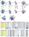
Figure 2. Porphobilinogen synthase (PBGS) is the prototype morpheein ensemble
(a) The equilibrium of pea PBGS quaternary structure forms is shown. For most species, the asymmetric unit of the crystal structure is an asymmetric homo-dimer. The hexamer and its asymmetric unit, the detached dimer (PDB code: 1PV8) are shown in shades of blue. The octamer and its asymmetric unit, the hugging dimer (PDB code: 1GZG) are shown in shades of pink. For the octamer, the dimers assemble at a 90° rotation around a central axis; for the hexamer, the dimers assemble at a 120° rotation around a central axis. The octamer contains a phylogenetically variable binding site for an allosteric magnesium ion which binds to the arm-to-barrel interface that is unique to the octamer; the allosteric magnesium binding site is not present in the hexamer (Breinig et al., 2003; Jaffe, 2003). The hexamer contains a surface cavity not present in the octamer and is the predicted small molecule binding site. The small molecule inhibitor (depicted as yellow balls) draws the equilibrium toward the hexamer. (b) Left - The small molecule binding site in the pea PBGS hexamer model contains components from three subunits, shown as ribbons. The GLIDE docking box is superimposed at the subunit interface. The box into which docked ligands must fit is in magenta, the box in which the center of the docked ligands must fit is in green. Right – The three subunits forming the docking site are shown as ribbons in the context of the hexamer with the remaining subunits shown as surfaces, subunit labels correspond to subunit colors. (c) A multiple sequence alignment of the regions of PBGS contained within the hexamer-specific inhibitor docking site of pea PBGS is shown with highly conserved residues shaded in grey. Organisms highlighted in peach, yellow, and green are representative metazoa, microbes, and plants, respectively. Sequence conservation was determined from an alignment of 33 PBGS sequences (not shown); a residue was defined as “highly conserved” if it was present in at least 32 sequences. Numbers correspond to the pea PBGS sequence. Residues highlighted in blue of the P. sativum PBGS are within 4 Å of docked morphlock-1; these residues are highlighted in light blue where they are conserved in other sequences. Residues marked with “A”, “B”, or “E” indicate a sidechain interaction between subunits A, B, or E of pea PBGS and docked morphlock-1. A closeup of the docked structure of morphlock-1 is shown in Fig. 3a.
Figure 3
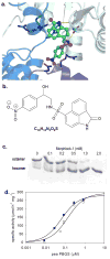
Figure 3. The oligomer-trapping inhibitor, morphlock-1, stabilizes the hexameric assembly of pea PBGS
(a) Morphlock-1 is illustrated as posed by GLIDE. Subunits A, B, and E are shown as ribbons using colors corresponding to Fig. 2b. Morphlock-1 is shown with carbon atoms in green (other atoms colored CPK) and illustrated as ball and stick. Side chains within 4.0 Å of morphlock-1 are shown as sticks, carbons are colored by chain (other atoms colored CPK). Hydrogen bonds to morphlock-1 are shown in yellow. (b) The chemical structure of morphlock-1. (c) Pea PBGS (1 mg/mL) resolves on native PAGE into its octameric and hexameric components. Pea PBGS was incubated with various concentrations of morphlock-1 (in a DMSO solution). Each concentration of morphlock-1 with a fixed amount of DMSO was incubated with protein for 30 minutes at 37 °C prior to resolution on 12.5% polyacrylamide native PhastGels. Morphlock-1 draws the pea PBGS morpheein equilibrium entirely to the hexamer in a dose dependent fashion. (d) The protein concentration-dependent pea PBGS specific activity is shown. PBGS (at varied concentrations) was incubated in the presence of DMSO (●) or 50 μM morphlock-1 in DMSO (○) for 30 minutes at 37 °C prior to assay. The final concentration of morphlock-1 in the inhibited assay was 5 μM, and the concentration of PBGS in the inhibited and control assays ranged from 0.05 – 50 μg/mL (0.014 – 1.4 μM).
Figure 4
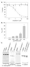
Figure 4. Species-specific effects of morphlock-1 on PBGS
(a)) Dose-response curves showing morphlock-1 inhibition of pea PBGS at 1 μg/ml (○) and human PBGS at 0.6 μg/ml (Δ) and 10 μg/ml (□). Proteins were incubated in the presence of 10X inhibitor for 30 minutes at 37 °C prior to assay. Concentrations on the X-axis represent the final concentration of morphlock-1 in the assay. (b) The IC50 for morphlock-1 is a function of pea PBGS concentration. IC50 values were determined as in (a) using the noted concentrations of pea PBGS. (c) Native PAGE evaluation of the effect of morphlock-1 on the quaternary structure equilibria of PBGS (~1 mg/ml) from D. melanogaster, P. aeruginosa, V. cholerae, and H. sapiens. Note that the charge/mass ratio is not the same for PBGS from different species. The position of the oligomeric equilibrium varies among PBGS from different species; thus, only one form is observed under these conditions for the PBGS from D. melanogaster, P. aeruginosa, V. cholerae. The lanes marked with an “X” contain unrelated compounds.
Figure 5
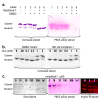
Figure 5. Morphlock-1 and substrate induced interconversion of PBGS quaternary structure assemblies
(a) Matched Coomassie (left) and PBGS activity (right) stained native PAGE (at 1 mg/ml PBGS) illustrates specific binding of morphlock-1 (at 2 mM) to the hexameric assembly. Enzyme-bound morphlock-1 prevents the in-gel transition of inactive hexamer to active octamer (Fig. S2). (b) Hexameric pea PBGS (1 mg/mL) was incubated with DMSO (left) or 500 μM morphlock-1 (right) for 30 minutes at 37 °C. ALA was added at the indicated concentrations, and the incubation was continued for 30 minutes prior to resolution on 12.5 % polyacrylamide native PhastGels. (c) Morphlock-1 induced stabilization of the pea PBGS hexameric assembly. Morphlock-1 concentrations are listed above each lane. Protein samples for the silver and activity stained gels were at 50 μg/ml (1.4 μM subunit) and 1 mM ALA. Samples for the Krypton Infrared stained gel were at 5 μg/ml (0.14 μM subunit) and 10 mM magnesium.

Similar articles

Cited by

References

    1. Baeyens KJ, De Bondt HL, Raeymaekers A, Fiers W, De Ranter CJ. The structure of mouse tumour-necrosis factor at 1.4 A resolution: towards modulation of its selectivity and trimerization. Acta Crystallogr D Biol Crystallogr. 1999;55(Pt 4):772–778. - PubMed
    1. Balch WE, Morimoto RI, Dillin A, Kelly JW. Adapting proteostasis for disease intervention. Science. 2008;319(5865):916–919. - PubMed
    1. Battersby AR. Tetrapyrroles: the pigments of life. Nat Prod Rep. 2000;17(6):507–526. - PubMed
    1. Boese QF, Spano AJ, Li JM, Timko MP. Aminolevulinic acid dehydratase in pea (Pisum sativum L.). Identification of an unusual metal-binding domain in the plant enzyme. J Biol Chem. 1991;266(26):17060–17066. - PubMed
    1. Bollivar DW, Clauson C, Lighthall R, Forbes S, Kokona B, Fairman R, Kundrat L, Jaffe EK. Rhodobacter capsulatus porphobilinogen synthase, a high activity metal ion independent hexamer. BMC Biochemistry. 2004;5:17. - PMC - PubMed

Publication types

LinkOut - more resources