. 2008 Aug 29;283(35):23721-30.
doi: 10.1074/jbc.M708360200. Epub 2008 Jun 17.

Insulin-like growth factor-1 receptor and ErbB kinase inhibitor combinations block proliferation and induce apoptosis through cyclin D1 reduction and Bax activation

Affiliations

Insulin-like growth factor-1 receptor and ErbB kinase inhibitor combinations block proliferation and induce apoptosis through cyclin D1 reduction and Bax activation

Julie L Wilsbacher et al. J Biol Chem. .

Abstract

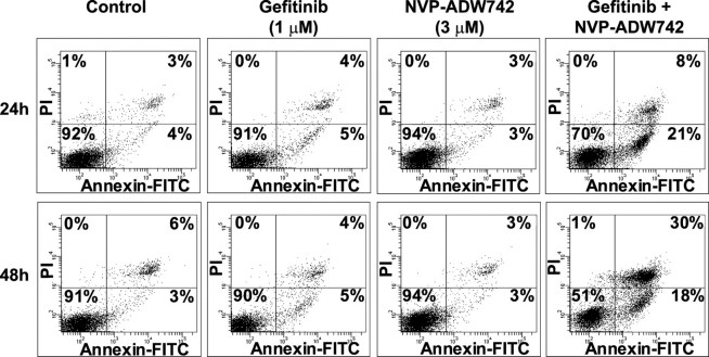
The insulin-like growth factor-1 receptor (IGF-1R) and ErbB family of receptors are receptor tyrosine kinases that play important roles in cancer. Lack of response and resistance to therapies targeting ErbB receptors occur and are often associated with activation of the IGF-1R pathway. Combinations of agents that inhibit IGF-1R and ErbB receptors have been shown to synergistically block cancer cell proliferation and xenograft tumor growth. To determine the mechanism by which targeting both IGF-1R and ErbB receptors causes synergistic effects on cell growth and survival, we investigated the effects of combinations of selective IGF-1R and ErbB kinase inhibitors on proliferative and apoptotic signaling. We identified A431 squamous cell carcinoma cells as most sensitive to combinations of ErbB and IGF-1R inhibitors. The inhibitor combinations resulted in not only blockade of A431 cell proliferation, but also induced apoptosis, which was not seen with either agent alone. Upon examining phosphorylation states and expression levels of proteins in the IGF-1R and ErbB signaling pathways, we found a correlation between the ability of combinations to inhibit proliferation and to decrease levels of phosphorylated Akt and cyclin D1. In addition, the massive cell death induced by combined IGF-1R/ErbB inhibition was associated with Mcl-1 reduction and Bax activation. Thus, targeting both IGF-1R and ErbB receptors simultaneously results in cell cycle arrest and apoptosis through combined effects on Akt, cyclin D1, and Bax activation.


Figures

FIGURE 1.
FIGURE 1.
Combinations of gefitinib and NVP-ADW742 synergistically inhibit proliferation and survival of and increase caspase-3 activation in A431 cells. Top, decrease in cell number after 72 h of treatment; middle, inhibition of BrdUrd incorporation after 24 h of treatment; bottom, activation of caspase-3 after 24 h of treatment of A431 cells with 1 or 3 μm gefitinib and 3 μm NVP-ADW742 alone or in combination. Caspase-3 activation data are expressed as -fold level of caspase-3 activity in the DMSO-treated samples. *, combinations of 1 μm gefitinib and 3 μm NVP-ADW742 are statistically different from single agents with p ≤ 0.002. **, combinations of 3μm gefitinib and 3μm NVP-ADW742 are statistically different from single agentswithp≤0.001.***, combinations of 1 μm gefitinib and 3μm NVP-ADW742 are statistically different from single agents with p < 0.01. Data are the average ± S.D. of duplicate samples and are representative of two or three independent experiments.
FIGURE 2.
FIGURE 2.
Combinations of gefitinib and NVP-ADW742 induce apoptosis of A431 cells. Shown is flow cytometry analysis of annexin V and PI staining of A431 cells treated with 1 μm gefitinib and 3 μm NVP-ADW742 alone or in combination. Cells were treated with compounds for 24 or 48 h before they were stained with annexin V-fluorescein isothiocyanate and PI. The percentages of cells in each quadrant are indicated. Data are representative of two independent experiments.
FIGURE 3.
FIGURE 3.
Effects of combinations of gefitinib and NVP-ADW742 on signaling and cell cycle proteins in A431 cells. A, levels of phosphorylated and total EGFR, IGF-1R, Akt, and ERK1/2 after a 6-h treatment of A431 cells with 1 or 3μm gefitinib and 3μm NVP-ADW742 alone or in combination followed by stimulation with 50 ng/ml IGF-1 for 10 min. Data in blots are representative of two independent experiments. B, quantification of effects of compounds on pEGFR, pIGF-1R, pAkt, and pERK from experiments performed as in A. The average ± S.E. for each analyte from densitometry measurements in the two different experiments are shown. Data for phosphorylation of EGFR, IGF-1R, Akt, and ERK were normalized to total protein for each analyte and expressed as -fold signal in the DMSO plus IGF-1 control. pEGFR levels in cells treated with the indicated compounds were statistically different from DMSO-treated cells with p < 0.02 (*). Levels of pIGF-1R in cells treated with 3 μm NVP-ADW742 alone or in combination with gefitinib were statistically different from DMSO- or gefitinib-treated cells with p < 0.001 (+). pAkt levels in cells treated with 1 μm gefitinib, 3 μm NVP-ADW742, or the combinations were statistically different from DMSO-treated cells with p < 0.005 (#), and pAkt levels in cells treated with the combinations were statistically different from levels in cells treated with the single agents with p ≤ 0.01 (▵). Levels of pERK in cells treated with gefitinib alone or in combination with NVP-ADW742 were statistically different from levels in cells treated with DMSO or NVP-ADW742 alone with p ≤ 0.002 (^). C, levels of phosphorylated and total EGFR, IGF-1R, Akt, ERK1/2, and Rb and levels of cyclin D1, p27, p16, and actin following overnight treatment of A431 cells with combinations of 1 or 3 μm gefitinib and 3 μm NVP-ADW742. Data are representative of four or five independent experiments. D, quantification of effects of compound combinations on phosphorylated Akt and Rb and total cyclin D1, p27, and p16 from experiments performed as in C. Data were normalized using phosphorylated protein/total protein for Akt and Rb and as total protein/actin for cyclin D1, p27, and p16. Data are expressed as the -fold DMSO control and are graphed as the average ± S.E. from the independent experiments. Levels of pAkt in cells treated with 1 μm gefitinib, 3 μm gefitinib, or 3 μm NVP-ADW742 were statistically different from DMSO-treated cells with p < 0.0001 (*), and pAkt levels in cells treated with combinations were statistically different from levels in cells treated with the single agents or DMSO with p ≤ 0.02 (**). Cyclin D1 levels in cells treated with both gefitinib concentrations were statistically different from DMSO-treated cells with p < 0.05 (+), and cyclin D1 levels in cells treated with the compound combinations were statistically different from levels in other treatment groups with p < 0.003 (++). Levels of p27 in cells treated with the compound combinations were statistically different from levels in other treatment groups with p < 0.05 (#). Levels of p16 in gefitinib- or combination-treated cells were statistically different from levels in DMSO or NVP-ADW742-treated cells with p < 0.05 (^). E, levels of phosphorylated and total EGFR, IGF-1R, Akt, ERK1/2, and Rb and levels of cyclin D1, p27, p16, and actin following 6- or 16-h treatment of A431 cells with combinations of 1 or 3 μm gefitinib and 3 μm NVP-ADW742. Data are representative of two independent experiments.
FIGURE 4.
FIGURE 4.
Expression of constitutively active Akt blocks the effects of gefitinib and NVP-ADW742 combinations on cell proliferation and apoptosis. A, decrease in cell number after a 72-h treatment and activation of caspase-3 after a 24-h treatment of A431-pBABE control (left) or A431-myr-Akt (right) cells with 1 or 3 μm gefitinib and 3μm NVP-ADW742 alone or in combination. Caspase-3 activation data are expressed as -fold caspase-3 activity in the DMSO-treated samples. Data are the average ± S.D. of triplicate samples and are representative of two independent experiments. B, flow cytometry analysis of annexin V and PI staining of A431-pBABE control (top) or A431-myr-Akt (bottom) cells treated with 1 μm gefitinib and 3 μm NVP-ADW742 alone or in combination. Cells were treated with compounds for 48 h before they were stained with annexin V-fluorescein isothiocyanate and PI. The percentages of cells in each quadrant are indicated. Data are representative of two independent experiments. C, levels of phosphorylated and total EGFR, IGF-1R, Akt, ERK1/2, and Rb and levels of cyclin D1, p27, p16, and actin following overnight treatment of A431-pBABE control or A431-myr-Akt cells with combinations of 1 or 3 μm gefitinib and 3 μm NVP-ADW742. Data are representative of two independent experiments.
FIGURE 5.
FIGURE 5.
Effects of combinations of gefitinib and NVP-ADW742 on apoptotic proteins in A431 cells. A, levels of active and total Bax, total and cleaved caspase-3, and actin following overnight treatment of A431 cells with combinations of 1 or 3 μm gefitinib and 3 μm NVP-ADW742. Data are representative of three independent experiments. B, levels of Bcl-2, Bcl-XL, Mcl-1, and actin following overnight treatment of A431 cells with combinations of 1 or 3 μm gefitinib and 3 μm NVP-ADW742. Data are representative of three independent experiments. The average ± S.E. for Mcl-1 levels normalized to actin from densitometry measurements in the three experiments are shown in the graph below the blots. Mcl-1 levels in the combination-treated cells were statistically different from levels in DMSO- or NVP-ADW742-treated cells with p ≤ 0.003 (*), and Mcl-1 levels in the 3 μm gefitinib plus 3 μm NVP-ADW742-treated cells were statistically different from levels in cells treated with either gefitinib concentration with p ≤ 0.02 (**). C, levels of BimEL, BimL, BimS, and Mcl-1 in Mcl-1 immunoprecipitates (IP) or in whole cell lysate samples (WCL) from A431 cells treated with gefitinib and NVP-ADW742, alone or in combination. A431 cells were treated with compounds as in A. Data are representative of two independent experiments. The average ± S.E. for Bim levels normalized to Mcl-1 in the Mcl-1 immunoprecipitates are shown in the graph. Bim levels in immunoprecipitates from the 1 μm gefitinib- and the combination-treated cells were statistically different from levels in DMSO- or NVP-ADW742-treated cells with p ≤ 0.03 (*).
FIGURE 6.
FIGURE 6.
Model illustrating how effects of combinations of IGF-1R and ErbB kinase inhibitors on signaling proteins could result in G1 arrest and apoptosis. A, IGF-1R and ErbB signaling results in proliferation and survival. Activation of IGF-1R and ErbB receptors by respective ligands or through receptor cross-talk leads to phosphorylation and activation of Akt and ERK. Activation of Akt and ERK results in increased cyclin D1 expression. Cyclin D1 sequesters p27, preventing it from inhibiting cyclin E/Cdk2 activity and allowing cell cycle progression and proliferation. Active ERK phosphorylates Bim and targets it for degradation. Active Akt causes stabilization of Mcl-1, so Mcl-1 is available to inhibit Bax activation and prevent apoptosis. B, inhibition of IGF-1R and ErbB receptor activity with small molecules causes cell cycle arrest and apoptosis. When cells are treated with the IGF-1R inhibitor, NVP-ADW742, and the EGFR inhibitor, gefitinib, activation and autophosphorylation of the two receptors are blocked. Akt and ERK are no longer phosphorylated and activated. As a result, levels of cyclin D1 are decreased and levels of p27 protein are increased. The increased p27 is available to bind to and inhibit cyclin E/Cdk2, leading to cell cycle arrest. In addition, when ERK is not activated, Bim remains stable and available to bind to Mcl-1. Mcl-1 expression is also lower because Akt is not active. Thus, the levels of Mcl-1 available to inhibit Bax are decreased, resulting in an increase in Bax activation and apoptosis.

Similar articles

Cited by

References

    1. Hofmann, F., and Garcia-Echeverria, C. (2005) Drug Discov. Today 10 1041–1047 - PubMed
    1. Yakar, S., Leroith, D., and Brodt, P. (2005) Cytokine Growth Factor Rev. 16 407–420 - PubMed
    1. Baserga, R. (2005) Expert Opin. Ther. Targets 9 753–768 - PubMed
    1. Hubbard, R. D., and Wilsbacher, J. L. (2007) Chem. Med. Chem. 2 41–46 - PubMed
    1. Bohula, E. A., Playford, M. P., and Macaulay, V. M. (2003) Anticancer Drugs 14 669–682 - PubMed

MeSH terms