. 2008 Aug;36(13):e79.
doi: 10.1093/nar/gkn377. Epub 2008 Jun 17.

Primase-based whole genome amplification

Affiliations

Primase-based whole genome amplification

Ying Li et al. Nucleic Acids Res. 2008 Aug.

Abstract

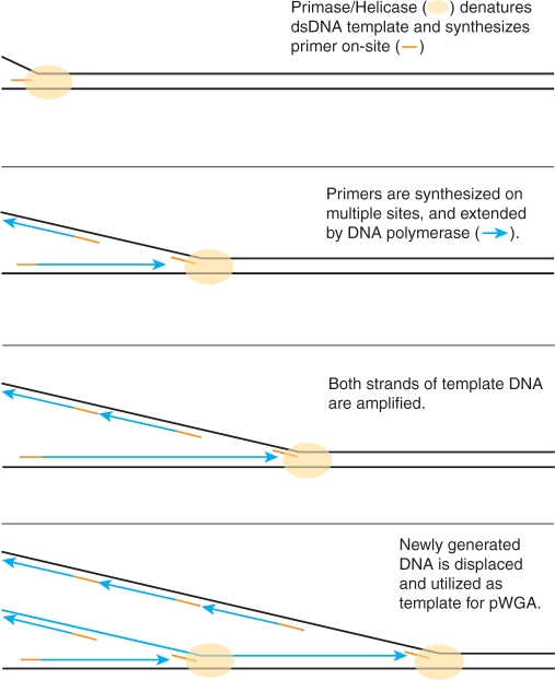
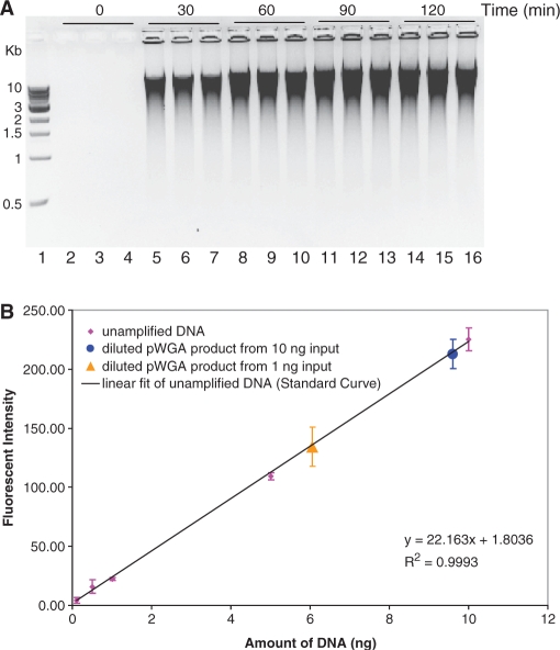
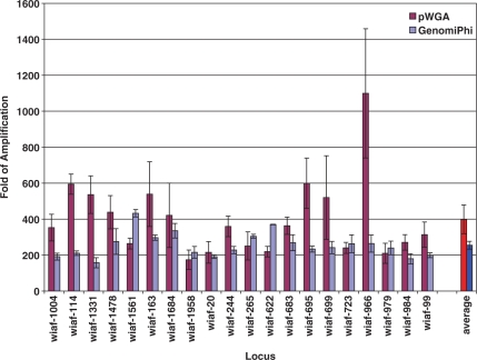
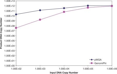
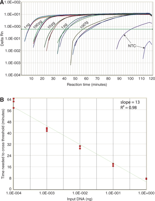
In vitro DNA amplification methods, such as polymerase chain reaction (PCR), rely on synthetic oligonucleotide primers for initiation of the reaction. In vivo, primers are synthesized on-template by DNA primase. The bacteriophage T7 gene 4 protein (gp4) has both primase and helicase activities. In this study, we report the development of a primase-based Whole Genome Amplification (pWGA) method, which utilizes gp4 primase to synthesize primers, eliminating the requirement of adding synthetic primers. Typical yield of pWGA from 1 ng to 10 ng of human genomic DNA input is in the microgram range, reaching over a thousand-fold amplification after 1 h of incubation at 37 degrees C. The amplification bias on human genomic DNA is 6.3-fold among 20 loci on different chromosomes. In addition to amplifying total genomic DNA, pWGA can also be used for detection and quantification of contaminant DNA in a sample when combined with a fluorescent reporter dye. When circular DNA is used as template in pWGA, 10(8)-fold of amplification is observed from as low as 100 copies of input. The high efficiency of pWGA in amplifying circular DNA makes it a potential tool in diagnosis and genotyping of circular human DNA viruses such as human papillomavirus (HPV).


Figures

Figure 1.
Figure 1.
Mechanism of pWGA reaction.
Figure 2.
Figure 2.
pWGA reaction time, product length, and product yield. (A) pWGA reactions containing 10 ng human genomic DNA were terminated after a specified incubation time [0 (lanes 2–4), 30 (lanes 5–7), 60 (lanes 8–10), 90 (lanes 11–13) or 120 (lanes 14–16) minutes], as indicated at the top of the gel, and 5 µl of each 25 µl reaction was analyzed on a 0.6% agarose gel. The reactions were performed in triplicate, and lane 1 shows 500 ng of 1-kb DNA ladder (NEB) with the sizes of bands indicated on the left of the gel. The lane numbers are indicated at the bottom of the gel. (B) Quantification of pWGA product yield by PicoGreen® assay. Fluorescent intensity of 0.1, 0.5, 1, 5 and 10 ng of un-amplified DNA (magenta diamonds) is plotted against the amount of DNA and is fitted to the linear equation shown at the bottom right of the plot (black line) as a standard curve for quantification. Triplets of pWGA product from 1 ng (orange triangle) and 10 ng (blue circle) were diluted 20 times. One microliter of the diluted products were assayed for PicoGreen® fluorescence and mapped on the standard curve according to their intensity. The ± SD over three experiments are shown as error bars on each data point.
Figure 3.
Figure 3.
Representative sequencing results using the pWGA amplification product as template. The template for sequencing was the pWGA amplification product from plasmid pET28a(+). The primer used for sequencing was the T7 terminator primer (S1271, NEB) that covers residues 66–84 on the plasmid and it is in the reverse direction of the plasmid. The sequencing experiment was performed by Dye-Terminator Sequencing method using AmpliTaq® DNA polymerase FS on an ABI 3130xl Genetic analyzer.
Figure 4.
Figure 4.
Fold of pWGA and GenomiPhi amplification over 20 SNP loci. The average fold of amplification of 10 ng input human genomic DNA for each locus from three pWGA (magenta) or GenomiPhi (light blue) experiments is plotted with standard deviation against the primer name used to amplify that locus. The average fold of amplification over the 20 loci among three pWGA (red) or GenomiPh (blue) reactions is also plotted.
Figure 5.
Figure 5.
Amplification of circular DNA. Various amounts of pCMV_GLuc plasmid were used as substrates for side-by-side pWGA (blue diamonds) and MDA reactions (magenta squares). The copy numbers of amplified plasmids DNA were determined by comparing with standard curves generated from templates of known copy numbers through real-time PCR using primers Luc-3F and Luc-3R.
Figure 6.
Figure 6.
Real-time quantitative pWGA. (A) Real-time amplification signal (Delta Rn) from triplets of 0 (NTC), 100 fg, 1 pg, 10 pg, 100 pg and 1 ng of input DNA is plotted against reaction time. The threshold is drawn as a green line. (B) Ct number from different amount of input template is plotted against the amount of input template with a linear fit shown as a green line. The slope and the goodness of fit (R2-value) are shown at the top right of the plot. The template amount on the x-axis is distributed on a log scale.

References

    1. Zhang L, Cui X, Schmitt K, Hubert R, Navidi W, Arnheim N. Whole genome amplification from a single cell: implications for genetic analysis. Proc. Natl Acad. Sci. USA. 1992;89:5847–5851. - PMC - PubMed
    1. Telenius H, Carter NP, Bebb CE, Nordenskjold M, Ponder BA, Tunnacliffe A. Degenerate oligonucleotide-primed PCR: general amplification of target DNA by a single degenerate primer. Genomics. 1992;13:718–725. - PubMed
    1. Dietmaier W, Hartmann A, Wallinger S, Heinmoller E, Kerner T, Endl E, Jauch KW, Hofstadter F, Ruschoff J. Multiple mutation analyses in single tumor cells with improved whole genome amplification. Am. J. Pathol. 1999;154:83–95. - PMC - PubMed
    1. Kittler R, Stoneking M, Kayser M. A whole genome amplification method to generate long fragments from low quantities of genomic DNA. Anal. Biochem. 2002;300:237–244. - PubMed
    1. Wells D, Delhanty JD. Comprehensive chromosomal analysis of human preimplantation embryos using whole genome amplification and single cell comparative genomic hybridization. Mol. Hum. Reprod. 2000;6:1055–1062. - PubMed

Publication types