. 2008 Sep 15;17(18):2766-75.
doi: 10.1093/hmg/ddn176. Epub 2008 Jun 17.

PTHR1 mutations associated with Ollier disease result in receptor loss of function

Affiliations

PTHR1 mutations associated with Ollier disease result in receptor loss of function

Alain Couvineau et al. Hum Mol Genet. .

Abstract

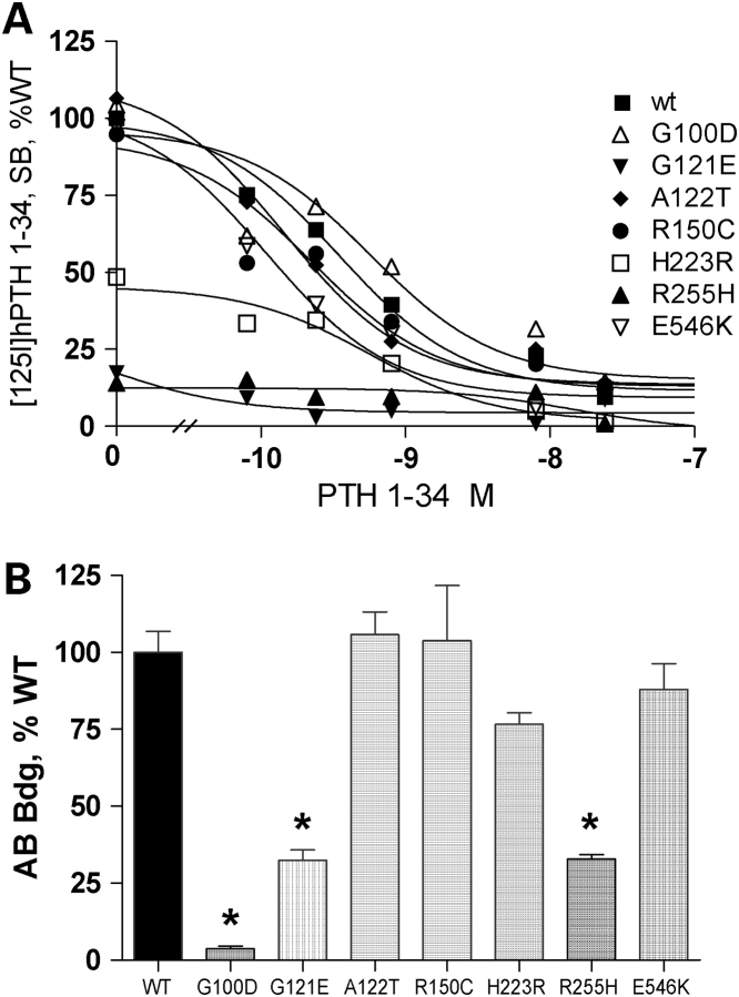
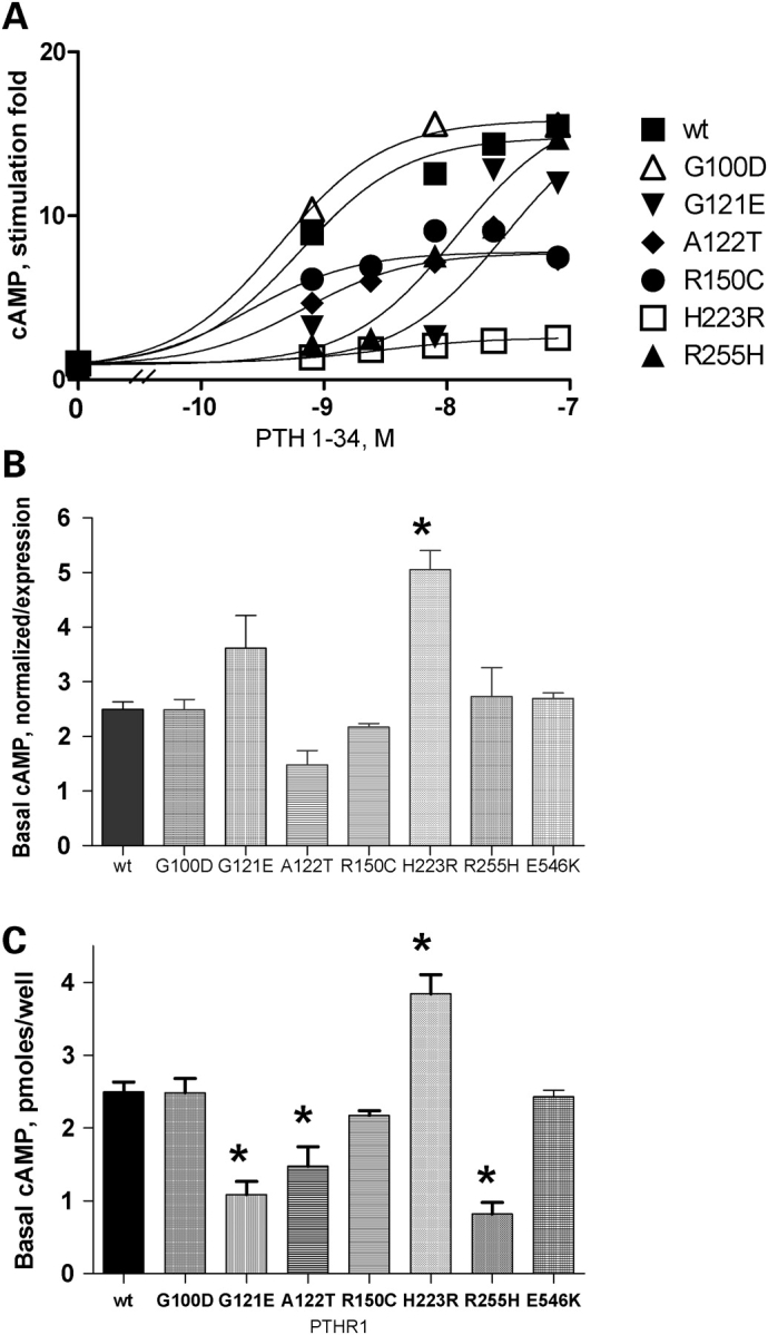
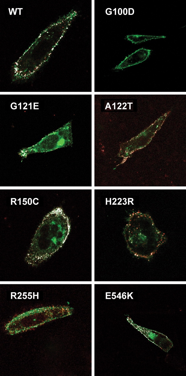
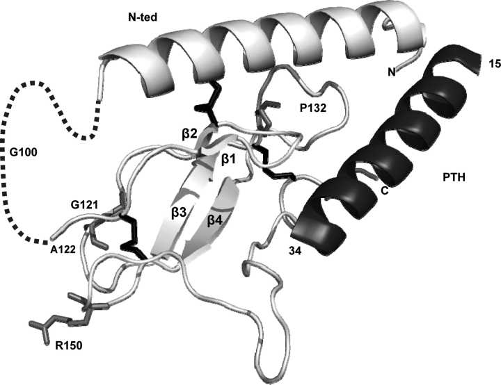
PTHR1-signaling pathway is critical for the regulation of endochondral ossification. Thus, abnormalities in genes belonging to this pathway could potentially participate in the pathogenesis of Ollier disease/Maffucci syndrome, two developmental disorders defined by the presence of multiple enchondromas. In agreement, a functionally deleterious mutation in PTHR1 (p.R150C) was identified in enchondromas from two of six unrelated patients with enchondromatosis. However, neither the p.R150C mutation (26 tumors) nor any other mutation in the PTHR1 gene (11 patients) could be identified in another study. To further define the role of PTHR1-signaling pathway in Ollier disease and Maffucci syndrome, we analyzed the coding sequences of four genes (PTHR1, IHH, PTHrP and GNAS1) in leucocyte and/or tumor DNA from 61 and 23 patients affected with Ollier disease or Maffucci syndrome, respectively. We identified three previously undescribed missense mutations in PTHR1 in patients with Ollier disease at the heterozygous state. Two mutations (p.G121E, p.A122T) were present only in enchondromas, and one (p.R255H) in both enchondroma and leukocyte DNA. Assessment of receptor function demonstrated that these three mutations impair PTHR1 function by reducing either the affinity of the receptor for PTH or the receptor expression at the cell surface. These mutations were not found in DNA from 222 controls. Including our data, PTHR1 functionally deleterious mutations have now been identified in five out 31 enchondromas from Ollier patients. These findings provide further support for the idea that heterozygous mutations in PTHR1 that impair receptor function participate in the pathogenesis of Ollier disease in some patients.


Figures

Figure 1.
Figure 1.
Functional evaluation of WT and mutant PTHR1 expressed in CHO cells. (A) Binding of [125I] PTH 1–34 by cells incubated with radio ligand only (maximal binding) and in the presence of increasing concentrations of unlabeled hPTH 1–34. The maximal binding measured in cells transfected with the WT-PTHR1 was set as 100%. Results were corrected for non-specific binding measured in the presence of 8 × 10⁻⁷ m PTH 1–34. (B) Immunological assessment of cell-surface expression of WT and mutant PTHR1 expressed in CHO cells. Transfected cells were incubated sequentially with polyclonal rabbit anti-human PTHR1 antibody and [125I] -labeled anti-rabbit immunoglobin antibody. Results are expressed as % [125I]-labeled anti-rabbit immunoglobin antibody bound in cells transfected with the WT-PTHR1. p.G100D substitution disrupts the epitope recognition by the PTHR1 antibody (see Results). Results are the mean ± SEM of at least three experiments performed in duplicate with two plasmid preparations. *P < 0.01 compared with WT. Statistical analysis was performed using one-way ANOVA and comparisons between mutant and WT PTHR1 receptors using a Dunnett’s test.
Figure 2.
Figure 2.
(A) cAMP accumulation in response to increasing concentrations of hPTH 1–34. Results are expressed as stimulation fold over basal. CHO cells were transfected with 1 µg plasmid DNA coding for the WT or mutant PTHR1, and functional studies were performed 48 h later as described in Materials and Methods. Results are the mean of at least three experiments performed in duplicate with two plasmid preparations. SB, specific binding. (B) Basal cAMP production in cells transfected with WT or mutant PTHR1. Basal cAMP accumulation is expressed as picomoles cAMP/well normalized to cell-surface expression as determined in Figure 1B. Expression of the mutant p.G100D PTHR1 was considered to be that of WT PTHR1 on the basis of results from radioligand binding and confocal immunofluorescence experiments (see Results). (C) Basal cAMP production in cells transfected with WT or mutant PTHR1 expressed as picomoles cAMP/well. CHO cells were transfected with 1 µg plasmid DNA coding for the WT or mutant PTHR1 and experiments performed 48 h later. Results are the mean ± SEM of at least three experiments performed in duplicate with two plasmid preparations. *P < 0.01 compared with WT. Statistical analysis was performed using one-way ANOVA and comparisons between mutant and WT PTHR1 receptors using a Dunnett’s test.
Figure 3.
Figure 3.
Confocal immunofluorescent microscopy analysis of WT and mutant PTHR1 tagged with GFP and using sequentially polyclonal rabbit anti-human PTHR1 antibody and rhodamine-labeled anti-rabbit immunoglobin antibody, without cellular permeabilization. Co-localization (originally yellow, and falsely colored in white for clarity) of green GFP-tagged PTHR1 and the rhodamine-labeled secondary antibody against the PTHR1 primary antibody (directed against the E2 extracellular epitope) indicates cell-surface expression. No co-localization was observed in cells transfected with the p.G100D PTHR1, indicating that the p.G100D mutation prevents PTHR1 recognition by the PTHR1 antibody (see Results). CHO cells were transfected with 1 µg plasmid DNA coding for the WT or mutant PTHR1, and confocal immunofluorescent microscopy analysis was performed as described in Material and Methods.
Figure 4.
Figure 4.
Representation of the recently resolved crystal structure of the PTH–PTHR1 N-ted complex (PDB ID code 3C4M) (28) and the position of the PTHR1 mutations identified in this and previous studies. The figures show ribbon representation of receptor N-ted. Light gray, PTHR1 N-ted main chain containing β-sheets (β1–β4); dashed line, exon E2 (residues 57–105); black, disulfide bonds between residues Cys48–Cys117, Cys108–Cys148 and Cys131–Cys170); dark gray, PTH. The mutated amino acids are indicated in medium gray. All mutated amino acids are located within the structured core.

References

    1. Fletcher C.D.M., Unni K., Mertens F., editors. World Health Organization Classification of Tumors. Pathology and Genetics. Tumors of Soft Tissue and Bone. Lyon: IARCPress; 2002.
    1. Maroteaux P., Le Merrer M. Les maladies osseuses de l’enfant. 4ème ed. Paris: Médecine-Sciences, Flammarion; 2002.
    1. Unni K.K. Cartilaginous lesions of bone. J. Orthop. Sci. 2001;6:457–472. - PubMed
    1. Whyte M. Acquired Disorders of Cartilage and Bone. 5th edn. Washington, DC: American Society for Bone and Mineral Research; 2003.
    1. Silve C., Juppner H. Ollier disease. Orphanet J. Rare Dis. 2006;1:37. - PMC - PubMed

Publication types

MeSH terms