

Theory of mind and schizophrenia: a positron emission tomography study of medication-free patients
- PMID: 18559406
- PMCID: PMC2632446
- DOI: 10.1093/schbul/sbn034
Theory of mind and schizophrenia: a positron emission tomography study of medication-free patients
Erratum in
- Schizophr Bull. 2009 Sep;35(5):1030. Calage, Chadi A [corrected to Calarge, Chadi A]
Abstract
Background: "Theory of mind" (TOM) refers to the ability to attribute mental states (ie, beliefs and goals) to one's self and others and to recognize that behaviors are guided by these mental states. This capacity, critical for social competence, is impaired in schizophrenia. We undertook a study of TOM in a group of patients with schizophrenia and healthy controls.
Method: We used positron emission tomography to identify the neural circuits recruited during a verbal task that required participants to attribute mental states to a character in a story of their creation. The comparison task consisted of reading aloud a neutral story, controlling for the speech component of the task.
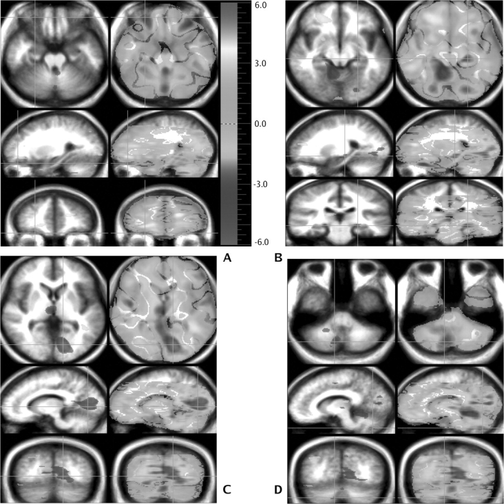
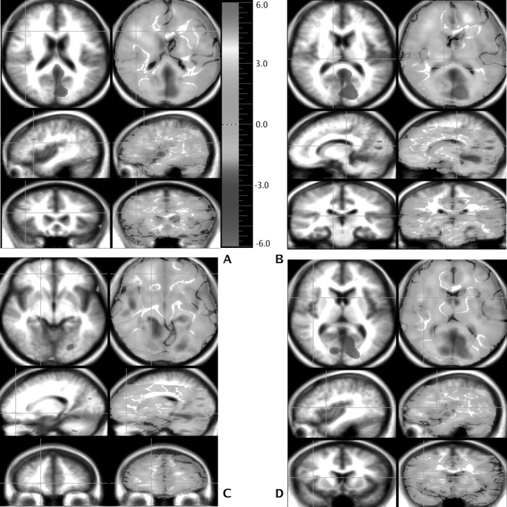
Results: Patients and controls generated the same percentage of TOM utterances. However, the two groups had markedly different patterns of brain activation. Compared with controls, patients had a lower blood flow in multiple regions in the left hemisphere including the frontal and visual association cortices, posterior hippocampus, and insula. The flow was also lower in contralateral areas in the lateral cerebellum and vermis, thalamus, and posterior insula. On the other hand, the flow was higher in the patients predominantly in the right hemisphere, including multiple frontal and parietal regions, insula, visual association cortex, and pulvinar.
Discussion: The areas of lower flow are consistent with previous studies indicating impairment in recruiting cortical-cerebellar circuitry in schizophrenia. The areas of higher flow may reflect a need to draw on the right hemisphere to compensate for deficits in left hemisphere networks that include frontal cortex, anterior cingulate, cerebellum, and thalamus.
Figures
References
-
- Bleuler E. Dementia Preacox or the Group of Schizophrenias. Madison, CT: International Universities Press, Inc; 1950.
-
- Dennett DC. Beliefs about beliefs. Behav Brain Sci. 1978;1(4):568–569.
-
- Frith CD, Frith U. Interacting minds—a biological basis. Science. 1999;286(5445):1692–1695. - PubMed
-
- Premack D, Woodruff G. Does the chimpanzee have a theory of mind? Behav Brain Sci. 1978;1(4):515–526.
-
- Wimmer H, Perner J. Beliefs about beliefs: representation and constraining function of wrong beliefs in young children's understanding of deception. Cognition. 1983;13(103–128) - PubMed
Publication types
MeSH terms
Substances
Grants and funding
LinkOut - more resources
Full Text Sources
Medical
