. 2008 Aug;28(16):5014-26.
doi: 10.1128/MCB.00640-08. Epub 2008 Jun 16.

MPYS, a novel membrane tetraspanner, is associated with major histocompatibility complex class II and mediates transduction of apoptotic signals

Affiliations

MPYS, a novel membrane tetraspanner, is associated with major histocompatibility complex class II and mediates transduction of apoptotic signals

Lei Jin et al. Mol Cell Biol. 2008 Aug.

Abstract

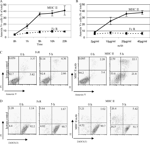
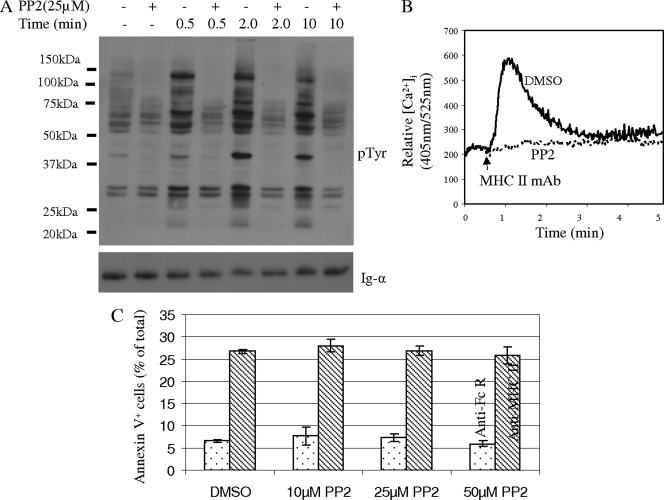
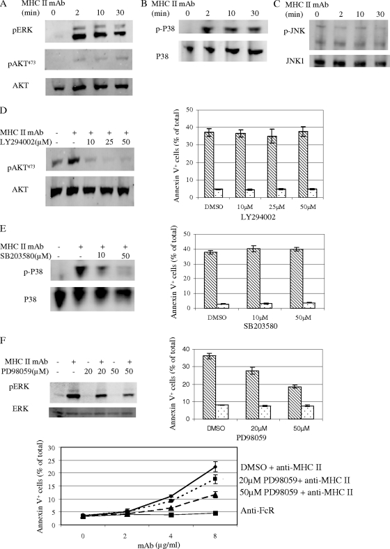
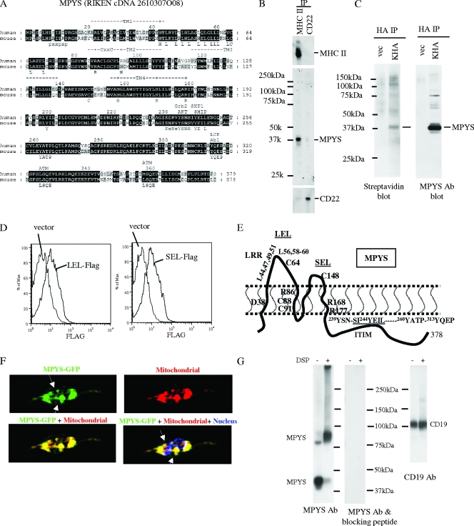
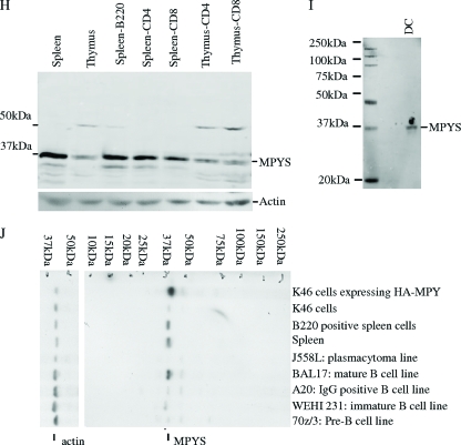
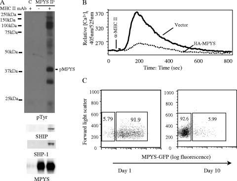
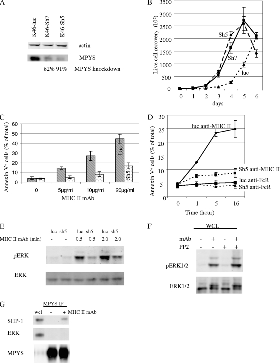
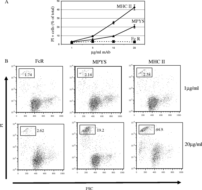
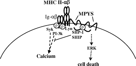
Although the best-defined function of type II major histocompatibility complex (MHC-II) is presentation of antigenic peptides to T lymphocytes, these molecules can also transduce signals leading alternatively to cell activation or apoptotic death. MHC-II is a heterodimer of two transmembrane proteins, each containing a short cytoplasmic tail that is dispensable for transduction of death signals. This suggests the function of an undefined MHC-II-associated transducer in signaling the death response. Here we describe a novel plasma membrane tetraspanner (MPYS) that is associated with MHC-II and mediates its transduction of death signals. MPYS is unusual among tetraspanners in containing an extended C-terminal cytoplasmic tail (approximately 140 amino acids) with multiple embedded signaling motifs. MPYS is tyrosine phosphorylated upon MHC-II aggregation and associates with inositol lipid and tyrosine phosphatases. Finally, MHC class II-mediated cell death signaling requires MPYS-dependent activation of the extracellular signal-regulated kinase signaling pathway.


Figures

FIG. 1.
FIG. 1.
MHC class II MAb induces time and dosage-dependent apoptosis of K46 B lymphoma cells. (A) K46 cells were treated with 15 μg/ml biotinylated anti-MHC class II (M5/114; rat IgG2b) MAb or its isotype control, biotinylated anti-mouse FcγRIIB MAb ((2.4G2; rat IgG2b), followed by 20 μg/ml avidin for the indicated time. Cell death was measured by annexin V/PI dual staining (n > 3). (B) Cells were treated as for panel A at the indicated Ab doses for 5 h. Cell death was measured as for panel A (n > 3). (C and D) Cells were treated with 15 μg/ml biotinylated M5/114 or 2.4G2 plus 20 μg/ml avidin for 5 h. Cell death was measured by annexin V/PI (C) or DiOC6/PI dual staining (D) (n > 3). Numerical annotation reflects the percentage of cells in each quadrant. Error bars represent standard deviations of triplicates.
FIG. 2.
FIG. 2.
MHC-II signaling of cell death does not require activation of Src family tyrosine kinases. (A) K46 cells were treated with 25 μM PP2 or dimethylsulfoxide (DMSO) for 30 min at 37°C and then stimulated with biotin-MHC-II (M5.114; 20 μg/ml) and avidin (20 μg/ml) for the indicated times. SDS-PAGE-fractionated whole-cell lysates were probed with the indicated Abs (n = 3). (B) The intracellular free calcium concentration ([Ca2+]i) was measured before and during K46 stimulation with biotin-MHC-II (M5.114; 10 μg/ml) and avidin (10 μg/ml). Cells were pretreated with DMSO or PP2 as described for panel (A) (n > 3). (C) K46 cells treated as for panel A were stimulated with biotin-anti-MHC-II (M5.114; 10 μg/ml; shaded bars) or biotin-anti-FcγRIIB (2.4G2; 10 μg/ml; open bars) and avidin (10 μg/ml) for 5 h, and death was measured by annexin V-PI staining as described in Materials and Methods (n > 3). Error bars represent standard deviations of triplicates.
FIG. 3.
FIG. 3.
ERK function is required for MHC-II-mediated cell death. (A to C) K46 cells were stimulated with biotin-MHC-II (M5/114; 20 μg/ml) plus avidin (20 μg/ml) for the indicated times and detergent lysates prepared. Transfers of SDS-PAGE-fractionated whole-cell lysates were probed with the indicated Abs (n = 4). (D to F) K46 cells were treated with the indicated doses of LY294002, SB203580, or PD98059 as described in Materials and Methods and then stimulated, lysed, and probed as for panels A to C. Cell death was stimulated and measured as for panel C of Fig. 2. Shaded bars denote biotin-anti-MHC-stimulated samples. Open bars denote biotin-anti-FcR-stimulated samples. Error bars represent standard deviations of triplicates (n > 3).
FIG. 4.
FIG. 4.
Identification of a novel MHC-II-associated membrane protein. MHC-II-associated proteins were isolated by coimmunoprecipitation from K46 cell CHAPS detergent lysates and analyzed using nano-LC-MS/MS. One candidate, designated MPYS, was identified based on detection of three peptides predicted by the DNA sequence. (A) Alignment of mouse and human MPYS orthologs with annotation of their predicted signaling motifs. (B) K46 cells were lysed in CHAPS buffer. MHC-II IP was accomplished using M5/114-coupled Sepharose beads; CD22 was immunoprecipitated using Cy34-coupled Sepharose beads. MPYS, MHC-II, and CD22 were detected with the indicated Abs (n > 3). (C) HA IP was performed using 1% NP-40 lysates of surface-biotinylated K46 cells (5 × 10⁶) expressing HA-MPYS (KHA) or vector using HA MAb 16B12 (vec). Electrophoretic transfers were probed with indicated Abs (n > 3). (D) K46 cells expressing LEL-Flag, SEL-Flag, or vector were stained using biotin-Flag (M2; Sigma) and streptavidin-PE (n = 2). (E) A cartoon illustrates plasma membrane disposition of MPYS and its four transmembrane domains. Key residues conserved between human and mouse MPYS are annotated. (F) K46 cells expressing MPYS-GFP were stained with dihydrorodamine 6G (red) and Hoescht (blue). Arrows indicate MPYS-GFP localization on the cell surface (n = 3). (G) K46 cells were treated with dithiobis(succinimidyl)propionate (DSP) (+) or DMSO (−) and lysed in radioimmunoprecipitation assay buffer as described previously (16). Whole-cell lysates were run on 5% nonreducing gels, transferred, and probed with MPYS Ab and then stripped and reprobed with MPYS Ab plus the blocking peptide. Afterward, the blot was probed again with anti-CD19 Ab. (H and I) Transfers of whole-cell detergent lysates from AutoMACS (Miltenyi Biotec)-selected CD4+, CD8+, B220+ cells, and cultured BMDC were probed with anti-MPYS Ab (n = 3). (J) Transfers of whole-cell lysates from various B-lineage lymphomas were probed with anti-MPYS Ab (n = 3).
FIG. 4.
FIG. 4.
Identification of a novel MHC-II-associated membrane protein. MHC-II-associated proteins were isolated by coimmunoprecipitation from K46 cell CHAPS detergent lysates and analyzed using nano-LC-MS/MS. One candidate, designated MPYS, was identified based on detection of three peptides predicted by the DNA sequence. (A) Alignment of mouse and human MPYS orthologs with annotation of their predicted signaling motifs. (B) K46 cells were lysed in CHAPS buffer. MHC-II IP was accomplished using M5/114-coupled Sepharose beads; CD22 was immunoprecipitated using Cy34-coupled Sepharose beads. MPYS, MHC-II, and CD22 were detected with the indicated Abs (n > 3). (C) HA IP was performed using 1% NP-40 lysates of surface-biotinylated K46 cells (5 × 10⁶) expressing HA-MPYS (KHA) or vector using HA MAb 16B12 (vec). Electrophoretic transfers were probed with indicated Abs (n > 3). (D) K46 cells expressing LEL-Flag, SEL-Flag, or vector were stained using biotin-Flag (M2; Sigma) and streptavidin-PE (n = 2). (E) A cartoon illustrates plasma membrane disposition of MPYS and its four transmembrane domains. Key residues conserved between human and mouse MPYS are annotated. (F) K46 cells expressing MPYS-GFP were stained with dihydrorodamine 6G (red) and Hoescht (blue). Arrows indicate MPYS-GFP localization on the cell surface (n = 3). (G) K46 cells were treated with dithiobis(succinimidyl)propionate (DSP) (+) or DMSO (−) and lysed in radioimmunoprecipitation assay buffer as described previously (16). Whole-cell lysates were run on 5% nonreducing gels, transferred, and probed with MPYS Ab and then stripped and reprobed with MPYS Ab plus the blocking peptide. Afterward, the blot was probed again with anti-CD19 Ab. (H and I) Transfers of whole-cell detergent lysates from AutoMACS (Miltenyi Biotec)-selected CD4+, CD8+, B220+ cells, and cultured BMDC were probed with anti-MPYS Ab (n = 3). (J) Transfers of whole-cell lysates from various B-lineage lymphomas were probed with anti-MPYS Ab (n = 3).
FIG. 5.
FIG. 5.
MPYS negatively regulates MHC-II signaling. (A) K46 cells were stimulated using biotinylated anti-MHC-II (M5/114, 20 μg/ml) and avidin (40 μg/ml) for 2 min. Cells were lysed in CHAPS buffer. MPYS was immunoprecipitated using MPYS polyclonal Ab and protein G. Normal rabbit serum IgGs and protein G were used as a control IP in the lane designated C. The blot was probed with the indicated Abs (n = 3). (B) K46 cells expressing vector alone or HA-MPYS were stimulated and calcium mobilization measured as for Fig. 2B (n > 3). (C) Sorted GFP-positive MPYS-GFP-expressing A20 cells were cultured and GFP expression levels monitored on the indicated days (n = 5).
FIG. 6.
FIG. 6.
Knockdown of MPYS expression in K46 cells inhibits MHC class II-mediated cell death and ERK activation. (A) K46 cells expressing the control shRNA targeting the luciferase gene (luc) or exon 5 of the mpys gene (sh5) or exon 7 of the mpys gene (sh7) were lysed, SDS-PAGE fractionated, transferred, and blotted with indicated Abs (n = 4). (B) K46 cells expressing different shRNA constructs were cultured, and the cell growth rate was measured using trypan blue (15250061; Invitrogen) (n = 3). (C) K46 cells expressing luc shRNA (luc) or mpys-sh5 shRNA (sh5) were stimulated with biotin-m5/114 and avidin for 5 h in cells and death measured by annexin V staining (n = 3). (D) Time course of cell death induced in shRNA-expressing populations by biotin-m5/114 (10 μg/ml) and avidin (20 μg/ml) (n = 3). Biotinylated anti-FcR MAb 2.4G2 (10 μg/ml) and avidin (20 μg/ml) were used as the isotype control (n = 3). (E) K46 cells expressing luc shRNA or sh5 shRNA were activated as for Fig. 3A. The blot was probed with the indicated Abs (n = 3). (F) Cells were treated with PP2 (10 μM) for 2 min as for Fig. 2 and were then stimulated and analyzed as for Fig. 3F. (G) MPYS IP was done as for Fig. 5A. The blot was probed with indicated Abs (n = 2). Error bars in all panels represent standard deviations of triplicate assays. Data shown are representative of at least three experiments.
FIG. 7.
FIG. 7.
Aggregation of surface MPYS induces K46 cell death. (A) K46 cells expressing the Flag-tagged-MPYS (LEL-MPYS) were stimulated for 15 h with different doses of 2.4G2 anti-FcR MAb (FcR), anti-Flag MAb (MPYS), or D3.137 anti-MHC-II MAb (MHC II). Cell death was measured by PI staining and forward light scatter properties. Shown are dead cells as a percentage of the total (n = 2). (B) Cytograms displaying PI staining as a function of forward light scatter of cells described for panel A (n = 2).
FIG. 8.
FIG. 8.
Model: MPYS and ERK mediate MHC-II signaling-induced cell death.

References

    1. Al-Daccak, R., N. Mooney, and D. Charron. 2004. MHC class II signaling in antigen-presenting cells. Curr. Opin. Immunol. 16108-113. - PubMed
    1. Bertho, N., V. M. Blancheteau, N. Setterblad, B. Laupeze, J. M. Lord, B. Drenou, L. Amiot, D. J. Charron, R. Fauchet, and N. Mooney. 2002. MHC class II-mediated apoptosis of mature dendritic cells proceeds by activation of the protein kinase C-delta isoenzyme. Int. Immunol. 14935-942. - PubMed
    1. Bobbitt, K. R., and L. B. Justement. 2000. Regulation of MHC class II signal transduction by the B cell coreceptors CD19 and CD22. J. Immunol. 1655588-5596. - PubMed
    1. Bridges, S. H., A. M. Kruisbeek, and D. L. Longo. 1987. Selective in vivo antitumor effects of monoclonal anti-I-A antibody on B cell lymphoma. J. Immunol. 1394242-4249. - PubMed
    1. Brown, K. S., D. J. Levitt, M. Shannon, and B. K. Link. 2001. Phase II trial of Remitogen (humanized 1D10) monoclonal antibody targeting class II in patients with relapsed low-grade or follicular lymphoma. Clin. Lymphoma 2188-190. - PubMed

Publication types

MeSH terms