. 2008 Jun 15;22(12):1690-703.
doi: 10.1101/gad.469108.

The anaphase-promoting complex/cyclosome (APC/C) is required for rereplication control in endoreplication cycles

Affiliations

The anaphase-promoting complex/cyclosome (APC/C) is required for rereplication control in endoreplication cycles

Norman Zielke et al. Genes Dev. .

Abstract

Endoreplicating cells undergo multiple rounds of DNA replication leading to polyploidy or polyteny. Oscillation of Cyclin E (CycE)-dependent kinase activity is the main driving force in Drosophila endocycles. High levels of CycE-Cdk2 activity trigger S phase, while down-regulation of CycE-Cdk2 activity is crucial to allow licensing of replication origins. In mitotic cells relicensing in S phase is prevented by Geminin. Here we show that Geminin protein oscillates in endoreplicating salivary glands of Drosophila. Geminin levels are high in S phase, but drop once DNA replication has been completed. DNA licensing is coupled to mitosis through the action of the anaphase-promoting complex/cyclosome (APC/C). We demonstrate that, even though endoreplicating cells never enter mitosis, APC/C activity is required in endoreplicating cells to mediate Geminin oscillation. Down-regulation of APC/C activity results in stabilization of Geminin protein and blocks endocycle progression. Geminin is only abundant in cells with high CycE-Cdk2 activity, suggesting that APC/C-Fzr activity is periodically inhibited by CycE-Cdk2, to prevent relicensing in S-phase cells.


Figures
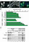
Figure 1.
Figure 1.
Expression of APC/C components in salivary glands. (A) Endocycle progression is not affected in rca12 mutant cells. Section of a third instar salivary gland containing cells mutant for rca12 that were generated in the embryonic salivary placode by the MARCM technique and were positively marked by GFP. DNA staining reveals no difference in nuclear size between rca12 mutant and adjacent control cells, indicating that Rca1 is not essential for endocycle progression in salivary glands. Bar, 50 μm. (B) qRT–PCR analysis of transcript levels in salivary glands. The relative expression of the indicated genes in feeding third instar salivary glands and embryos (4–8 h AED) was determined by the ΔΔCT method. GAPDH was used as endogenous control to normalize expression between both cDNA preparations. In salivary glands all tested transcripts were expressed at lower levels when compared with embryos. However, expression levels of mitotic genes (G2–M) were dramatically reduced compared with genes involved in G1–S control (e.g., CycE, Cdk2), which are known to be essential for endoreplication. APC/C components, including the activator protein Fzr, were expressed at levels comparable with those of the G1–S genes. (C) Fzr protein is present in larval salivary glands. Extracts from embryos and salivary glands of indicated time after egg deposition were analyzed by Western blotting. The blot was probed with antibodies against Fzr and Tubulin. Ponceau S staining is shown as a reference of total protein levels. Several dilutions were tested to compare signal intensities. The number of embryos or salivary glands per lane is indicated. Lanes 2, 7, and 10 show roughly similar signal intensities of tubulin and Ponceau S. In these lanes, similar levels of Fzr were detectable, indicating that embryos and larval salivary glands contain similar concentrations of Fzr protein.
Figure 2.
Figure 2.
Expression of Rca1 impairs endoreplication in larval salivary glands. Spatially and temporally expression of HA-Rca1 was achieved by ptc-Gal4 and the TARGET system that is based on a temperature-sensitive form of the Gal4 inhibitor Gal80 (Gal80ts). Ptc-Gal4 activates expression specifically in the salivary gland (sg) but not in the neighboring fat body (fb). (A) At restrictive temperature for Gal80ts (18°C), salivary development occurred normally and neither HA-Rca1 nor the coexpressed GFP was detectable. (B) At 29°C, Gal80ts is inactive, allowing Gal4 to activate UAS-HA-Rca1 (and UAS-GFP) resulting in very small salivary glands compared with controls. (C) When expression was triggered 10–18 h AED by a shift from permissive to restrictive temperature, salivary glands with reduced size were still observed. (D) Even when the temperature shift was performed at 40–48 h AED salivary gland development was severely impaired. When expression was induced at the two latter time points, cells had already passed the transition from mitotic to endoreplication cycles, thus demonstrating that HA-Rca1 overexpression perturbs endocycle progression and not initiation. Bar, 100 μm. (E) Overexpression of HA-Rca1 in individual salivary gland cells using the flp-out technique. Expression was induced at 40–48 h AED. HA-Rca1-expressing cells were marked by coexpression of GFP and HA staining. Overexpression of HA-Rca1 results in smaller cells with reduced DNA content compared with the neighboring control cells. Bar, 25 μm.
Figure 3.
Figure 3.
APC/C depletion blocks DNA replication in salivary glands. To visualize DNA replication larvae were fed for 24 h with BrdU-containing medium. Expression of the indicated constructs was induced 10–18 h AED. BrdU was added to the medium 60 h post-induction. For conditional expression ptc-Gal4 was applied in combination with tub-Gal80ts. (A) In wild type, BrdU was detectable throughout the salivary gland, suggesting that all nuclei underwent at least one S phase during the labeling period. (B) Expression of HA-Rca1 results in small salivary glands, and no BrdU can be detected within salivary gland nuclei but in adjacent fat body cells, demonstrating that DNA replication has been halted. (C) Overexpression of Cyclin E results in the same phenotype as Rca1 overexpression, suggesting that both genes affect similar downstream pathways. (D) Cdc16 depletion in salivary glands interferes with endocycle progression. BrdU incorporation is highly reduced in the majority of Cdc16-depleted cells. Most cells show a punctuate BrdU pattern that is reminiscent of late DNA replication (arrowheads). However, individual cells were found that appear to undergo normal S phases (arrows). The weaker phenotype compared with the Rca1 overexpression is likely intrinsic to the RNAi technique. Nonetheless, the similarity of the phenotypes suggests that the Rca1 overexpression phenotype is due to inhibition of APC/C activity. Bar, 100 μm.
Figure 4.
Figure 4.
Compromised APC/C–Fzr activity increases CycE–Cdk2 activity. The MPM-2 antibody recognizes a CycE–Cdk2 phosphorylated protein that assembles into the histone locus body. Conditional overexpression of the indicated constructs was achieved by using ptc-Gal4 together with the TARGET system. Expression was induced 10–18 h AED. (A) In control salivary glands derived from wandering larvae that have terminated endoreplication, CycE activity has ceased and nuclei were devoid of MPM-2 staining. (B) Upon continuous expression of CycE MPM-2-positive subnuclear spheres (filled arrowheads) can be detected in virtually all nuclei of the salivary gland. In addition, brightly stained speckles (open arrowheads), which likely represent heterochromatin, were visible on the DNA. (C,D) MPM-2-positive spheres as well as DNA regions with intense Hoechst staining appear after overexpression of HA-Rca1 or knockdown of Cdc16. This indicates that compromised APC/C–Fzr activity results in increased CycE–Cdk2 activity. It is also important to note that the differences in nuclear size were clearly visible in the high-magnification images. Bar, 10 μm.
Figure 5.
Figure 5.
Rca1 overexpression results in E2F1 accumulation and subsequent activation of its target genes. The indicated UAS constructs were overexpressed in individual salivary gland cells using the flp-out method. Expression was induced 40–48 h AED. (A) Overexpression of HA-Rca1 results in higher levels of CycE protein. (B) A reporter construct comprised of the CycE regulatory region fused to the lacZ gene (CycE16,4kb-lacZ) shows that CycE transcription is enhanced after HA-Rca1 overexpression. (C) The CycE-lacZ reporter construct is also up-regulated after CycE overexpression, indicating the presence an autoregulatory feedback loop. (D) qRT–PCR reveals that E2F1 target genes were up-regulated in salivary glands overexpressing either CycE or HA-Rca1. Data obtained from wild-type salivary glands was set as a reference point and the relative quantity is shown as a log10. Transcript levels of CycE, Rnr2, and PCNA were up-regulated after overexpression of HA-Rca1 or CycE, while E2F1 transcript levels were not increased. (E,F) E2F1 protein levels were up-regulated in cells overexpressing either HA-Rca1 or CycE, indicating that CycE accumulation is due to increased E2F1-dependent transcription. (G) Overexpression of Geminin disturbs endocycle progression. The GFP-marked cells in which Geminin was overexpressed displayed a markedly reduced DNA content compared with neighboring control nuclei. (H,I) Overexpression of Geminin results in elevated levels of E2F1 and CycE protein, demonstrating that E2F1 and its target genes generally accumulate in cells with impaired DNA replication. Bar, 50 μm.
Figure 6.
Figure 6.
APC/C–Fzr activity is crucial for Geminin oscillation during endoreplication. (A,B) Geminin is up-regulated after overexpression of HA-Rca1 or CycE. Individual cells overexpressing HA-Rca1 or CycE were generated by the flp-out technique 40–48 h AED. (C–F) Geminin protein levels fluctuate during salivary gland endocycles. Wild-type salivary glands derived from early third instar larvae (72–74 h AED) were stained with antibodies against Geminin and either CycE (C), E2F1 (D), or PCNA-GFP (E). As a reference, PCNA-GFP-expressing salivary glands of the same age were labeled for 1 h with BrdU (F). Costaining with these cell cycle markers allowed us to categorize individual cells into G phase, late G phase/early S phase (open arrowheads), mid-S phase (closed arrowheads), or late S phase (brackets). Cells in G phase display high levels of E2F1 protein. Late G-phase/early S-phase cells are positive for PCNA-GFP, contain moderate levels of CycE but are negative for BrdU. Mid-S-phase cells show high levels of BrdU, CycE, and PCNA-GFP, but lack E2F1 protein. In late S-phase cells, PCNA-GFP is only present at trace amounts and only residual levels of BrdU were visible. Geminin can be detected in mid to late S-phase cells. (F) RNAi-mediated knockdown of Cdc16 in salivary glands results in stabilization of Geminin protein levels in all cells throughout the salivary gland. The appearance of brightly stained DNA regions (arrows) indicates that Cdc16 depletion was effective. Expression of Cdc16-RNAi was induced 10–18 h AED. Conditional expression was achieved by using ptc-Gal4 together with the TARGET system. Bar, 50 μm.
Figure 7.
Figure 7.
Oscillating APC/C–Fzr activity is essential for rereplication control in endoreplicating cells. The endocycle is driven by oscillating activities of CycE–Cdk2 and the transcription factor E2F1. During G phases, CycE transcription is activated by E2F1. CycE–Cdk2 enhances its own activity by inhibiting the E2F1 repressor Rbf. This peak of CycE-dependent kinase activity eventually triggers S-phase entry and simultaneously inhibits APC/C–Fzr activity allowing accumulation of Geminin protein. High levels of Geminin prevent relicensing by sequestering dup/Cdt1, which is an essential factor for the assembly of the pre-RC. The initiation of DNA replication activates a negative feedback loop that down-regulates E2F1 activity and simultaneously promotes the degradation of dup/Cdt1. The subsequent decrease of CycE–Cdk2 activity releases the APC/C–Fzr complex, which in turn mediates Geminin degradation. This enables dup/Cdt1 to promote pre-RC formation during the next G phase, thereby preparing the next S phase. Thus, this mechanism allows temporal separation of licensing and firing of replication origins, two processes that are mutually exclusive.

Similar articles

Cited by

References

    1. Arias E.E., Walter J.C. Strength in numbers: Preventing rereplication via multiple mechanisms in eukaryotic cells. Genes & Dev. 2007;21:497–518. - PubMed
    1. Asano M., Nevins J.R., Wharton R.P. Ectopic E2F expression induces S phase and apoptosis in Drosophila imaginal discs. Genes & Dev. 1996;10:1422–1432. - PubMed
    1. Baker N.E., Yu S.Y. The EGF receptor defines domains of cell cycle progression and survival to regulate cell number in the developing Drosophila eye. Cell. 2001;104:699–708. - PubMed
    1. Bell S.P., Dutta A. DNA replication in eukaryotic cells. Annu. Rev. Biochem. 2002;71:333–374. - PubMed
    1. Blow J.J., Dutta A. Preventing re-replication of chromosomal DNA. Nat. Rev. Mol. Cell Biol. 2005;6:476–486. - PMC - PubMed

Publication types

MeSH terms